Nearly one out of every four patients with Long Covid (LC) had cardiac involvement findings on positron emission tomography/magnetic resonance imaging (PET/MRI) that were suggestive of myocarditis, according to a new prospective study.
For the study, recently published in the Journal of Nuclear Medicine, researchers examined imaging data for 98 patients (median age of 48.5) who had persistent cardiopulmonary symptoms nine to 12 months after initial COVID-19 infection. The study authors noted that 91 patients had PET/MRI scans, and 70 patients had dual-energy computed tomography (DECT). The study included a nine-patient control group who had a history of severe acute respiratory syndrome (SARS) with COVID-19 infection but did not have cardiopulmonary symptoms.
The study authors noted that 57 percent of patients had abnormal PET/MRI findings with 30 percent demonstrating aortic or pulmonary vascular uptake. In contrast to the control group that had no myocardial, pericardial or periannular uptake on PET/MRI scans, 24 percent of those with PET/MRI abnormalities had cardiac involvement suggestive of myocarditis and 22 percent had uptake suggesting possible pericarditis, according to the study authors. They added that 11 percent of the cohort had periannular uptake on PET/MRI scans.
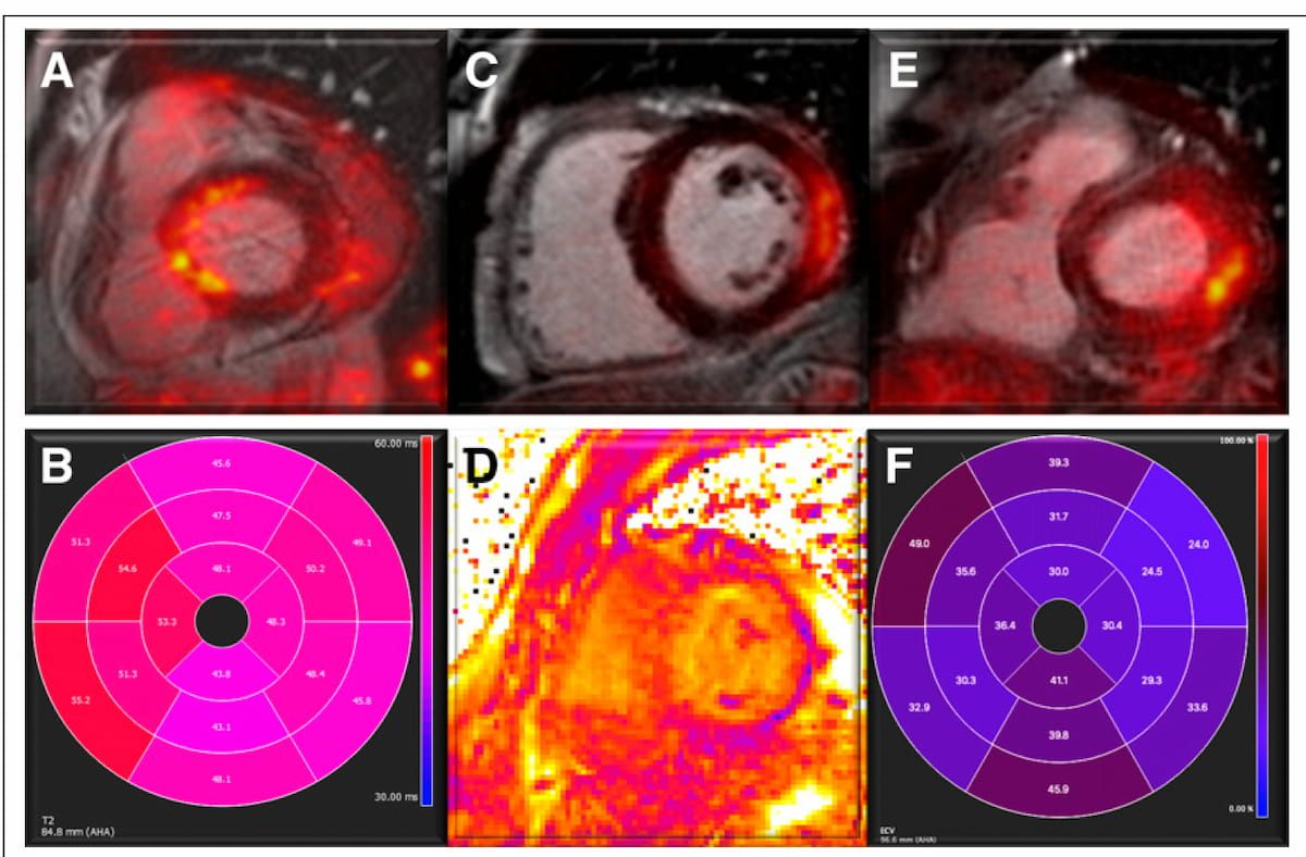
Here one can see findings from hybrid, fused PET/MRI that are suggestive of myocarditis. In a new study of people with persistent cardiopulmonary symptoms nine to 12 months after initial COVID-19 infection, 24 percent of the cohort had PET/MRI findings that revealed cardiac involvement suggestive of myocarditis. (Images courtesy of the Journal of Nuclear Medicine.)
“The imaging findings correlated with different serum biomarker profiles between LC and control subjects. This observation suggests that LC patients with long-term cardiopulmonary symptoms have a distinct plasma protein profile. Moreover, significant differences in plasma protein levels were observed between LC patients with normal and those with abnormal PET/MRI, supporting the hypothesis of specific humoral and immune mechanisms underlying pleiotrophic LC manifestation,” wrote lead study author Maria Giovanna Trivieri, M.D., an associate professor of cardiology, heart failure and cardiac transplantation with the Mount Sinai Health System in New York, and colleagues.
The researchers pointed out that 90 percent of the cohort had abnormalities on DECT. Pulmonary infiltrates were evident on DECT for 67 percent of patient and 59 percent demonstrated abnormal perfusion on DECT, according to the study authors. They noted a higher prevalence of these findings among patients with myocardial and pericardial involvement.
“Abnormal DECT was found in 100% of participants with myocardial involvement and periannular uptake and in 79% and 73% of subjects with pericardial involvement and vascular uptake. Subjects with myocardial involvement had the highest rates of infiltrates (71%) and abnormal perfusion (65%) followed by the group with periannular uptake, where 60% had pulmonary infiltrates and 60% had abnormal perfusion,” added Trivieri and colleagues.
Three Key Takeaways
- Cardiac Involvement common with Long Covid. Approximately 24 percent of patients with Long Covid and persistent cardiopulmonary symptoms showed PET/MRI findings suggestive of myocarditis, and 22 percent showed possible pericarditis, highlighting a significant rate of cardiac involvement.
- High prevalence of DECT abnormalities. Ninety percent of patients demonstrated abnormalities on dual-energy CT (DECT), particularly pulmonary infiltrates and perfusion defects. These were most common in those with myocardial or pericardial involvement.
- Potential link between imaging findings and heart failure risk. Patients with abnormal PET/MRI findings had a trend toward increased heart failure risk (odds ratio 3.4), suggesting these imaging markers may serve as potential risk indicators for heart failure in Long Covid, though the study was underpowered to confirm statistical significance.
Clinical follow-up revealed that nine percent of the cohort had heart failure (HF) with myocardial and pericardial involvement frequently noted in abnormal PET/MRI findings that occurred in 78 percent of these patients.
“These results suggest a trend for future HF events based on abnormal PET/MRI with an odds ratio of 3.4, although statistical significance was not reached in this cohort … These findings additionally indicate that the presence of myocardial and pericardial involvement on PET/MRI could serve as a potential risk marker for HF in the LC population,” posited Trivieri and colleagues.
(Editor’s note: For related content, see “New Meta-Analysis Details Most Common Brain MRI Findings with COVID-19,” “New CT Angiography Study Shows Impact of COVID-19 on Coronary Inflammation and Plaque” and “MRI Long Covid Study Reveals Link Between Lower Pulmonary Gas Exchange and Cognitive Dysfunction.”)
In regard to study limitations, the authors acknowledged the relatively small cohort and a lack of statistical power to assess possible connections between cardiopulmonary findings and post-COVID symptoms. They also conceded a possible patient selection bias with the majority of the cohort being self-referred to a pulmonologist or cardiologist.