Sample Page
SC makes key administrative reshuffle to boost institutional efficiency
Written by
admin
in
1. Pakistan
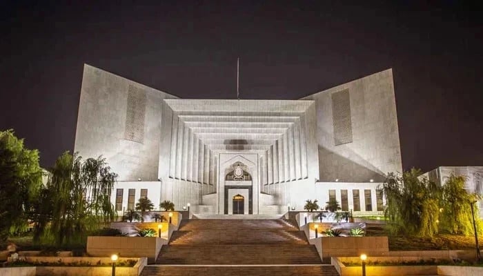
A view of Supreme Court building in Islamabad. — SC Website/File
ISLAMABAD: As part of…
Continue Reading
←
Researchers issue warning after discovering overlooked factor that could increase risk for ALS: ‘Potentially related’
Differentiated thyroid cancer outcomes comparable in immunocompromised patients
→
More posts
We issued a Marine Beach Warning Advisory at Thea’s Park, Jack Hyde Park, and Jerisich Dock
December 23, 2025
Nine mental-health tips for a happier 2026
December 23, 2025
Rogers Selected 2025 Walter Camp D2 All-American
December 23, 2025
Summary of Governing Council deliberations: Fixed announcement date of December 10, 2025
December 23, 2025