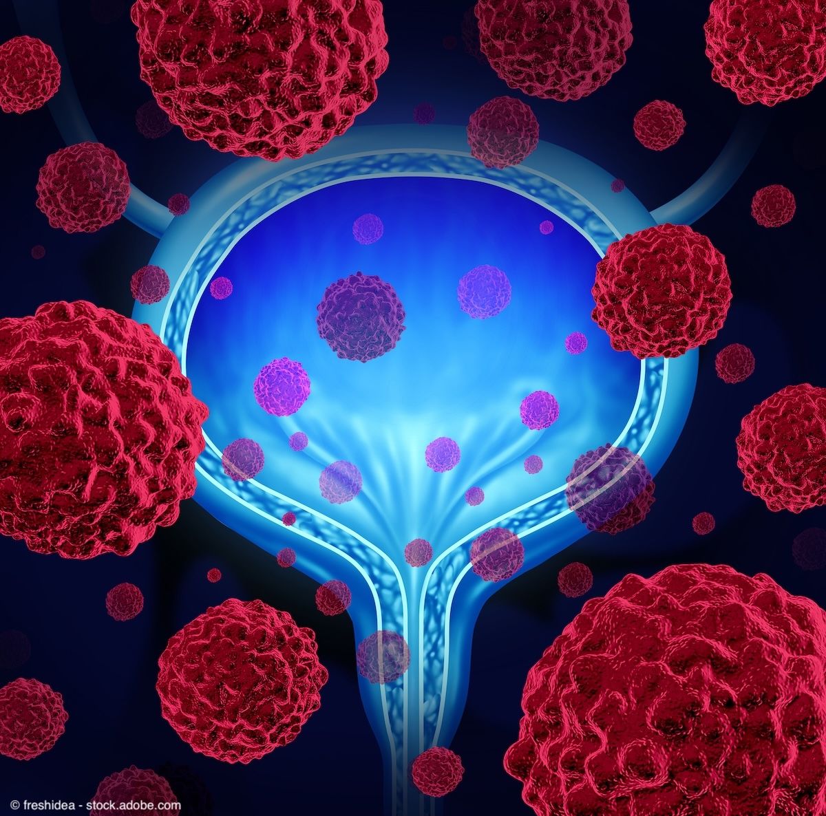
Detalimogene voraplasmid (formerly EG-70) in an investigational non-viral gene therapy for patients with high-risk, non-muscle invasive bladder cancer (NMIBC). The agent is currently being assessed in the phase 2 LEGEND trial (NCT04752722), which includes a pivotal cohort in patients with BCG-unresponsive NMIBC with carcinoma in situ (CIS), with or without concomitant papillary disease.
As new agents such as nadofaragene firadenovec (Adstiladrin), nogapendekin alfa inbakicept-pmln (Anktiva), and the gemcitabine intravesical system (Inlexzo; formerly TAR-200) have joined the treatment landscape, options for patients with BCG-unresponsive disease have expanded considerably. There are even more agents in development, including detalimogene, which are poised to further reshape management in this space. Yet questions remain around how to best select and sequence these therapies as well as the long-term implications of bladder-sparing approaches, according to Yair Lotan, MD.
In a recent interview with Urology Times®, Lotan discussed how new agents such as detalimogene may fit into this evolving treatment landscape and highlighted the ongoing need for longer-term data and biomarkers to guide treatment selection. He also emphasized the importance of cost considerations, in addition to safety and efficacy, as these new agents come to market.
Lotan is the chief of urologic oncology and a professor of urology at UT Southwestern Medical Center in Dallas, Texas.
Urology Times: Detalimogene voraplasmid is currently being explored in the LEGEND trial. How is this trial designed? What have interim data from the study shown so far?
Lotan: This trial includes various cohorts. There is the pivotal cohort, which [includes] patients with BCG-unresponsive non–muscle invasive bladder cancer with CIS with or without papillary disease. They have some additional cohorts in patients who are BCG-naïve or BCG-exposed.
The pivotal cohort is similar to many of the other drugs that have used the single-arm pathway that the FDA has provided for approval of these agents in patients with CIS. The benefit from a development standpoint is that, since there is no gold standard for patients with BCG-unresponsive disease, drugs can be evaluated for their complete response and durability of response without being randomized to several different agents. Placebo doesn’t make sense in this setting [because] these patients have aggressive disease. So, the cohort is just including patients with unresponsive CIS, and [patients are] monitored every 3 months to look for response.
The cohort so far has included 21 patients. [This is] a fairly typical population, with a median age of 74 [years] and mostly men. [Overall,] 71% of the patients had CIS alone, and about a third had Ta or T1 with CIS. The CR rate at 3 months is 67%, and any time CR rate is 71%,1 so quite favorable. There were very few treatment-related adverse events, and no grade 3 toxicity so far. The cohort has already accrued 100 patients, so hopefully sometime next year we’ll have more mature data for the entire cohort of patients.
Urology Times: This study includes the pivotal cohort, as you mentioned, in high-risk BCG-unresponsive NMIBC, a disease space that’s seen rapid growth over the past couple of years. How do you see this agent fitting into the current treatment landscape, if approved?
Lotan: There are several aspects that people are looking for. There’s obviously efficacy. If it maintains somewhere around 70% CR any time, it would be certainly comparable to many of the other approved treatments. There is some benefit in the sense that it’s easy to administer; it’s only 4 treatments that are intravesical. The drug is well-tolerated. There are no grade 3 toxicities. From that standpoint, it has a very favorable profile, and it’s relatively convenient. It’s also easy to store, so it’s not something that would be challenging for urologists to obtain and administer.
Other aspects that we have to wait and see are going to be how favorable it is in terms of cost and some of the other logistical aspects. Overall, if it has good efficacy and very good tolerability, I think it should definitely be considered among the available treatments for patients with BCG-unresponsive CIS.
Urology Times: What role do you foresee for gene therapy in the treatment landscape moving forward?
Lotan: There are various ways of looking at gene therapy. If you look at a drug like Adstiladrin, that is a virus that’s been modified to stimulate interferon production, and so it’s a form of gene therapy. In general, having a plasmid or a viral delivery system makes sense. Looking at other drugs, Anktiva is an IL-15 stimulant, and detalimogene has IL-12 stimulation, plus RIG-1. Even a viral drug like cretostimogene stimulates the immune system and has an oncolytic component. So, a lot of these drugs are focused on simulating the immune system, but I think the real question is going to be, what pathways does it make sense to stimulate? Is there a better approach or is there another approach to stimulate the immune system?
Companies have become much more sophisticated in terms of how to stimulate the immune system. The question is what parts of the immune system would lead to the most efficacious results, especially considering the fact that BCG is also a very good immune treatment. We’re taking patients where BCG was insufficient to get the immune system to control the disease. So, what other pathways would make sense to stimulate in a patient where BCG, which is a non-specific immune stimulant, didn’t work?
Urology Times: How else do you see the therapeutic landscape for BCG-unresponsive NMIBC evolving over the next several years?
Lotan: We’ve had so many changes since pembrolizumab was approved about 3 to 4 years ago. Then we had Adstiladrin, then Anktiva, and very recently, TAR-200. [There are others coming,] such as cretostimogene and detalimogene. There are also several other drugs in development such as NDV-01, which is sustained release of gem/doce, and TARA-200. There are a lot of potential new agents.
One of the things that we need to consider is that we don’t really have biomarkers to personalize the treatments. How do we select among these drugs? How do we sequence these drugs? When is it too much? When we should do a cystectomy, when the risk of progression might increase? These are all challenges that we face, because patients will have a number of choices. We don’t have very good data, other than the efficacy and safety data right now, to personalize the treatment. I’m hopeful that we can develop some biomarkers, or maybe AI tools, that will help us select patients.
We also need better data on what the natural history might be in terms of how safe it is to continue to do bladder-sparing approaches. Ultimately, removing the bladder is the best way to prevent a cancer from recurring in the bladder. We do know that some patients do progress on these drugs, and that those patients will potentially have worse outcomes.
Urology Times: Is there anything else that you wanted to add?
Lotan: I think one of the big issues for patients is accessibility. There is going to be a cost component that is going to affect patients’ ability to get [these drugs.] Many of these drugs do have early access programs, which is great, but the costs are pretty significant. If patients have co-pays, they may not be able to afford these treatments.
We’ve made tremendous advances in being able to offer new drugs. Ten years ago, removal of bladder was the main option for patients who didn’t respond to BCG. The only other drug was valrubicin, which was not effective in most patients and had very poor durability. Now that we have a lot of drugs, being able to give them to the patients is going to be a high priority. Figuring out how to make it affordable for patients is going to be important.
REFERENCE
1. Taylor III JA, Joshi S, Satkunasivam R, et al. Preliminary results from LEGEND: A phase 2 study of detalimogene voraplasmid (EG-70), a novel, non-viral intravesical gene therapy for patients with BCG-unresponsive non-muscle invasive bladder cancer (NMIBC) with carcinoma in situ (CIS). J Clin Oncol. 2025;43(suppl 5). doi.org/10.1200/JCO.2025.43.5_suppl.802.