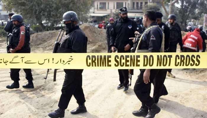
A thorough knowledge of brain function and malfunction requires imaging technologies that balance high spatiotemporal resolution, vast fields of view, and good sensitivity. Despite the rapid development of imaging technologies in neuroscience, no single method can fully meet these requirements, yet functional ultrasound imaging offers an unprecedented balance among these key parameters.
Image Credit: Iconeus
Ultrafast functional Doppler ultrasound (fUS) is based on the principles of conventional Doppler ultrasonography but operates at an extremely fast frame rate. This significantly improves spatiotemporal resolution and sensitivity while maintaining standard Doppler techniques’ deep penetration and usability.
Like conventional Doppler ultrasound, functional Doppler ultrasonography, captures echoes from moving scatterers within tissues, such as red blood cells moving through the cerebral vasculature.
These echoes are then combined to determine the cerebral blood volume (CBV) at any point in time. Unlike standard ultrasonography, fUS uses planar ultrasound waves to capture echoes from an entire 2D plane at once rather than from discrete regions sequentially.
High-frequency plane-wave ultrasonic pulses are transmitted to the tissue, with the angle increasing incrementally over a timescale of around 1 ms.
Echoes from the entire scanning region are monitored and merged at 500 Hz to create a sequence of compound images. Several echoes are merged following signal filtering to create a single highly resolved frame.
Montage visualization of the mouse brain vasculature with 100 μm in-plane resolution, and showing automatic registration of a transcranial 3D scan to the Allen Mouse Brain Reference Atlas. Image Credit: Iconeus
The principle of functional ultrasound. (1) High-frequency plane-wave ultrasound pulses are incident upon the subject, with the angle varying incrementally on a timeframe of ~1 ms. (2) Echoes from across the whole scanning region are simultaneously monitored and combined to form a sequence of compound images. (3) Following signal filtering, multiple echoes are combined into a single frame. The mean intensity of this signal (the ‘Power Doppler’ value) is proportional to the number of red blood cells in a unit volume, which can hence can be used to infer the cerebral blood volume (CBV). Image Credit: M. Tanter
By leveraging this hemodynamic response, fUS achieves a high signal-to-noise ratio, which significantly improves both sensitivity and spatiotemporal resolution. This makes it possible to visualize brain function in remarkable detail, capturing rapid and localized CBV dynamics at scales and speeds previously unattainable with other imaging modalities.
These technical features enable fUS to fulfill various purposes and applications. It has previously been demonstrated that neuronal activation generated by continuous stimuli or spontaneous activity causes an increase in blood volume in the region of activation, which serves to supply neuronal cells with nutrition and oxygen.
Neurovascular coupling (NVC) enables functional ultrasound (fUS) to quantify brain activity indirectly by detecting localized changes in cerebral blood volume (CBV) associated with neuronal activation.
Unlike fMRI, fUS provides a direct, high-speed view of how blood flow dynamically responds to neural activity, making it ideal for studying stimulus-based tasks, drug-induced hemodynamic responses, and spontaneous variations in resting conditions.
The high-resolution visualization of the vessels provided by fUS makes this technology suitable for the study of cerebral vasculature, as well as the real-time detection of vascular changes in disease models such as stroke and dementia.
The adaptability of fUS extends beyond the number of different applications to the variety of experimental settings and animal models. The technique’s minimum invasiveness ensures its usability in both anesthetized and awake animals, even those that are freely moving.
Iconeus One, a functional ultrasonic imaging platform, combines all these features and characteristics. An all-in-one solution that, unlike other technologies (e.g., fMRI, optical imaging), strikes a balance between sensitivity, high spatiotemporal resolution, and wide fields of view.
Iconeus One offers:
- 100 µm resolution for functional imaging and 5 µm for vascular analysis with contrast agent
- Temporal resolution between 100 and 400 ms to record fast hemodynamic responses
- Dedicated probes allow for fast imaging of the entire mouse brain and huge volumes in other animal models, with penetration depths up to 4 cm
- The platform is suitable for anesthetized, awake, head-fixed, or freely moving animals
- An integrated and user-friendly software suite for real-time analysis, with integrated mouse and rat atlases for precise and reproducible processes
Image Credit: Iconeus
Iconeus One’s unique combination of sensitivity, speed, and resolution has allowed it to lead the field in preclinical studies on awake animals, and it is currently being considered for use in clinical research as well.
While proof of concept for fUS in humans is currently available, Iconeus is working hard to make it a common bedside imaging technology in pediatric medicine and a reality in intraoperative hospital settings.
Iconeus aims for excellence and works hard to ensure that it is a part of the future of neuroimaging.
About Iconeus
Iconeus is a Paris-based company, founded by the inventors of functional ultrasound, who have invented an easy-to-use functional ultrasound system for imaging cerebral blood flow and microvasculature. Its unique combination of sensitivity, speed and high resolution has enabled it to lead the field in preclinical studies on awake animals, and it is now being proposed for applications in clinical research too.
Iconeus is introducing functional ultrasound neuro imaging: a breakthrough imaging modality for brain activity monitoring based on blood flow imaging with ultra-high sensitivity.
For preclinical research, the portability and high versatility of their technology enables the study of brain activity at unprecedented scales and in a large variety of subject states: awake, behaving, freely-moving, resting-state and asleep conditions.
Iconeus’ technology can be easily combined with other complementary modalities such as EEG headstage or optogenetics which are inherently difficult to combine with FMRI. Iconeus also offers key applications such as functional connectivity assessment between brain structures (connectomics) or stroke 4D monitoring. Building on years of preclinical expertise and research, they’re now investing and supporting exciting new clinical applications.
Sponsored Content Policy: News-Medical.net publishes articles and related content that may be derived from sources where we have existing commercial relationships, provided such content adds value to the core editorial ethos of News-Medical.Net which is to educate and inform site visitors interested in medical research, science, medical devices and treatments.