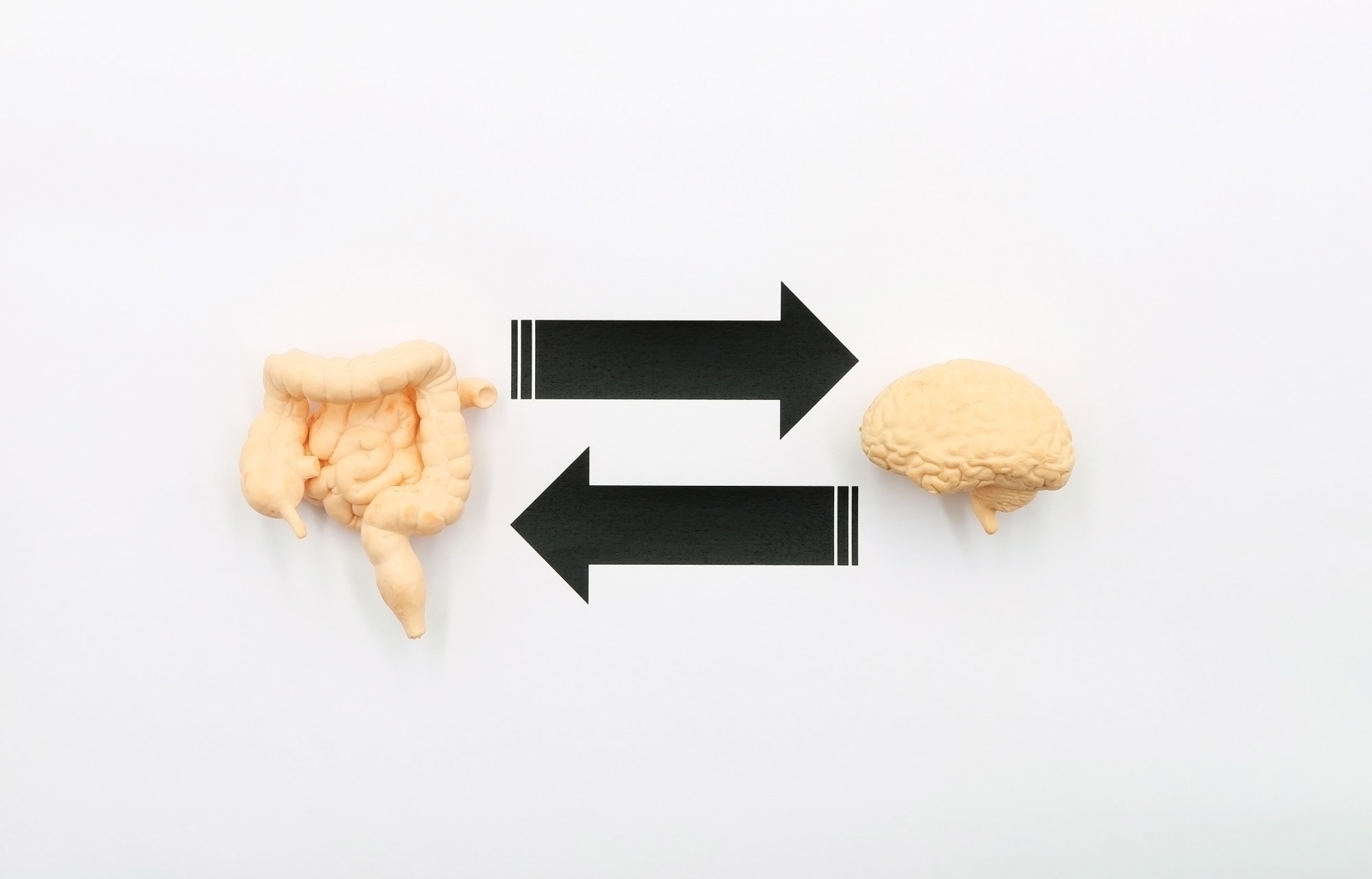
In a first-of-its-kind lab study, scientists prove that probiotic bacteria stimulate immediate and measurable changes in brain cell function, hinting at a new direct communication between your gut and your mind
Study: An in vitro neurobacterial interface reveals direct modulation of neuronal function by gut bacteria. Image credit: TopMicrobialStock/Shutterstock.com
Researchers developed a neuron-bacteria interface that reveals that the gut bacterial community can directly interact with brain neurons and alter their activities in vitro. This study, published in Scientific Reports, provides new insights into possible mechanisms within the gut-brain axis.
Background
The gut-brain axis, a bidirectional communication network between the gut microbiota and central nervous system, has gained significant attention in the scientific community because of its substantial involvement in normal physiological functions and disease pathogenesis.
Alterations in gut microbiota composition and functionality, referred to as dysbiosis, have been linked to several neurological diseases, including Alzheimer’s disease, autism spectrum disorders, and depression.
It is already well-established in the literature that the gut microbial communities and their metabolites directly influence the gut-brain axis. However, information on direct communications and bidirectional information exchange between bacteria and brain neurons is not widely available.
In the current study, researchers developed a neuro-bacterial interface model using the foodborne probiotic bacterium Lactiplantibacillus plantarum and rat cortical neural cultures to explore how nerve cells respond to the bacterial presence at the morphological, functional, and transcriptional levels.
It is important to note that the study was conducted entirely in vitro using rat embryonic cortical neurons, which do not directly encounter gut bacteria in vivo. The use of these neurons was intended to test whether central nervous system neurons can respond to bacterial contact, rather than to model a real-life gut-brain scenario.
Key findings
The assessment of physical interaction between the bacteria and nerve cells revealed that the bacteria adhere to the surface of nerve cells without entering the neuronal cytoplasm.
Regarding functional response of nerve cells following bacterial exposure, the study found a significant increase in calcium signaling when nerve cells encounter the bacteria. This increased signaling was dependent on bacterial concentrations and active metabolism.
Nerve cells exposed to a high concentration of metabolically active bacteria exhibited the highest calcium signaling. Similarly, nerve cells exposed to heat-killed bacteria (normal cellular integrity but no metabolic activity) showed the second highest calcium signaling at the same concentration. However, nerve cells exposed to a very low concentration of active bacteria exhibited similar intensity signaling as unexposed cells. These observations indicate that bacterial load and membrane-to-membrane contact are essential for inducing a neuronal response, and neuronal activation is significantly greater when nerve cells interact with live bacteria rather than metabolically inactive bacteria.
Regarding bacteria-mediated altering of neural activity, the study found significant changes in the expression of key proteins involved in neuroplasticity, including phosphorylated cyclic-AMP-responsive element-binding protein (pCREB; a marker of early neural activity), and Synapsin I (SYN I; a cytoplasmic marker related to synaptic connections).
Specifically, the study found a significant reduction in pCREB expression and a significant increase in SYN I expression in nerve cells exposed to bacteria. These findings indicate bacteria-mediated functional changes in nerve cells. The study also verified that these functional changes are not due to cytotoxic effects, as no reduction of neuronal viability or induction of nerve cell death was observed following bacterial exposure.
Transcriptional changes associated with the neuro-bacterial interactions revealed significant restructuring of the transcriptional landscape of nerve cells in response to bacteria, affecting vital biological processes related to neuroplasticity, gene expression regulation, signaling pathways, and stress response. A detailed transcriptional analysis identified a prominent role for several bioelectricity-related genes in underlying neuro-bacterial interactions.
Study significance
The study provides valuable experimental evidence on direct interactions between brain and bacterial cells, leading to multiple downstream changes at the structural, functional, and transcriptional levels.
The study reveals that the live bacteria, when they come in direct contact with nerve cells, significantly affect nuclear factors relevant to gene expression and increase synaptic connections between nerve cells. A set of potential bioelectricity-related genes involved in neural responses has also been identified, including the brain-derived neurotrophic factor gene Bdnf, the adrenomedullin gene, Adm, and the potassium and chloride ion channel genes Kcna1 and Clcn1, respectively.
The bioelectrical profile is increasingly considered a functional property of bacterial cells, as it correlates with relevant physiological events regardless of causality. Overall, the study provides a promising platform to decode the direct effects of bacteria on brain cells and generate new knowledge on the biological–biophysical interaction between highly divergent cells.
The study model is based on a two-dimensional culture of rat-derived cortical neurons, which lacks the structural and cellular complexity of a physiological neural environment. The cortical neurons were selected as an experimental cell type to explore whether brain neurons can respond directly to bacterial presence instead of enteric or sensory neurons. However, these cortical neurons may not reflect a biologically plausible scenario, as there is currently no evidence for direct interactions between gut microbiota and cortical neurons under normal conditions.
The study model includes a single bacterial strain and a single timepoint (30 minutes) for neuro-bacterial interaction. These factors may restrict the generalizability of study findings. Despite these limitations, the study provides a fundamental framework for developing novel neuroactive bacterial therapeutics or bioelectronic interventions targeting brain–microbiota interactions. Future studies will use more physiologically relevant systems, including co-cultures with enteric neurons and organ-on-chip platforms.
Download your PDF copy now!
