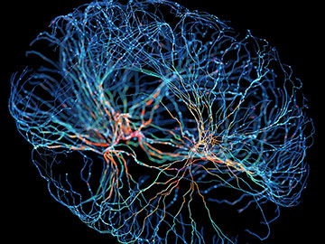
Using the optical technique TEMPO, researchers have discovered two new types of brain waves. [Image: Andriy Onufriyenko/ Getty Images]
Scientists first detected electrical “brain waves” a century ago, but seeing large-scale patterns of these neural oscillations has remained a challenge. Now, researchers have developed an optical technique for capturing these waves up to frequencies of roughly 100 Hz.
The technique, dubbed TEMPO (for “trans-membrane electrical measurements performed optically”), incorporates an optical-fiber sensor that can detect electrical activity in the brains of mice while they are awake and moving, rather than sedated (Cell, doi:10.1016/j.cell.2025.06.028). TEMPO also uses an optical “mesoscope,” an imaging device with a wide field of view and high spatial resolution. “This technology allows us to look at multiple brain areas at once and see the brain waves sweeping across the cortex with cell-type specificity,” said researcher Mark J. Schnitzer.
By examining mice with TEMPO, the cross-disciplinary team at Stanford University, USA, discovered two types of brain waves not previously observed. Researchers hope the devices will help humans learn more about neural interactions in both healthy brains and those with cognitive impairments. “There are a lot of very important applications in the field of neuroscience for understanding pathology and different dynamics in the brain,” said lead author Simon Haziza.
Ultra-sensitive fiber sensors
The sensors in TEMPO, named uSMAART (ultra-sensitive measurement of aggregate activity in restricted cell types), contain four separate modules. In the first, beams from two lasers, one with a wavelength of 561 nm and the other with a wavelength of 488 nm, pass through a series of mirrors and beam splitters before combining to produce low-noise illumination that is fed into optical fiber. A second module provides active decoherence of the laser light; this avoids mode-hopping noise in the fiber path. Other modules provide phase-sensitive demodulation and low-noise sensing via avalanche photodiodes.
The Stanford researchers say their illumination is roughly 10 times more stable than existing voltage-sensing photometry systems.
The Stanford researchers say their illumination is roughly 10 times more stable than existing voltage-sensing photometry systems. This type of stability is key to picking up the signals from genetically encoded voltage indicators (GEVIs), or proteins that fluoresce in response to voltage differences across cell membranes.
The mesoscope
The TEMPO mesoscope gets its illumination from two high-power LEDs chosen to excite the two GEVIs used to highlight certain frequencies of brain waves. Its high-numerical-aperture lens provides a field of view of up to 8 mm. The research team equipped two detection pathways with one CMOS camera each to track the fluorescent emissions. The cameras operated at 130 fps in wide-field mode and 300 fps over narrower fields of view.
The scientists say the development of new GEVI substances could improve the sensitivity of the TEMPO technique, along with multi-fiber probes and graphene-based electrodes. More research needs to be done, but the team believes that the new technology could open up many avenues for neuroscience as well as development of bio-inspired AI models.
