AI assistance can improve the ability of clinicians to identify misplaced endotracheal tubes on chest x-rays, according to a study published July 28 in Critical Care.
The finding is from a reader study involving 18 clinical readers of varying seniority and 14,400 image interpretations, with AI especially useful for identifying critically misplaced tubes, noted lead author Alex Novak, MD, of Oxford University in Oxford, England, and colleagues.
“Studies have demonstrated the potential for AI-led algorithms to detect [endotracheal tube] placement on chest x-ray images; however, their effect on clinician accuracy remains unexplored,” the group wrote.
More than 60% of critically ill patients require endotracheal intubation, with studies reporting tube misplacement in up to 17% of cases, the authors explained. These misplacements can lead to serious clinical harm, and endotracheal tube (ETT) position is typically confirmed via chest x-ray.
Yet interpretation of the ETT position on x-rays can be challenging, depending on such factors as the experience of the clinician, the quality of the image, characteristics of the patient, or type of ETT, the authors added. Hence, in this study, the group evaluated the impact of AI-assisted image interpretation on the ability of critical and acute care clinicians to accurately identify ETT misplacement.
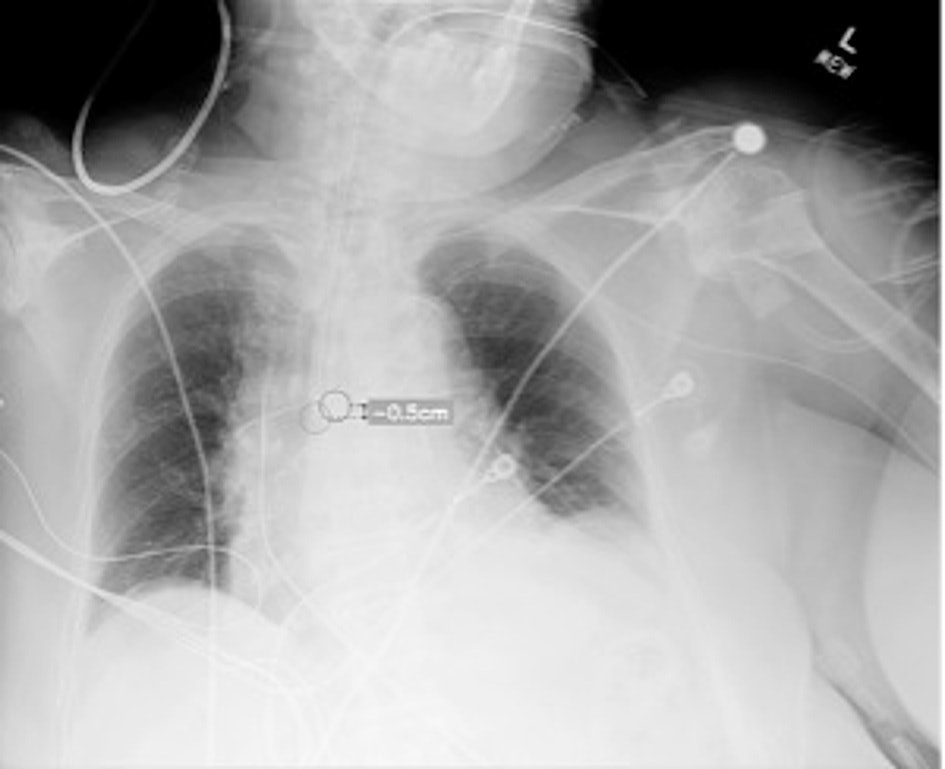
The researchers gathered 400 chest x-rays of intubated adult patients from three hospitals from a range of clinical settings, including the intensive care units and emergency departments. A panel of thoracic radiologists classified each image by tube placement as correct, too low (distal), or too high (proximal), with correct ETT position defined as the tip located 3 to 7 cm above the carina, in line with established guidelines. Out of the 400 images, 209 were classified as well-positioned, 56 as too high, and 134 as too low.
Next, they recruited 18 clinical readers of varying seniority from six clinical specialties (adult intensive care, anesthetics, emergency medicine, anesthetic advanced care practitioners, radiology, and radiography). The participants completed reads of the images via an online platform over a three-week period alone and then again with the assistance of AI after a four-week washout period. The AI tool is an algorithm included in GE HealthCare’s Critical Care Suite.
According to the results, accurate classification of tube placement across the whole group increased from 73.6% without AI assistance to 77.4% (p = 0.002) with AI assistance. In subgroup analyses, accuracy for identifying critically misplaced tubes increased significantly from 79.3% to 89% (p = 0.001). In addition, reader confidence improved with AI assistance, and there was no change in mean interpretation time at 36 seconds per image, the group reported.
“The significant improvement seen in accurate detection of critically misplaced tubes is an important finding, as this arguably represents a key application for the algorithm in clinical practice,” the researchers wrote.
Nonetheless, future prospective studies are required to generate definitive evidence in this regard and to determine the magnitude of any potential clinical and health economic impact, the group concluded.
The full study can be found here.