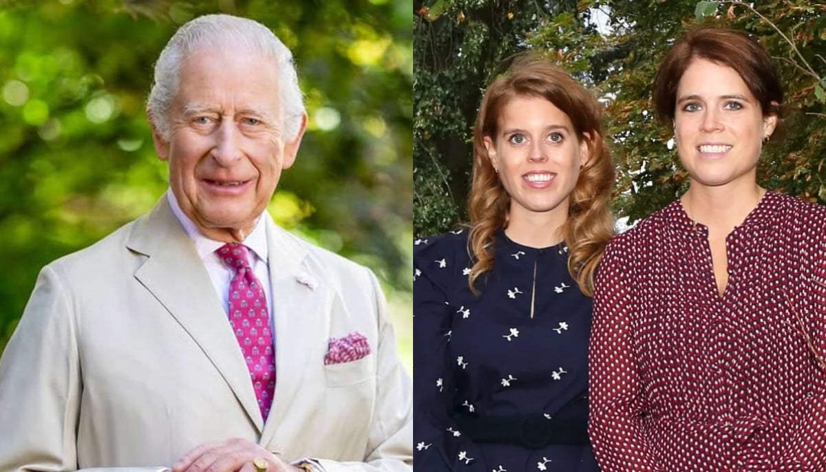
DALLAS – July 03, 2025 – By using a genetic technique developed at UT Southwestern Medical Center that forces cells to rid themselves of mitochondria, researchers are gaining new insights into the function of these critical organelles. Their findings, published in Cell, add to fundamental knowledge about the role of mitochondria in cells and evolution and could eventually lead to new treatments for patients with mitochondrial diseases such as Leigh syndrome and Kearns-Sayre syndrome, which can affect numerous organ systems.
“Our new tool allows us to study how changes in mitochondrial abundance and the mitochondrial genome affect cells and organisms,” said Jun Wu, Ph.D., Associate Professor of Molecular Biology at UT Southwestern. Dr. Wu co-led the study with Daniel Schmitz, Ph.D., a former graduate student in the Wu Lab who is now a postdoctoral fellow at the University of California, Berkeley.
Mitochondria are organelles found in the cells of most eukaryotic organisms, including animals, plants, and fungi, whose cells contain a membrane-bound nucleus and other membrane-bound organelles. They have their own genetic material, passed down exclusively through females of a species. Mitochondria are thought to have originated as prokaryotic cells – which lack membrane-bound organelles – and to have invaded ancestral eukaryotic cells and formed a symbiotic relationship with them.
Researchers have long known that these organelles serve as cells’ powerhouses, generating the energetic molecule adenosine triphosphate that fuels all cellular operations. However, recent studies have shown mitochondria play direct roles in regulating cell death, differentiating stem cells into other cell types, transmitting molecular signals, aging, and developmental timing.
Although mitochondria appear to perform many of these roles through “crosstalk” with the DNA in a cell’s nucleus, how they perform this function – and what happens if this crosstalk ceases – has been unknown.
To help answer these questions, Dr. Wu, Dr. Schmitz, and their colleagues took advantage of a pathway called mitophagy that cells normally use to dispose of old or damaged mitochondria. Using genetic engineering, the researchers forced cells to degrade all their mitochondria – a process known as “enforced mitophagy.”
The researchers used this process on human pluripotent stem cells (hPSCs), a type of cell typically formed early in development that can differentiate into other cell types. Although this alteration caused the cells to stop dividing, the researchers unexpectedly found that the mitochondria-depleted cells could survive in petri dishes up to five days. They had similar results with different types of mouse stem cells and hPSCs harboring a pathogenic mitochondrial DNA mutation, suggesting enforced mitophagy can be a viable tool for depleting mitochondria across species and cell types.
To determine how removing mitochondria affected the hPSCs, the researchers assessed nuclear gene expression. They found that 788 genes became less active and 1,696 became more active. An analysis of the affected genes showed the hPSCs appeared to retain their ability to form other cell types and that they could partially compensate for the lack of mitochondria, with proteins encoded by nuclear genes taking over energy production and certain other functions typically performed by the missing organelles.
Then the researchers, in an attempt to better understand crosstalk between mitochondria and the cell nucleus, fused hPSCs with pluripotent stem cells (PSCs) from humans’ closest primate relatives – including chimpanzee, bonobo, gorilla, and orangutan. This formed “composite” cells with two nuclear genomes and two sets of mitochondria, one from each species. These composite cells selectively removed all non-human primate mitochondria, leaving behind only human mitochondria.
Next, using enforced mitophagy, the scientists created hPSCs devoid of human mitochondria and fused them to non-human primate PSCs, again creating cells carrying nuclear genomes from both species, but this time only non-human mitochondria. An analysis of composite cells containing either human or non-human mitochondria showed that the mitochondria were largely interchangeable despite millions of years of evolutionary separation, causing only subtle differences in gene expression within the composite nucleus.
Interestingly, the genes that differed in activity among cells harboring human and non-human mitochondria were mostly linked to brain development or neurological diseases. This raises the possibility that mitochondria may play a role in the brain differences between humans and our closest primate relatives. However, Dr. Wu said, more research – especially studies comparing neurons made from these composite PSCs – will be needed to better understand these differences.
Finally, the researchers studied how depleting mitochondria might affect development in whole organisms. They used a genetically encoded version of enforced mitophagy to reduce the amount of mitochondria in mouse embryos, then implanted them into surrogate mothers to develop. Embryos missing more than 65% of their mitochondria failed to implant in their surrogate’s uterus. However, those missing about a third of their mitochondria experienced delayed development, catching up to normal mitochondrial numbers and a typical developmental timeline by 12.5 days after fertilization.
Together, the researchers say, these results serve as starting points for new lines of research into the different roles mitochondria play in cellular function, tissues and organ development, aging, and species evolution. They plan to use enforced mitophagy to continue studying these organelles in a variety of capacities.
Other UTSW researchers who contributed to this study are Peter Ly, Ph.D., Assistant Professor of Pathology and Cell Biology; Daiji Okamura, Ph.D., Visiting Assistant Professor of Molecular Biology; Seiya Oura, Ph.D., and Leijie Li, Ph.D., postdoctoral researchers; Yi Ding, Ph.D., Research Associate; Rashmi Dahiya, Ph.D., Senior Research Associate; Emily Ballard, B.S., graduate student researcher; and Masahiro Sakurai, Ph.D., Research Scientist.
Dr. Wu is a Virginia Murchison Linthicum Scholar in Medical Research. Drs. Wu and Ly are members of the Harold C. Simmons Comprehensive Cancer Center.
About UT Southwestern Medical Center
UT Southwestern, one of the nation’s premier academic medical centers, integrates pioneering biomedical research with exceptional clinical care and education. The institution’s faculty members have received six Nobel Prizes and include 25 members of the National Academy of Sciences, 23 members of the National Academy of Medicine, and 14 Howard Hughes Medical Institute Investigators. The full-time faculty of more than 3,200 is responsible for groundbreaking medical advances and is committed to translating science-driven research quickly to new clinical treatments. UT Southwestern physicians provide care in more than 80 specialties to more than 140,000 hospitalized patients, more than 360,000 emergency room cases, and oversee nearly 5.1 million outpatient visits a year.