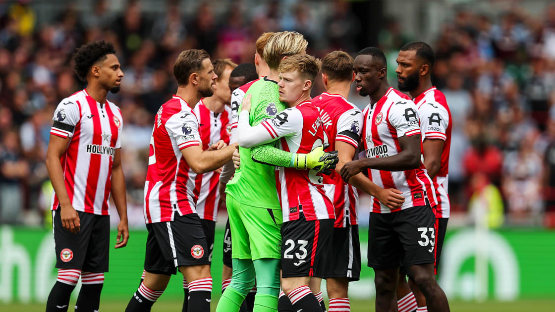
Inferior vena cava septic thrombophlebitis: A severe complication of p

Division of Infectious Diseases and Tropical Medicine, Department of Internal Medicine, Tri-Service General Hospital, National Defense Medical University, Taipei, Taiwan

Correspondence: Yung-Chih Wang, Division of Infectious Diseases and Tropical Medicine, Department of Internal Medicine, Tri-Service General Hospital, National Defense Medical University, No. 325, Section 2, Chenggong Road, Taipei, 11490, Taiwan, Tel +886 2 87927257, Fax +886 2 87927258, Email [email protected]

Abstract: Paraspinal steroid injections, while widely employed for the treatment of back pain, carry risks of serious complications including infection. We describe a 54-year-old immunocompetent woman who developed a disseminated methicillin-resistant Staphylococcus aureus (MRSA) infection following a lumbar paraspinal steroid injection for back pain. She presented with fever, dyspnea, and acute low back pain, progressing to hypoxic respiratory failure requiring intubation. Diagnostic workup revealed MRSA bacteremia complicated by a lumbar spinal epidural abscess, bilateral psoas and quadratus lumborum pyomyositis, inferior vena cava (IVC) septic thrombophlebitis, pulmonary septic emboli, and meningoencephalitis. The patient received intravenous vancomycin and anticoagulation therapy with apixaban. Clinical improvement was observed with resolution of bacteremia and complete radiological resolution of the IVC thrombus after completing an 8-week treatment course. This case underscores the devastating potential of MRSA to transform a localized infection into widespread metastatic disease, driven by its potent virulence factors. It points out the importance of early recognition of systemic complications, including vascular thrombosis and central nervous system involvement, following paraspinal interventions. Moreover, it demonstrates the need for prompt imaging, timely administration of targeted antibiotics, and multidisciplinary management. Given the increasing use of corticosteroid injections and rising concerns about antibiotic-resistant pathogens, this case serves as a reminder of potential iatrogenic complications and the critical importance of infection control and vigilant monitoring. Clinicians should maintain a high index of suspicion for deep-seated infections in patients presenting with systemic symptoms after steroid injection, even in the absence of overt immunosuppression.

Keywords: Staphylococcus aureus, corticosteroid injection, meningoencephalitis, septic thrombophlebitis, spinal epidural abscess

Introduction

Paraspinal muscle steroid injections are a common therapeutic modality for alleviating back pain.1 Despite their effectiveness in pain management, these procedures carry risks, including hematoma and infection.2 Although rare, infection may arise from patient immunosuppression, prolonged steroid use, inadequate aseptic technique during the procedure, or improper medication storage3–5 and escalate into severe or even life-threatening conditions.3 Staphylococcus aureus, the most frequently implicated pathogen in post-injection infections,6,7 can cause a wide range of infections, from localized abscesses to more serious conditions such as bacteremia, infective endocarditis, osteomyelitis, and septic thrombophlebitis.8

This report describes a 54-year-old immunocompetent woman who presented with fever, shortness of breath, acute low back pain, and an inability to walk following a recent lumbar paraspinal muscle steroid injection. Her clinical presentation, characterized by systemic inflammation and neurological symptoms, underscores the critical need for rapid diagnostic evaluation and management in such complex scenarios. This study was approved by the Institutional Review Board of the Tri-Service General Hospital (TSGHIRB No.: C202515066), and written informed consent was provided by the patient to have the case details and any accompanying images published. We aim to highlight the diagnostic approach, discuss potential complications of interventional pain procedures, and emphasize the pivotal role of multidisciplinary care in optimizing patient outcomes.

Case Presentation

A 54-year-old woman with a past medical history of hyperlipidemia and migraine presented to the emergency department with shortness of breath, acute low back pain, and progressive inability to ambulate over the preceding week (Figure 1). She reported lifting heavy objects one week prior and had received a lumbar paraspinal muscle steroid injection three days before admission. Following the injection, she developed headache and neck stiffness. Upon presentation, her vital signs were notable for a body temperature of 37°C, heart rate of 120 beats per minute, respiratory rate of 22 breaths per minute, and blood pressure of 115/69 mmHg. Neurological examination revealed intact cranial nerves and preserved muscle strength. Initial laboratory investigations demonstrated leukocytosis (white blood cell count of 14,600/µL; reference range: 4500–11,000/µL), markedly elevated inflammatory markers (C-reactive protein: 44 mg/dL; reference range: <0.8 mg/dL, procalcitonin: 5.49 ng/mL; reference range: <0.05 ng/mL), impaired renal function (blood urea nitrogen: 36 mg/dL; reference range: 7–25 mg/dL, creatinine: 2.4 mg/dL; reference range: 0.5–0.9mg/dL), and an elevated D-dimer (11.36 µg/mL; reference range: <0.5 µg/mL).

Figure 1 Disease course timeline of the case.

The patient was admitted to the intensive care unit (ICU) and, on hospital day 1, developed hypoxic respiratory failure. Arterial blood gas analysis revealed respiratory acidosis with hypoxemia (pH of 7.204, PaO2 of 86 mmHg) while on 100% oxygen via a non-rebreathing mask. Emergent endotracheal intubation was performed. Contrast-enhanced computed tomography (CT) of the chest and abdomen revealed multifocal, wedge-shaped ground-glass opacities in both lungs, consistent with septic emboli (Figure 2), and a 1.5 cm thrombus within the inferior vena cava (IVC) (Figure 3). Abdominal magnetic resonance imaging (MRI) demonstrated bilateral pyomyositis involving the psoas and quadratus lumborum muscles (Figure 4), as well as a spinal epidural abscess (Figure 5). MRI of the brain revealed gyral swelling and leptomeningeal enhancement suggestive of meningoencephalitis (Figure 6).

Figure 2 Contrast-enhanced computed tomography of the chest demonstrating multifocal, wedge-shaped ground-glass opacities (arrows) in both lungs, consistent with septic emboli.

Figure 3 Contrast-enhanced computed tomography of the abdomen revealing a 1.5 cm thrombus (arrow) within the inferior vena cava.

Figure 4 T2-weighted magnetic resonance imaging of the lumbar spine demonstrating bilateral pyomyositis, with abscess formation involving the psoas (arrow) and quadratus lumborum muscles (arrowhead).

Abbreviations: A, anterior; P, posterior; R, right side; L, left side.

Figure 5 Sagittal T2-weighted magnetic resonance imaging of the lumbar spine demonstrating a spinal epidural abscess (arrow).

Abbreviations: H, head; F, foot.

Figure 6 Magnetic resonance imaging of the brain revealing gyral swelling (arrowhead) and leptomeningeal enhancement (arrow), suggestive of meningoencephalitis.

Blood cultures repeatedly yielded positive results using the BACTEC FX automated blood culture detection system (Becton Dickinson, Sparks, MD, USA). Staphylococcus aureus was identified via Matrix-assisted laser desorption/ionization time-of-flight mass spectrometry (MALDI-TOF MS) using the VITEK MS system (bioMérieux, Marcy l’Étoile, France). Antimicrobial susceptibility testing, performed with the VITEK 2 automated system (bioMérieux, Marcy l’Étoile, France), revealed resistance to oxacillin, confirming the isolate as methicillin-resistant S. aureus (MRSA) (Table 1). Both transthoracic and transesophageal echocardiography revealed no evidence of valvular vegetation. A diagnosis of disseminated MRSA infection was established, encompassing bacteremia, bilateral pyomyositis, spinal epidural abscess, IVC septic thrombophlebitis, pulmonary septic emboli, and meningoencephalitis. Empirical antimicrobial therapy with vancomycin was initiated on the day of admission. Anticoagulation with apixaban was started to manage the IVC thrombus and pulmonary emboli. Blood cultures became sterile by hospital day 13. The patient completed an 8-week course of targeted antimicrobial treatment and recovered fully. A follow-up abdominal CT performed three weeks after treatment completion demonstrated resolution of the IVC thrombus.

Table 1 Antimicrobial Susceptibility of the S. aureus Isolate

Discussion

This case highlights the potentially devastating and widespread complications of MRSA infection following a seemingly routine lumbar paraspinal steroid injection, including bacteremia, bilateral pyomyositis, spinal epidural abscess, septic IVC thrombophlebitis, pulmonary emboli, and meningoencephalitis. The patient was managed with an 8‐week course of targeted antibiotic therapy in conjunction with anticoagulation and ultimately recovered without any neurological sequelae.

Paraspinal steroid injections are widely used for the management of low back pain.9 Although generally safe, this intervention carries a small but significant risk of complications, including epidural hematoma, procedural pain, neurovascular injury, and infection.10 Post-injection infection occurs in approximately 0.12% of cases,1 most commonly manifesting as paraspinal muscle or epidural abscesses, which may extend into the spinal canal and lead to pyogenic spondylitis.2 Potential sources of infection include contaminated steroid preparations, breaches in aseptic technique, inadequate hand hygiene, or patient‐related factors such as immunosuppression.2,4,5

S. aureus is the most frequently isolated pathogen in 50–75% of infections following paraspinal procedures.2,4,6,7 Its array of virulence factors facilitates robust colonization of host tissues, often culminating in localized abscess formation. Additionally, S. aureus produces enzymes such as staphylokinase that degrade tissue barriers and promote local dissemination.11 If the organism breaches the bloodstream, hematogenous spread may ensue, giving rise to severe systemic complications—including bacteremia, infective endocarditis, and septic thrombophlebitis.12 S. aureus strains resistant to methicillin (MRSA) are often associated with greater disease severity, morbidity, and mortality compared to those susceptible to methicillin (MSSA). Their enhanced virulence reflects a spectrum of factors that promote tissue invasion, immune evasion, and systemic dissemination.13 Management of MRSA infections—especially when complicated by central nervous system involvement, such as meningoencephalitis—typically requires prolonged, targeted antibiotic therapy lasting four to eight weeks.14 Although several antimicrobial agents exhibit activity against MRSA, recent studies have identified promising compounds that target essential bacterial proteins. These novel drug candidates may offer a potential solution to the growing challenge of antibiotic resistance in S. aureus.15–17

Septic thrombophlebitis is characterized by venous thrombosis, inflammation, and a purulent response secondary to bacteremia or fungemia.18 Common affected sites include peripheral and pelvic veins, the superior vena cava, the internal jugular veins— seen as in Lemierre’s syndrome19—and the portal venous system.20 Suppurative thrombophlebitis of the IVC represents a severe clinical entity associated with significant morbidity and mortality.21 This condition should be strongly suspected in patients presenting with persistent fever, indications of sepsis, or positive blood cultures, especially when accompanied by radiographic evidence of IVC thrombus.22 While prolonged central venous catheterization or the presence of an IVC filter are frequent etiologies,21 septic IVC thrombophlebitis may also result from the extension of infection from adjacent pathological processes, such as retroperitoneal or subhepatic abscesses, or pyelonephritis.22 Diagnostic imaging— particularly contrast-enhanced CT scan— is essential and may reveal hallmark features such as a gas-containing mural thrombus, suggestive of an infected clot.23 MRI can further characterize thrombus size and location, while CT-guided aspiration may assist in identifying the causative organisms.21 S. aureus is the most frequently implicated pathogen, owing to its potent virulence factors and its propensity to seed metastatic foci via the bloodstream.24 The mainstay of treatment includes eradication of infection source— such as removal of infected catheters or drainage of abscesses—combined with prolonged systemic antimicrobial therapy tailored to the identified organism.22 While the role of systemic anticoagulation remains debated, it is often advocated to mitigate the risk of pulmonary emboli and avert long-term post-thrombotic sequelae, especially in instances of thrombus propagation despite antibiotic therapy. Notably, complete resolution with antibiotic therapy alone has been reported in select cases.25 In cases of persistent sepsis, recurrent septic emboli, or progressive thrombus despite appropriate antimicrobial and anticoagulant therapy, percutaneous mechanical thrombectomy may be warranted. Surgical thrombectomy is another option in acute or refractory cases; however, it carries a high risk of re-thrombosis and is often limited by anatomical constraints.26 These considerations highlight the diagnostic and therapeutic complexities associated with septic IVC thrombophlebitis.

According to the IDSA guidelines, vancomycin or linezolid is recommended for patients with spinal epidural abscess and pneumonia.27 For MRSA bacteremia and infective endocarditis, vancomycin or daptomycin is advised. In this case, MRSA infection involved multiple sites, including bacteremia, spinal epidural abscess, and septic pulmonary emboli; therefore, vancomycin was selected for treatment. The duration of therapy varies depending on disease severity and the sites of infection, with a minimum 8-week course recommended for MRSA osteomyelitis. Although there was no evidence of bone involvement, we extended the use of vancomycin due to the severity of the infection. The patient responded well to treatment and experienced no significant adverse drug reactions.

In our patient, the lumbar paraspinal steroid injection likely served as the nidus for disseminated MRSA bacteremia, culminating in a spinal epidural abscess and septic thrombophlebitis of the IVC. This case highlights the aggressive pathogenic potential of MRSA and its ability to produce widespread metastatic complications from an initially localized presentation, such as back pain. Clinicians should therefore maintain a high index of suspicion for deep spinal or central nervous system infection in any patient with severe back pain accompanied by systemic inflammatory signs or neurological symptoms. The rapid clinical progression underscores the potent virulence of MRSA and its ability to transform a localized insult into a life-threatening, multi-site disease.

Conclusion

This case underscores the aggressive nature of MRSA bacteremia and the critical need to recognize thrombotic and metastatic complications early. What began as back pain and systemic inflammation after a steroid injection rapidly progressed to complex, disseminated disease. Successful management depended on prompt initiation of targeted antimicrobial therapy, early anticoagulation, and intensive supportive care. Clinicians must therefore maintain a high index of suspicion for infection—and its vascular sequelae—in any patient who develops systemic signs of infection following paraspinal steroid administration.

Abbreviations

SA, Staphylococcus aureus; MRSA, Methicillin-Resistant Staphylococcus aureus; IVC, inferior vena cava; CT, computed tomography; MRI, magnetic resonance imaging.

Ethical Approval and Informed Consent

The study has reviewed and approved by the Institutional Review Board of the Tri-Service General Hospital (TSGHIRB No.: C202515066) and was conducted in accordance with the principles of the Declaration of Helsinki. Case details may be published without additional hospital approval. Written informed consent to have the case details and any accompanying potentially identifiable images or data published has been obtained by the patient.

Funding

This work was supported by a grant from Tri-Service General Hospital [TSGH-E-114278] to Y.C.W. The funder had no role in study design, data collection and analysis, decision to publish, or preparation of the manuscript.

Disclosure

None of the authors have anything to disclose.

References

1. Santiago K, Cheng J, Jivanelli B, et al. Infections following interventional spine procedures: a systematic review. Pain Physician. 2021;24(2):101–116.

2. Hooten WM, Kinney MO, Huntoon MA. Epidural abscess and meningitis after epidural corticosteroid injection. Mayo Clin Proc. 2004;79(5):682–686. doi:10.4065/79.5.682

3. Tong SY, Davis JS, Eichenberger E, et al. Staphylococcus aureus infections: epidemiology, pathophysiology, clinical manifestations, and management. Clin Microbiol Rev. 2015;28(3):603–661. doi:10.1128/CMR.00134-14

4. Gaul C, Neundörfer B, Winterholler M. Iatrogenic (para-) spinal abscesses and meningitis following injection therapy for low back pain. Pain. 2005;116(3):407–410. doi:10.1016/j.pain.2005.05.032

5. Paul G, Meißner A, Neuneier J, et al. Outbreak of Pseudomonas aeruginosa infections after CT-guided spinal injections. J Hosp Infect. 2021;116:1–9. doi:10.1016/j.jhin.2021.07.004

6. Vakili M, Crum-Cianflone NF. Spinal epidural abscess: a series of 101 cases. Am J Med. 2017;130(12):1458–1463. doi:10.1016/j.amjmed.2017.07.017

7. Radcliffe R, Meites E, Briscoe J, et al. Severe methicillin-susceptible Staphylococcus aureus infections associated with epidural injections at an outpatient pain clinic. Am J Infect Control. 2012;40(2):144–149. doi:10.1016/j.ajic.2011.03.007

8. Gordon RJ, Lowy FD. Pathogenesis of methicillin-resistant Staphylococcus aureus infection. Clin Infect Dis. 2008;46(Suppl 5):S350–9. doi:10.1086/533591

9. Armon C, Narayanaswami P, Potrebic S, et al. Epidural steroids for cervical and lumbar radicular pain and spinal stenosis systematic review summary: report of the AAN Guidelines Subcommittee. Neurology. 2025;104(5):e213361. doi:10.1212/WNL.0000000000213361

10. Chang A, Ng AT. Complications associated with lumbar transforaminal epidural steroid injections. Curr Pain Headache Rep. 2020;24(11):67. doi:10.1007/s11916-020-00900-9

11. Kobayashi SD, Malachowa N, DeLeo FR. Pathogenesis of Staphylococcus aureus abscesses. Am J Pathol. 2015;185(6):1518–1527. doi:10.1016/j.ajpath.2014.11.030

12. Thomer L, Schneewind O, Missiakas D. Pathogenesis of Staphylococcus aureus Bloodstream Infections. Annu Rev Pathol. 2016;11:343–364. doi:10.1146/annurev-pathol-012615-044351

13. Wu H, Jia C, Wang X, et al. The impact of methicillin resistance on clinical outcome among patients with Staphylococcus aureus osteomyelitis: a retrospective cohort study of 482 cases. Sci Rep. 2023;13(1):7990. doi:10.1038/s41598-023-35111-w

14. Tong SYC, Fowler VG, Skalla L, et al. Management of Staphylococcus aureus Bacteremia: a Review. JAMA. 2025. doi:10.1001/jama.2025.4288

15. Kumari R, Dalal V. Identification of potential inhibitors for LLM of Staphylococcus aureus: structure-based pharmacophore modeling, molecular dynamics, and binding free energy studies. J Biomol Struct Dyn. 2022;40(20):9833–9847. doi:10.1080/07391102.2021.1936179

16. Dalal V, Dhankhar P, Singh V, et al. Structure-based identification of potential drugs against FmtA of Staphylococcus aureus: virtual screening, molecular dynamics, MM-GBSA, and QM/MM. Protein J. 2021;40(2):148–165. doi:10.1007/s10930-020-09953-6

17. Kumari R, Rathi R, Pathak S, et al. Structural-based virtual screening and identification of novel potent antimicrobial compounds against YsxC of Staphylococcus aureus. J Mol Struct. 2022;1255:132476. doi:10.1016/j.molstruc.2022.132476

18. Koton Y, Or Z, Bisharat N. Septic thrombophlebitis with persistent methicillin-resistant staphylococcus aureus bacteremia and de novo resistance to vancomycin and daptomycin. Infect Dis Rep. 2017;9(2):7008. doi:10.4081/idr.2017.7008

19. Osifo SE, Umolu AG, Perera WR, et al. Lemierre’s Syndrome: a Cloaked Dagger. Cureus. 2023;15(3):e36087. doi:10.7759/cureus.36087

20. Befurt L, Ghadim Khani A, Malzfeldt EJ, et al. Septic thrombophlebitis in the portal veins: a case of pylephlebitis linked to colo-venous fistula and diverticulitis. Am J Case Rep. 2025;26:e946107. doi:10.12659/AJCR.946107

21. Talebi-Taher M, Naghavi B, Hajsadeghi S, et al. Suppurative thrombophlebitis of the inferior vena cava resolved with intravenous antibiotic therapy: a case report. J Tehran Heart Cent. 2018;13(3):132–135.

22. Burgmans MC, Rommes JH, Spronk PE, et al. Septic thrombosis of the inferior vena cava treated with percutaneous mechanical thrombectomy. J Vasc Interv Radiol. 2006;17(10):1697–1702. doi:10.1097/01.RVI.0000240734.93460.83

23. Van Breda A, Rubin BE, Druy EM. Detection of inferior vena cava abnormalities by computed tomography. J Comput Assist Tomogr. 1979;3(2):164–169. doi:10.1097/00004728-197904000-00003

24. Mermel LA, Allon M, Bouza E, et al. Clinical practice guidelines for the diagnosis and management of intravascular catheter-related infection: 2009 Update by the Infectious Diseases Society of America. Clin Infect Dis. 2009;49(1):1–45. doi:10.1086/599376

25. Shi W, Dowell JD. Etiology and treatment of acute inferior vena cava thrombosis. Thromb Res. 2017;149:9–16. doi:10.1016/j.thromres.2016.07.010

26. McAree BJ, O’Donnell ME, Fitzmaurice GJ, et al. Inferior vena cava thrombosis: a review of current practice. Vasc Med. 2013;18(1):32–43. doi:10.1177/1358863X12471967

27. Liu C, Bayer A, Cosgrove SE, et al. Clinical practice guidelines by the infectious diseases society of america for the treatment of methicillin-resistant Staphylococcus aureus infections in adults and children: executive summary. Clin Infect Dis. 2011;52(3):285–292. doi:10.1093/cid/cir034

Continue Reading