Unlock the Editor’s Digest for free
Roula Khalaf, Editor of the FT, selects her favourite stories in this weekly newsletter.
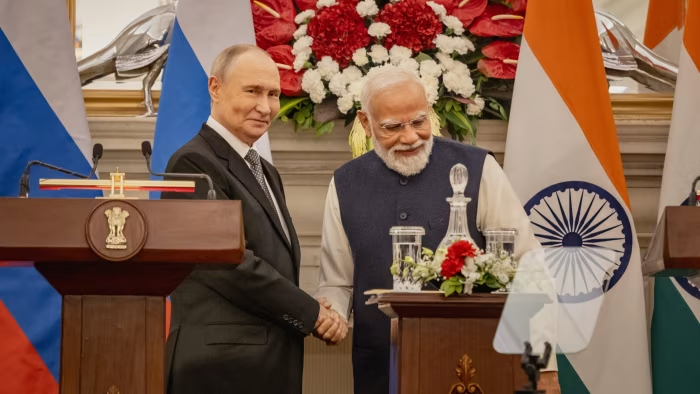
Russia’s President Vladimir Putin and India’s Prime Minister Narendra Modi have vowed to deepen their co-operation in…
Unlock the Editor’s Digest for free
Roula Khalaf, Editor of the FT, selects her favourite stories in this weekly newsletter.
Russia’s President Vladimir Putin and India’s Prime Minister Narendra Modi have vowed to deepen their co-operation in…