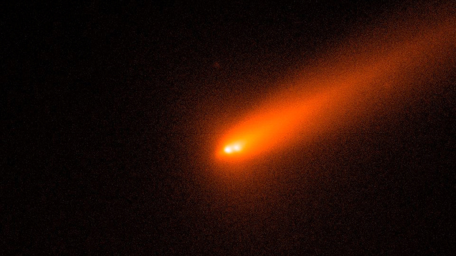
‘Habitat’ AI model shows promise for stratifying lung nodule disease risk on LDCT

A “habitat” AI model shows promise for quantifying spatial heterogeneity between lung lesions and could help clinicians better stratify disease risk from subsolid nodules (SSNs) identified on lung cancer screening, researchers have reported.

The model has an edge on its 2D and radiomics alone counterparts, according to study senior author Jieke Liu, MD, of Sichuan Clinical Research Center for Cancer in Chengdu, China. Liu and colleagues’ findings were published August 21 in the American Journal of Roentgenology.

“The ternary-classification habitat model for invasiveness and grade of lung adenocarcinoma presenting as a subsolid nodule on low-dose chest CT (LDCT) performed significantly better than the 2D model,” he said in a statement released by the journal. “Its performance was not significantly different from radiomic and combined models.”

Habitat imaging and models based on it are an “emerging approach” for identifying spatial heterogeneity within lesions by dividing them into “consistently defined subregions based on a shared characteristic” (such as signal intensity), the group noted. It shows promise for addressing the interobserver variability that can arise from radiologists’ use of subjective techniques for identifying solid components within subsolid nodules.

The study included 747 patients with 834 resected lung adenocarcinomas that presented as subsolid nodules on LDCT between July 2018 and May 2023. The authors categorized 440 adenocarcinomas as a training set, 189 as an internal test set, and 205 as an external test set. The group classified the adenocarcinomas as noninvasive, grade 1 invasive adenocarcinoma, or grade 2 or 3 invasive adenocarcinoma. They tested the following models:

  • 2D model: Diameter and consolidation-to-tumor ratio;
  • Habitat model: Volume and volume ratio of attenuation-based subregions;
  • Radiomic model: Extracted radiomic features; and
  • Combined model: Habitat and radiomic features.

The habitat model overall average AUC bested the 2D model’s, and the combined model had the highest average overall AUC, the group reported.

Model performance (AUC) in external test set for classifying invasiveness and grade of lung cancer presenting as a subsolid nodule on LDCT
Model type Macro-average Noninvasive adenocarcinoma Grade 1 invasive adenocarcinoma Grade 2/3 invasive adenocarcinoma
2D 0.87 0.95 0.79 0.88
Attenuation-based habitat 0.92 0.96 0.86 0.94
Radiomic 0.92 0.96 0.86 0.95
Combined (habitat and radiomic features) 0.93 0.96 0.86 0.96

53-year-old woman with pure ground-glass nodule, diagnosed postoperatively as minimally invasive adenocarcinoma. Left panel shows source axial image with total nodule and solid component measured; middle shows segmentation of nodule on single slice; right shows habitats. CTR is 0, given absence of solid component. Habitats 1, 2, 3, and 4 have volumes of 921.8, 374.4, 45.1, and 0 mm³; and volume ratios of 68.7%, 27.9%, 3.4%, and 0%, respectively.Images and caption courtesy of the AJR.

“Habitat imaging provides a novel interpretable approach for quantifying intralesional spatial heterogeneity that may aid noninvasive risk stratification of SSNs detected during lung cancer screening,” Liu and colleagues concluded.

The complete study can be found here.

Continue Reading