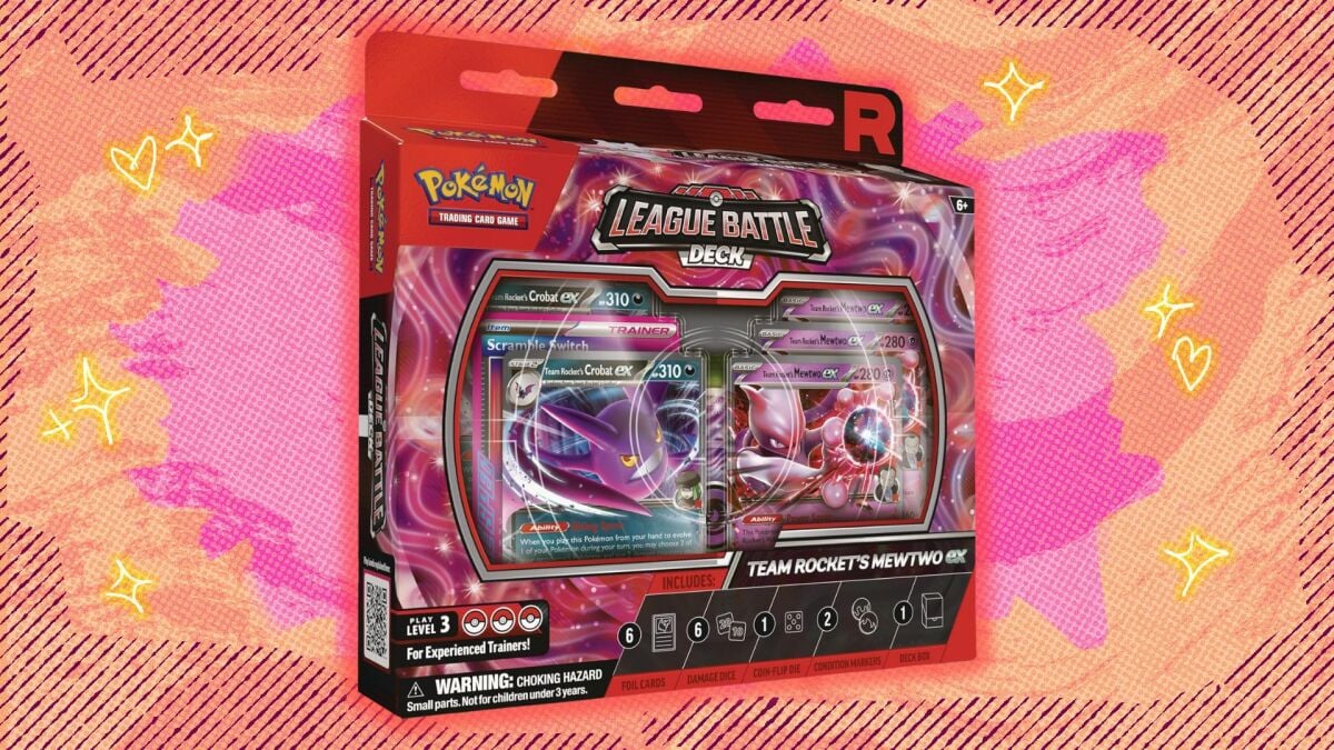
Department of General Practice, Union Hospital, Tongji Medical College, Huazhong University of Science and Technology, Wuhan, Hubei, People’s Republic of China
Abstract: Low skeletal muscle mass is an important feature and foundation of sarcopenia, which can affect body function and increase the risk of falls and fractures in older adults, while being associated with multiple systemic diseases. Helicobacter pylori is a common gastrointestinal pathogen associated with various systemic gastrointestinal and non-gastrointestinal diseases. Although extensive studies have been conducted on each of these, the direct link between them has not been fully explored. Clarifying the relationship between Helicobacter pylori infection and low muscle mass is important for the prevention and management of these diseases.
Keywords: inflammation, hormones, metabolism, nutrients, gastrointestinal microbiome
Introduction
Sarcopenia is an age-related condition characterized by the reduction of muscle mass, decline in muscle strength, and deterioration of physical function.1 Low skeletal muscle mass is a crucial feature and foundation of sarcopenia, defined as a muscle mass below normal levels. This not only significantly impacts physical performance but also greatly increases the risk of falls and fractures among the elderly.2 Low skeletal muscle mass is closely linked to inflammatory responses and immune function and is associated with diseases such as cardiovascular disease, diabetes, osteoporosis, and cognitive dysfunction.3 These conditions not only diminish individual quality of life but also impose a heavy burden on health-care systems worldwide.
Helicobacter pylori (H. pylori) is a spiral-shaped, gram-negative gastric pathogen that is extremely common in human populations, with a high global infection rate.4 The rising prevalence of H. pylori infections has become a prominent global concern. This bacterium is strongly associated with various gastrointestinal diseases, including chronic gastritis, peptic ulcers, gastric cancer, and gastric lymphoma.5 Moreover, current research suggests that it also interacts with extragastric diseases such as cardiovascular disorders, metabolic syndromes, dermatological issues, hematological diseases, neurological conditions, and autoimmune diseases.6
Although substantial research exists on H. pylori infection and low skeletal muscle mass globally, their direct relationship and associated risk factors have not been thoroughly explored. Clarifying the connection and underlying mechanisms between H. pylori infection and low skeletal muscle mass is crucial for the prevention and management of these conditions, thereby enabling the development of more comprehensive and individualized treatment plans and public health strategies. This article summarizes the epidemiology, risk factors, potential pathogenesis, diagnosis, and treatment of H. pylori infection and low skeletal muscle mass, as well as the relationship between H. pylori infection and low skeletal muscle mass.
Advances in Low Skeletal Muscle Mass Research
Epidemiology and Pathogenesis
Numerous studies have focused on the prevalence of sarcopenia across various populations, finding it particularly common among the elderly, where it is closely associated with increased risks of falls, hospitalization, and mortality.7 International research has predominantly explored the pathophysiological mechanisms of low skeletal muscle mass, focusing on aspects such as inflammatory responses, hormonal changes, malnutrition, and physical inactivity. Interventions like high-protein diets, vitamin D supplementation, and resistance training have demonstrated significant benefits.8–11 Domestic scholars are also investigating its mechanisms, emphasizing the impact of nutrition and lifestyle, and studying the effects of nutritional supplements, physical exercise, and traditional Chinese medicine in improving muscle mass and function.12 Research into biomarkers for frailty and sarcopenia11 has revealed that pro-inflammatory cytokines like IL-1, IL-6, and TNF-α may contribute to muscle mass and strength loss during aging through synergistic actions. Additionally, anemia can reduce tissue oxygenation, leading to increased fatigue, frailty, and deterioration of muscle mass and function. Some studies13 have suggested that growth hormone (GH) directly affects target tissues, including skeletal muscle and bone, whereas insulin-like growth factor 1 (IGF-1) released by the liver binds to IGF receptors within cells, promoting cell growth and inhibiting cell death. Dysregulation of GH and IGF-1 may lead to reduced muscle and bone mass, with more than 30% of older adults having circulating IGF-1 levels below those of younger individuals. Scott, D et al found a positive correlation between low serum vitamin D levels and decreased lean body mass, leg strength, and leg muscle mass.14 A Mendelian randomization genetic association study by Tingting Sha et al in the UK Biobank validated a nonlinear relationship between inadequate 25-hydroxyvitamin D levels and sarcopenia risk, indicating the need for randomized clinical trials in participants with insufficient 25-hydroxyvitamin D levels to confirm potential causal links.15
Diagnosis and Treatment
The definition and diagnostic criteria for low skeletal muscle mass are relatively consistent, primarily based on recommendations from the European Working Group on Sarcopenia in Older People (EWGSOP) and the Asian Working Group for Sarcopenia (AWGS).16 AWGS recommends using Dualenergy X-ray absorptiometry (DXA) or Bio-impedance analysis (BIA) to measure muscle mass,17 with diagnostic cut-off values for sarcopenia being: for DXA-analyzed appendicular skeletal muscle mass index (ASMI): males <7.0 kg/m², females <5.4 kg/m²; or for BIA: males <7.0 kg/m², females <5.7 kg/m².2
Treatment of low skeletal muscle mass typically involves comprehensive, multidimensional intervention strategies, including nutritional supplementation, physical exercise, and pharmacotherapy. In particular, increased protein intake is crucial for muscle synthesis and repair. Supplements rich in leucine, vitamin D, calcium, and Omega-3 fatty acids can help improve muscle mass.9,18,19 Resistance training is the most effective method for enhancing muscle mass and strength,20 recommended two–three times per week, involving exercises with dumbbells, barbells, resistance bands, or fitness machines. Aerobic activities such as walking, swimming, and cycling benefit cardiovascular health and complement the overall fitness improvements. Balance and flexibility exercises, such as yoga and tai chi, enhance agility and balance and reduce fall risk. Although there are currently no specific drugs specifically used to treat low skeletal muscle mass, some drugs are used in clinical practice to supplement treatment, such as hormone replacement therapy, which can help increase muscle mass and strength in men with low testosterone,21 and appropriate anti-inflammatory therapy may also help improve muscle condition.22 Psychological support and education can enhance patients’ understanding of the disease, boosting motivation and adherence to treatment.23 Ideally, personalized treatment plans should be developed based on individual circumstances, incorporating multiple aspects of intervention, requiring collaboration from a multidisciplinary team, including general practitioners, dietitians, rehabilitation therapists, and endocrinologists, to maximize improvements in muscle mass and function. Through personalized treatment plans and multidisciplinary management, patient quality of life can be effectively enhanced, and related complications can be reduced (Figure 1).
Figure 1 Diagnosis and treatment of low skeletal muscle mass. Created with BioGDP.com. Abbreviations: AWGS, the Asian Working Group for Sarcopenia; ASMI, appendicular skeletal muscle mass index; DXA, Dualenergy X-ray absorptiometry; BIA, Bio-impedance analysis.
|
Advances in Helicobacter pylori Research
Epidemiology and Pathogenesis
The increasing prevalence of H. pylori infections has become a significant global concern. It is estimated that over 50% of the global population is colonized by H. pylori, with higher incidence rates observed in developing countries and less economically developed regions.24 According to China’s White Paper on Infection Control,12 the global infection rate of H. pylori is approximately 50%, ranging from 18.9% to 88.7%, while in China, it approaches 50%, with different groups showing infection rates between 35.4% and 66.4%. Rural areas exhibit higher rates than urban areas, and adults are more affected than children and are characterized by high infection rates, substantial disease burden, and high resistance rates.
The risk factors for H. pylori infection are multifaceted, encompassing environmental, lifestyle, and individual characteristics.25 Firstly, residing in areas with poor sanitation, such as rural or developing regions, significantly increases the risk of H. pylori infection due to water contamination and inadequate public health facilities that facilitate bacterial transmission. Secondly, dietary habits play a crucial role as the consumption of raw or undercooked foods may serve as a vector for infection.26 Furthermore, practices such as communal dining and sharing utensils can further promote the spread of the bacterium. In terms of individual characteristics, children and the elderly are at heightened risk because of their relatively weak immune systems. Additionally, unhealthy lifestyle choices, including smoking and excessive alcohol consumption, are associated with the occurrence of H. pylori infection. Therefore, a comprehensive understanding of these risk factors is essential for developing effective prevention and control strategies to reduce the prevalence of H. pylori infection.
The pathogenic mechanisms of H. pylori infection are complex and primarily involve the production of various active toxins that disrupt the gastric mucosal barrier, leading to chronic inflammation. Notably, cytotoxin-associated protein (CagA) and vacuolating cytotoxin (VacA) are key toxins implicated in pathogenesis, with long-term inflammation resulting in gastritis, peptic ulcers, and gastric cancer.27 H. pylori also survives in acidic gastric conditions by producing urease, which hydrolyzes urea to ammonia, and its flagella enhance motility through gastric mucus, further facilitated by urease, reducing mucin viscosity.28 Recent studies have linked H. pylori to cardiovascular, metabolic, dermatological, hematological, neurological, and autoimmune diseases.29–34
Diagnosis and Treatment
In clinical practice, H. pylori detection primarily involves invasive and non-invasive tests. Invasive methods include the rapid urease test and histological examination, whereas non-invasive tests include the urea breath test, fecal antigen testing, and serology. Given its simplicity and broad acceptance, the 13C or 14C urea breath test is commonly used to diagnose H. pylori infection. Standard triple therapy is a widely used treatment option for eradicating H. pylori, consisting of a proton pump inhibitor (PPI) and two antibiotics. The commonly prescribed PPIs include omeprazole, lansoprazole, pantoprazole, and rabeprazole. The antibiotics that are often used are amoxicillin, clarithromycin, levofloxacin, and metronidazole. Typically, this regimen is maintained for 10–14 days to achieve complete eradication of the H. pylori infection. In regions where resistance to clarithromycin is high, bismuth quadruple therapy is recommended as first-line treatment for H. pylori. This therapy included a PPI, bismuth, and two antibiotics. However, the gradual increase in drug resistance poses a great challenge to effective H. pylori eradication treatment worldwide (Figure 2).27,35,36
![]() |
Figure 2 Diagnosis and treatment of Helicobacter pylori. Created with BioGDP.com.
|
Discussion
Clinical Studies on the Correlation Between Low Skeletal Muscle Mass and H. pylori
Studies indicate that H. pylori infection may directly or indirectly lead to low skeletal muscle mass through inflammatory responses, hormonal alterations, dysbiosis, nutritional malabsorption, or systemic metabolic disruptions. A retrospective cohort study conducted at the First Affiliated Hospital of Wenzhou Medical University37 demonstrated an increased risk of low skeletal muscle mass in women aged >40 years who were currently infected with H. pylori. A cross-sectional study in Seoul, Korea,38 showed that elderly women undergoing H. pylori eradication treatment had a reduced risk of low skeletal muscle mass, although prospective longitudinal studies are needed to confirm the long-term effects on sarcopenia. Another study involving 3,453 Americans aged 60 and above4 suggested that serum markers of H. pylori infection, including antibodies against H. pylori and cytotoxin-associated gene antibodies, were associated with sarcopenia and low skeletal muscle mass. Therefore, patients at high risk for low skeletal muscle mass who test positive for H. pylori may consider eradication therapy, which offers the dual benefits of treating bacterial infection and mitigating skeletal muscle degradation.
The clinical association between H. pylori infection and low skeletal muscle mass has garnered increasing attention, particularly in elderly populations. Studies indicate that H. pylori infection may trigger chronic inflammatory responses,39 which not only impair gastrointestinal function but also release pro-inflammatory cytokines into the systemic circulation, thereby negatively impacting muscle quality and strength. Chronic inflammation is recognized as one of the key pathological mechanisms underlying sarcopenia,40 as prolonged inflammatory states can accelerate muscle protein degradation and exacerbate loss of muscle mass. Furthermore, H. pylori infection may exacerbate the risk of low skeletal muscle mass by influencing hormonal levels. Specifically, H. pylori has been shown to interfere with the secretion of ghrelin and leptin, two hormones integral to appetite regulation and energy metabolism.41 Patients with low skeletal muscle mass typically exhibit lower levels of ghrelin and abnormal leptin levels.42 Thus, these hormones may serve as critical links between H. pylori infection and low skeletal muscle mass. As individuals age, particularly after women enter menopause, declining estradiol levels may diminish the protective effects of sex hormones against H. pylori infection. Such hormonal changes could render elderly women more susceptible to H. pylori infection, subsequently increasing the risk of low skeletal muscle mass. Additionally, certain steroid hormones may possess inhibitory effects on H. pylori growth; however, this protective effect may weaken with age, further intensifying the potential impact of infection on muscle quality.43 Moreover, H. pylori infection is closely associated with the onset of metabolic syndrome, a significant risk factor for low skeletal muscle mass. Metabolic syndrome is characterized by a cluster of vascular risk factors, including insulin resistance, hypertension, abdominal obesity, dyslipidemia, and impaired glucose metabolism.44 These factors may promote muscle mass loss through mechanisms involving inflammation and oxidative stress. H. pylori also disrupts gastrointestinal function,45 leading to malabsorption of essential nutrients and particularly affecting the intake of key nutrients such as iron, calcium, vitamin B12, and vitamin D. This nutritional deficiency not only hinders growth and development in children but also contributes to muscle mass deterioration in older adults. Variations in dietary habits and H. pylori prevalence across different regions may lead to divergent clinical outcomes,26 necessitating rigorous experimental studies to establish the role of geography as an influencing factor. Finally, H. pylori infection may induce dysbiosis of gut microbiota, which in turn affects skeletal muscle status via the “gut-muscle axis”.46 The diversity of gut microbiota is closely linked to the host’s metabolic health, and dysbiosis may lead to impaired nutrient absorption and metabolic pathways, ultimately resulting in diminished muscle quality.
Future research should focus on in-depth clinical studies to further explore the relationship between H. pylori infection and low skeletal muscle mass, along with related risk factors. While preliminary cross-sectional and cohort studies suggest an association, more longitudinal studies and randomized controlled trials are needed to clarify mechanisms and causality. For example, large-scale, multicenter prospective cohorts targeting elderly individuals, patients with chronic gastritis, malnutrition, or metabolic syndrome could track muscle mass changes over time while controlling for confounders such as age, sex, physical activity, and diet. Randomized trials assessing muscle mass, grip strength, and gait speed before and after H. pylori eradication, as well as combined interventions with protein, vitamin D, or probiotics supplementation, would be valuable. In addition, an in-depth investigation of the relationship between H. pylori infection and low skeletal muscle mass, along with its underlying mechanisms, is crucial for the development of effective prevention and treatment strategies.
Potential Mechanisms of Interplay Between Low Skeletal Muscle Mass and H. pylori
First, H. pylori infection initiates the release of various active toxins that compromise the gastric mucosal barrier, subsequently triggering the release of multiple pro-inflammatory cytokines and chemokines in the host, including IL-6, IL-8, IFN-γ, TNF-α, and NF-κB (Figure 3A).47 Notably, the levels of several inflammatory cytokines, particularly IL-8, CXCL1, and CXCL2, are significantly upregulated in gastric organ samples. These cytokines are rapidly secreted post-infection, leading to the infiltration of neutrophils and other immune cells, thereby establishing a localized inflammatory response. Studies have indicated that CagA-positive strains elicit greater IL-8 secretion than CagA-negative strains, suggesting that specific virulence factors of H. pylori can amplify the inflammatory response.48 H. pylori also suppresses the host immune response by interfering with the IFN-γ signaling pathway. Specifically, in H. pylori-infected cells, the JAK/STAT signaling pathway is inhibited, resulting in decreased levels of phosphorylated STAT1. This mechanism allows H. pylori to evade host immune surveillance while inducing an inflammatory response in nearby uninfected epithelial cells, creating a microenvironment conducive to its survival.49 Moreover, H. pylori stimulates the sonic hedgehog (Shh) signaling pathway in gastric wall cells, a process dependent on the activation of the NF-κB signaling pathway. Studies have demonstrated that, shortly after H. pylori infection, NF-κB activation promotes Shh expression. Pre-treatment with NF-κB inhibitors significantly weakens Shh signaling activation, indicating that NF-κB plays a crucial role in H. pylori-induced Shh signaling, potentially influencing gastric mucosal regeneration and inflammatory responses.50 H. pylori infection may also facilitate inflammation-related gastric cancer through the activation of the STAT3 signaling pathway. Specifically, cytokines, such as IL-6 and IL-11, are upregulated following H. pylori infection, activating STAT3 via the JAK signaling pathway. The activation of STAT3 correlates with the increased expression of intestinal metaplasia-related genes. These cytokine actions suggest that H. pylori may facilitate pathological changes in the gastric mucosa through STAT3-dependent mechanisms, thereby elevating the risk of gastric cancer.51 Through these specific mechanisms, H. pylori effectively incites host inflammatory responses, potentially leading to chronic gastritis and other related diseases. In the inflammatory mechanisms of sarcopenia, key inflammatory factors include TNF-α, IL-6, IL-1 and various chemokines. The elevation of these factors during chronic low-grade inflammation directly affects the health and function of the muscle tissue.52 TNF-α activates multiple signaling pathways by binding to its receptor (TNFR), leading to the activation of the transcription factor NF-κB,53 which subsequently induces the expression of a range of inflammatory genes. The products of these genes include additional inflammatory factors and chemokines that create a positive feedback loop that exacerbates the inflammatory response. Furthermore, TNF-α promotes apoptosis in muscle cells and enhances the degradation of muscle proteins, resulting in muscle atrophy. IL-6 serves as a multifunctional cytokine, acting both as a pro-inflammatory agent and, in certain contexts, as exerting anti-inflammatory agent.54 Elevated levels of IL-6 are associated with diminished muscle mass in cases of sarcopenia, as it promotes protein degradation in muscle cells while inhibiting protein synthesis via the JAK/STAT signaling pathway. IL-1, a potent pro-inflammatory cytokine, mediates its effects primarily through IL-1 receptors, leading to NF-κB activation and promoting the expression of inflammatory genes.54 IL-1 can also induce apoptosis in muscle cells, further diminishing muscle quality. They attract immune cells such as macrophages and lymphocytes to the site of inflammation, thereby intensifying local inflammatory reactions. The influx of immune cells triggers the release of additional inflammatory factors, thereby perpetuating a vicious cycle that exacerbates muscle damage. NF-κB is a central transcription factor in inflammatory responses, and its activation not only drives the expression of the aforementioned inflammatory factors but also regulates genes involved in cell survival, proliferation, and apoptosis. Persistent activation of NF-κB ultimately leads to muscle cell apoptosis and a decline in muscle quality, culminating in sarcopenia. These pro-inflammatory factors disseminate throughout the systemic circulation, causing long-term chronic inflammation that negatively affects skeletal muscle mass and strength, thereby accelerating the onset of sarcopenia.
![]() |
Figure 3 Potential Mechanisms of Interplay between Low Skeletal Muscle Mass and H. pylori. (A) H. pylori triggers chronic systemic inflammation, which promotes muscle catabolism and impairs protein synthesis, thereby negatively affecting skeletal muscle mass. (B) H. pylori infection may disrupt the balance of ghrelin and leptin, impair hormone signaling pathways, and thereby reduce anabolic stimuli essential for muscle maintenance. (C) Metabolic syndrome is closely linked to muscle loss, and H. pylori infection may exacerbate metabolic dysregulation through mechanisms such as inflammation, oxidative stress, and mitochondrial dysfunction, thereby impairing skeletal muscle mass. (D) Chronic gastritis and malabsorption caused by H. pylori may lead to protein and micronutrient deficiencies, compromising muscle health. (E) H. pylori alters gut microbiome composition, impairing short-chain fatty acid production and gut-muscle axis communication. Created with BioGDP.com. Abbreviations: CagA, cytotoxin-associated protein; VacA, vacuolating cytotoxin; DCs, dendritic cell; MS, metabolic syndrome; ROS, reactive oxygen species; Ca, calcium; Fe, ferrum; VitB12, Vitamin B12; VitD, Vitamin D; IGF-1, insulin-like growth factor 1.
|
Second, H. pylori infection influences the energy balance by affecting the secretion of ghrelin and leptin (Figure 3B). Ghrelin stimulates appetite and promotes the release of growth hormone, aiding in the maintenance of muscle quality, while leptin is associated with inflammatory responses, insulin sensitivity, appetite regulation, and fat deposition.55 Patients with low skeletal muscle quality typically exhibit reduced ghrelin levels, and leptin levels may also be closely linked to muscle aging; thus, these two hormones may serve as critical links between H. pylori infection and low skeletal muscle mass.56 The impact of sex hormones on H. pylori infection is complex. Studies have suggested that estrogen may have protective effects against H. pylori infections. Estrogen enhances the barrier function and antioxidant capacity of the gastric mucosa, potentially reducing gastritis and related lesions caused by H. pylori infection.43 In contrast, testosterone promotes H. pylori infection. Males may be more prone to develop gastritis or other related diseases after H. pylori infection due to lower immune responses.57 Estrogen is recognized for its immunomodulatory effects; it can enhance the functions of certain immune cells,58 such as macrophages and T cells, thereby bolstering the host’s resistance to H. pylori infection. Concurrently, estrogen can induce the expression of anti-inflammatory factors,58 mitigating the inflammatory responses that contribute to the gastric mucosal damage caused by H. pylori. Estrogen reduces oxidative stress levels by enhancing antioxidant enzyme activity,59 thereby alleviating cellular damage induced by H. pylori infection. Testosterone impairs the healing of ulcers by causing a reduction in blood flow at the ulcer periphery, elevating plasma concentrations of IL-1β and TNF-α, and, specifically for gastric ulcers, increasing gastric acid production.60 Physiological states such as the menstrual cycle, pregnancy, and menopause lead to fluctuations in estrogen and progesterone, which may influence H. pylori infection status and the incidence of associated complications. The effects of sex hormones on low skeletal muscle mass predominantly involve the estrogen and testosterone levels. Estrogen promotes muscle protein synthesis while inhibiting muscle degradation, thereby assisting in maintaining muscle quality in women.61 By binding to estrogen receptors, estrogen activates downstream signaling pathways such as the PI3K/Akt pathway, which plays a critical role in regulating muscle cell growth and survival.62 Testosterone is vital for maintaining male skeletal muscle quality, with low testosterone levels associated with low skeletal muscle mass. Testosterone helps sustain muscle quality by promoting muscle synthesis and inhibiting degradation, primarily through the activation of androgen receptors (AR) in the muscles. The activation of the AR occurs via ligand-independent pathways including IGF-I induced MAPK-ERK1/2, p38, and JNK phosphorylation in C2C12 muscle cells.63 In the elderly population, regardless of gender, declining levels of sex hormones with age may result in diminished skeletal muscle mass. The loss of protective effects of sex hormones, coupled with weakened muscle synthesis capabilities and increased inflammation and oxidative stress, accelerates the decline in muscle mass. Therefore, fluctuations in sex hormones not only directly affect skeletal muscle health but are also closely related to chronic diseases, nutritional status, and overall quality of life. Collectively, sex hormones modulate immune responses that affect the severity of H. pylori infection, whereas the presence of H. pylori can trigger chronic inflammation and nutrient absorption impairments, directly or indirectly affecting skeletal muscle health. A dynamic network of interactions among sex hormones, H. pylori infection, and low skeletal muscle mass warrants further investigation to elucidate its potential clinical significance.
Third, metabolic syndrome is characterized by a cluster of vascular risk factors, including insulin resistance, hypertension, abdominal obesity, dysglycemia, and dyslipidemia.44 These metabolic disorders are closely related to muscle loss, potentially exacerbating sarcopenia through mechanisms involving inflammatory factors, oxidative stress, and mitochondrial dysfunction. Inflammatory responses and factors produced during H. pylori infection are significant etiologies of insulin resistance and metabolic syndrome (Figure 3C). Long-term chronic inflammation and immune dysfunction coexisting with metabolic syndrome may represent susceptibility factors for H. pylori infection.64 Furthermore, H. pylori may aggravate these metabolic disturbances, indicating a potential link to low skeletal muscle mass.
Fourth, H. pylori can disrupt gastrointestinal function, leading to impaired nutrient absorption, particularly affecting the uptake of essential nutrients such as iron, calcium, vitamin B12, and vitamin D.45 Research indicates that H. pylori induces chronic gastritis and alters gastric acid secretion, thereby interfering with normal digestive processes and resulting in poor nutrient absorption (Figure 3D). For instance, H. pylori infection may damage gastric parietal cells, reducing the synthesis of intrinsic factors that are crucial for vitamin B12 absorption and potentially leading to deficiency. Furthermore, there is a correlation between H. pylori infection and iron-deficiency anemia, as it can reduce gastric acid secretion, subsequently affecting the absorption of non-heme iron.65 In children, nutrient absorption impairments during growth and development stages can result in stunted growth and limited cognitive development, and in older adults, nutritional deficiencies can accelerate the loss of skeletal muscle mass, increasing the risk of sarcopenia. However, variations in dietary habits across different regions as well as differences in H. pylori prevalence may significantly influence nutrient absorption outcomes,26 thus affecting skeletal muscle mass. Diets high in salt and fat may favor the growth and transmission of H. pylori, whereas diets rich in antioxidants and dietary fiber may help reduce the risk of infection. Additionally, lifestyle factors, sanitation conditions, and drinking water safety in various geographical areas also affect the epidemiological characteristics of H. pylori infections. For example, in some developing countries, poor sanitary conditions result in a higher prevalence of H. pylori infection, adversely affecting the nutritional status of the local populations. Conversely, in developed countries, improved public health standards have led to lower rates of H. pylori infections and enhanced nutrient absorption among residents. Therefore, regional factors represent critical variables influencing H. pylori infection and its effects on nutrient absorption, ultimately affecting skeletal muscle mass, warranting rigorous and systematic experimental studies to further elucidate these relationships.
Fifth, H. pylori infection disrupts the balance of the gut microbiota, altering the pH levels within the stomach, and consequently affecting the environment of the small and large intestines. Such changes may promote the growth of certain bacteria while inhibiting the proliferation of beneficial organisms, such as lactic acid bacteria and bifidobacteria, leading to dysbiosis.66 Treatment of H. pylori infection typically involves antibiotics, which may further disturb the equilibrium of gut microbiota, resulting in gastrointestinal health issues.36 A healthy gut microbiome produces short-chain fatty acids (SCFAs), maintaining stability within the intestinal environment (Figure 3E). SCFAs are primarily produced by the gut microbiota through the fermentation of indigestible carbohydrates such as dietary fiber. The common SCFAs include acetate, propionate, and butyrate. SCFAs enhance muscle protein synthesis by activating insulin signaling pathways, which increases amino acid uptake in muscle cells. For instance, SCFAs stimulate the expression of IGF-1, a key regulator of muscle growth. Additionally, SCFAs have been shown to activate the mammalian target of rapamycin (mTOR) signaling pathway, which is crucial for cell growth and muscle synthesis. By stimulating this pathway, SCFAs contribute to improved rates of muscle protein synthesis.67 Moreover, SCFAs possess anti-inflammatory properties that can lower the levels of pro-inflammatory factors, thereby reducing the detrimental effects of chronic inflammation on the muscle tissue. A low-inflammation environment protects muscle cells and decreases protein degradation rates. SCFAs also enhance the barrier function of intestinal epithelial cells, improving nutrient absorption, which is vital for maintaining muscle mass.68 Importantly, SCFAs are not confined to the gut; SCFAs can enter the bloodstream and exert systemic effects. Research has indicated that SCFAs, as metabolites of the gut microbiota, influence systemic inflammatory responses, glucose metabolism, and lipid metabolism. SCFAs play a significant role in preserving skeletal muscle quality through various mechanisms, including the promotion of muscle synthesis, inhibition of muscle breakdown, and regulation of inflammation. SCFAs levels are influenced by the gut microbiota, and H. pylori infection can disrupt this balance. Studies suggest that H. pylori infection leads to dysbiosis, potentially affecting skeletal muscle status through the “gut-muscle axis” ultimately resulting in muscle loss.69 As we delve into the conclusion, it is imperative to consider these interconnected factors in understanding the broader impacts of H. pylori on health, particularly concerning low skeletal muscle mass.
Conclusion
Overall, understanding the complex relationship between H. pylori infection and low skeletal muscle mass remains an important area for future research. Potential mechanisms underlying this association may include chronic inflammation, hormonal dysregulation, oxidative stress, mitochondrial dysfunction, malnutrition, and gut dysbiosis. This study underscores the clinical and public health significance of considering H. pylori screening in patients at risk of low skeletal muscle mass. Eradication therapy might offer dual benefits by controlling infection and preserving muscle health. Further longitudinal and mechanistic studies are essential to elucidate their interplay, ultimately enabling personalized treatment approaches and informed health policies.
Acknowledgments
We express our gratitude to all of the researchers who participated in this study related to low skeletal muscle mass and Helicobacter pylori infection. Additionally, we would like to thank BioGDP.com for providing the tools used to create the images included in this review. Their support has been instrumental in enhancing the clarity and visual presentation of our work.
Disclosure
The authors declare that this research was conducted in the absence of any commercial or financial relationships that could be construed as potential conflicts of interest.
References
1. Cruz-Jentoft AJ, Sayer AA. Sarcopenia. Lancet. 2019;393(10191):2636–2646. doi:10.1016/s0140-6736(19)31138-9
2. Moon SW, Kim K-J, Lee HS, et al. Low muscle mass, low muscle function, and sarcopenia in the urban and rural elderly. Sci Rep. 2022;12(1). doi:10.1038/s41598-022-18167-y
3. Kim D, Lee J, Park R, Oh CM, Moon S. Association of low muscle mass and obesity with increased all-cause and cardiovascular disease mortality in US adults. J Cachexia, Sarcopenia Muscle. 2024;15(1):240–254. doi:10.1002/jcsm.13397
4. Wu S-E, Chen W-L. Detrimental relevance of Helicobacter pylori infection with sarcopenia. Gut Pathog. 2021;13(1). doi:10.1186/s13099-021-00464-y
5. Lim MCC, Jantaree P, Naumann M. The conundrum of Helicobacter pylori-associated apoptosis in gastric cancer. Trends Cancer. 2023;9(8):679–690. doi:10.1016/j.trecan.2023.04.012
6. Gravina AG, Zagari RM, De Musis C, Romano L, Loguercio C, Romano M. Helicobacter pylori and extragastric diseases: a review. World J Gastroenterol. 24(29):3204–3221. doi:10.3748/wjg.v24.i29.3204
7. Wu X, Li X, Xu MH, Zhang ZF, He LX, Li Y. Sarcopenia prevalence and associated factors among older Chinese population: findings from the China health and retirement longitudinal study. Article PLoS One. 2021;16(3):
8. Ghayomzadeh M, Hackett D, SeyedAlinaghi S, Gholami M, Hosseini Rouzbahani N, Azevedo Voltarelli F. Combined training improves the diagnostic measures of sarcopenia and decreases the inflammation in HIV-infected individuals. J Cachexia Sarcopenia Muscle. 2022;13(2):1024–1035. doi:10.1002/jcsm.12926
9. Gielen E, Beckwée D, Delaere A, De Breucker S, Vandewoude M, Bautmans I. Nutritional interventions to improve muscle mass, muscle strength, and physical performance in older people: an umbrella review of systematic reviews and meta-analyses. Nutr Rev. 79(2):121–147. doi:10.1093/nutrit/nuaa011
10. Luo J, Quan Z, Lin S, Cui L. The association between blood concentration of 25- hydroxyvitamin D and sarcopenia: a meta-analysis. Asia Pac J Clin Nutr. 2018;27(6):1258–1270. doi:10.6133/apjcn.201811_27(6).0013
11. Picca A, Coelho-Junior H, Calvani R, Marzetti E, Vetrano D. Biomarkers shared by frailty and sarcopenia in older adults: a systematic review and meta-analysis. Ageing Res Rev. 2022;73:101530. doi:10.1016/j.arr.2021.101530
12. Institute for Infectious Disease Control and Prevention. Chinese center for disease control and prevention. White Paper on the Prevention and Control of Helicobacter pylori Infection in China 2024-7-5, Available from: https://icdc.chinacdc.cn/zxxx/xwdt/202306/t20230603_266504.html.
13. Perrini S, Laviola L, Carreira MC, Cignarelli A, Natalicchio A, Giorgino F. The GH/IGF1 axis and signaling pathways in the muscle and bone: mechanisms underlying age-related skeletal muscle wasting and osteoporosis. J Endocrinol. 2010;205(3):201–210. doi:10.1677/joe-09-0431
14. Scott D, Blizzard L, Fell J, Ding C, Winzenberg T, Jones G. A prospective study of the associations between 25-hydroxy-vitamin D, sarcopenia progression and physical activity in older adults. Clin Endocrinol. 2010;73(5):581–587. doi:10.1111/j.1365-2265.2010.03858.x
15. Sha T, Wang Y, Zhang Y, et al. Genetic variants, serum 25-hydroxyvitamin d levels, and sarcopenia. JAMA Netw Open. 2023;6(8):e2331558. doi:10.1001/jamanetworkopen.2023.31558
16. Cruz-Jentoft AJ, Bahat G, Bauer J, et al. Sarcopenia: revised European consensus on definition and diagnosis. Age Ageing. 2019;48(1):16–31. doi:10.1093/ageing/afy169
17. Keith Yu-Kin C, Simon Kwoon-Ho C, Vivian Wing-Yin H, et al. Diagnosis of sarcopenia by evaluating skeletal muscle mass by adjusted bioimpedance analysis validated with dual-energy X-ray absorptiometry. J Cachexia, Sarcopenia Muscle. 2021;12(6). doi:10.1002/jcsm.12825
18. Prado CM, Purcell SA, Laviano A. Nutrition interventions to treat low muscle mass in cancer. J Cachexia, Sarcopenia Muscle. 2020;11(2):366–380. doi:10.1002/jcsm.12525
19. Nunes EA, Colenso-Semple L, McKellar SR, et al. Systematic review and meta-analysis of protein intake to support muscle mass and function in healthy adults. J Cachexia, Sarcopenia Muscle. 2022;13(2):795–810. doi:10.1002/jcsm.12922
20. Fyfe JJ, Hamilton DL, Daly RM. Minimal-dose resistance training for improving muscle mass, strength, and function: a narrative review of current evidence and practical considerations. Sports Med. 2022;52(3):463–479. doi:10.1007/s40279-021-01605-8
21. Tian X, Lou S, Shi R. From mitochondria to sarcopenia: role of 17β-estradiol and testosterone. Front Endocrinol. 2023;14:1156583. doi:10.3389/fendo.2023.1156583
22. Batsis JA, Villareal DT. Sarcopenic obesity in older adults: aetiology, epidemiology and treatment strategies. Nat Rev Endocrinol. 2018;14(9):513–537. doi:10.1038/s41574-018-0062-9
23. Piotrowicz K, Gąsowski J, Michel JP, Veronese N. Post-COVID-19 acute sarcopenia: physiopathology and management. Aging Clin Exp Res. 2021;33(10):2887–2898. doi:10.1007/s40520-021-01942-8
24. Malfertheiner P, Camargo MC, El-Omar E, et al. Helicobacter pylori infection. Nat Rev Dis Primers. 2023;9(1). doi:10.1038/s41572-023-00431-8
25. Xie L, Liu GW, Liu YN, et al. Prevalence of Helicobacter pylori infection in China from 2014-2023: a systematic review and meta-analysis. World J Gastroenterol. 30(43):4636–4656. doi:10.3748/wjg.v30.i43.4636
26. Soares GAS, Moraes FAS, Ramos A, et al. Dietary habits and Helicobacter pylori infection: is there an association? Therap Adv Gastroenterol. 2023;16:17562848231160620. doi:10.1177/17562848231160620
27. Xu Y, Walduck AK, Pan H. Editorial: the pathogenesis and treatment of Helicobacter pylori-induced diseases. Front Cell Infect Microbiol. 2023;13. doi:10.3389/fcimb.2023.1219503.
28. Li H, Wang R, Sun H. Systems approaches for unveiling the mechanism of action of bismuth drugs: new medicinal applications beyond Helicobacter pylori infection. Acc Chem Res. 52(1):216–227. doi:10.1021/acs.accounts.8b00439
29. Franceschi F, Zuccalà G, Roccarina D, Gasbarrini A. Clinical effects of Helicobacter pylori outside the stomach. Nat Rev Gastroenterol Hepatol. 2014;11(4):234–242. doi:10.1038/nrgastro.2013.243
30. Kim HS. Microbiota in Rosacea. Am J Clin Dermatol. 2020;21(Suppl 1):25–35. doi:10.1007/s40257-020-00546-8
31. Sun L, Zheng H, Qiu M, et al. Helicobacter pylori infection and risk of cardiovascular disease. Helicobacter. 2023;28(3):e12967. doi:10.1111/hel.12967
32. Virgilio H-R, Claire R-B, Hugo VC, et al. Association between Helicobacter pylori infection and incident risk of dementia: the AMI cohort. J Am Geriatr Soc. 2024;72(4). doi:10.1111/jgs.18748
33. Wang L, Cao ZM, Zhang LL, et al. Helicobacter pylori and autoimmune diseases: involving multiple systems. Front Immunol. 2022;13:833424. doi:10.3389/fimmu.2022.833424
34. Ye J, Feng T, Su L, Li J, Gong Y, Ma X. Interactions between Helicobacter pylori infection and host metabolic homeostasis: a comprehensive review. Helicobacter. 2023;28(6):e13030. doi:10.1111/hel.13030
35. Rokkas T, Gisbert JP, Malfertheiner P, et al. Comparative effectiveness of multiple different first-line treatment regimens for Helicobacter pylori infection: a network meta-analysis. Gastroenterology. 2021;161(2):495–507.e4. doi:10.1053/j.gastro.2021.04.012
36. Evariste T-K, Yoshio Y. Helicobacter pylori infection and antibiotic resistance – from biology to clinical implications. Nat Rev Gastroenterol Hepatol. 2021;18(9). doi:10.1038/s41575-021-00449-x
37. Xu X, Qian Y, Jin K, et al. The impact of Helicobacter pylori infection on low skeletal muscle mass risk in Chinese women over 40: a cross-sectional analysis. Front Cell Infect Microbiol. 2023;13:1289909. doi:10.3389/fcimb.2023.1289909
38. Baeg MK, Choi MG, Ko SH, et al. Elderly women who received Helicobacter pylori-eradicating therapy have reduced risk of low skeletal muscle mass. Clin Interv Aging. 2015;10:1771–1777. doi:10.2147/cia.S95007
39. Wang X, Zhao G, Shao S, Yao Y. Helicobacter pylori triggers inflammation and oncogenic transformation by perturbing the immune microenvironment. Biochim Biophys Acta Rev Cancer. 2024;1879(5):189139. doi:10.1016/j.bbcan.2024.189139
40. Pan L, Xie W, Fu X, et al. Inflammation and sarcopenia: a focus on circulating inflammatory cytokines. Exp Gerontol. 154:111544. doi:10.1016/j.exger.2021.111544
41. Kasai C, Sugimoto K, Moritani I, et al. Changes in plasma ghrelin and leptin levels in patients with peptic ulcer and gastritis following eradication of Helicobacter pylori infection. BMC Gastroenterol. 16(1):119. doi:10.1186/s12876-016-0532-2
42. Mendes C, Carvalho M, Bravo J, Martins S, Raimundo A. Possible interaction between physical exercise and leptin and ghrelin changes following roux-en-y gastric bypass in sarcopenic obesity patients-a pilot study. Nutrients. 16(22). doi:10.3390/nu16223913
43. Fong P, Wang QT. Protective effect of oral contraceptive against Helicobacter pylori infection in US adult females: NHANES 1999-2000. Epidemiol Infect.
44. Silveira Rossi JL, Barbalho SM, Reverete de Araujo R, Bechara MD, Sloan KP, Sloan LA. Metabolic syndrome and cardiovascular diseases: going beyond traditional risk factors. Diabetes Metab Res Rev. 2022;38(3):e3502. doi:10.1002/dmrr.3502
45. Ford AC, Mahadeva S, Carbone MF, Lacy BE, Talley NJ. Functional dyspepsia. Lancet. 396(10263):1689–1702. doi:10.1016/s0140-6736(20)30469-4
46. Massironi S, Viganò C, Palermo A, et al. Inflammation and malnutrition in inflammatory bowel disease. Lancet Gastroenterol Hepatol. 2023;8(6):579–590. doi:10.1016/s2468-1253(23)00011-0
47. de Brito BB, da Silva FAF, Soares AS, et al. Pathogenesis and clinical management of Helicobacter pylori gastric infection. World J Gastroenterol. 25(37):5578–5589. doi:10.3748/wjg.v25.i37.5578
48. Sebrell TA, Hashimi M, Sidar B, et al. A novel gastric spheroid co-culture model reveals chemokine-dependent recruitment of human dendritic cells to the gastric epithelium. Cell Mol Gastroenterol Hepatol. 2019;8(1):157–171.e3. doi:10.1016/j.jcmgh.2019.02.010
49. Morey P, Pfannkuch L, Pang E, et al. Helicobacter pylori depletes cholesterol in gastric glands to prevent interferon gamma signaling and escape the inflammatory response. Gastroenterology. 2018;154(5):1391–1404.e9. doi:10.1053/j.gastro.2017.12.008
50. Schumacher MA, Feng R, Aihara E, et al. Helicobacter pylori-induced Sonic Hedgehog expression is regulated by NFκB pathway activation: the use of a novel in vitro model to study epithelial response to infection. Helicobacter. 2015;20(1):19–28. doi:10.1111/hel.12152
51. Ernst M, Najdovska M, Grail D, et al. STAT3 and STAT1 mediate IL-11-dependent and inflammation-associated gastric tumorigenesis in gp130 receptor mutant mice. J Clin Invest. 2008;118(5):1727–1738. doi:10.1172/jci34944
52. Jimenez-Gutierrez GE, Martínez-Gómez LE, Martínez-Armenta C, Pineda C, Martínez-Nava GA, Lopez-Reyes A. molecular mechanisms of inflammation in sarcopenia: diagnosis and therapeutic update. Cells. 11(15). doi:10.3390/cells11152359
53. Liu D, Wang S, Liu S, Wang Q, Che X, Wu G. Frontiers in sarcopenia: advancements in diagnostics, molecular mechanisms, and therapeutic strategies. Mol Aspects Med. 2024;97:101270. doi:10.1016/j.mam.2024.101270
54. Bennett JL, Pratt AG, Dodds R, Sayer AA, Isaacs JD. Rheumatoid sarcopenia: loss of skeletal muscle strength and mass in rheumatoid arthritis. Nat Rev Rheumatol. 2023;19(4):239–251. doi:10.1038/s41584-023-00921-9
55. Romo-González C, Mendoza E, Mera RM, et al. Helicobacter pylori infection and serum leptin, obestatin, and ghrelin levels in Mexican schoolchildren. Pediatr Res. 2017;82(4):607–613. doi:10.1038/pr.2017.69
56. Lin YL, Wang CH, Lai YH, Kuo CH, Syu RJ, Hsu BG. Negative correlation between leptin serum levels and sarcopenia in hemodialysis patients. Int J Clin Exp Pathol. 2018;11(3):1715–1723.
57. Lin XK, Wang WL. Analysis of high risk factors for chronic atrophic gastritis. Saudi J Gastroenterol. 2023;29(2):127–134. doi:10.4103/sjg.sjg_383_22
58. Hoffmann JP, Liu JA, Seddu K, Klein SL. Sex hormone signaling and regulation of immune function. Immunity. 56(11):2472–2491. doi:10.1016/j.immuni.2023.10.008
59. Chainy GBN, Sahoo DK. Hormones and oxidative stress: an overview. Free Radic Res. 2020;54(1):1–26. doi:10.1080/10715762.2019.1702656
60. Machowska A, Brzozowski T, Sliwowski Z, et al. Gastric secretion, proinflammatory cytokines and epidermal growth factor (EGF) in the delayed healing of lingual and gastric ulcerations by testosterone. Inflammopharmacology. 2008;16(1):40–47. doi:10.1007/s10787-007-1600-6
61. Geraci A, Calvani R, Ferri E, Marzetti E, Arosio B, Cesari M. Sarcopenia and menopause: the role of estradiol. Front Endocrinol. 2021;12:682012. doi:10.3389/fendo.2021.682012
62. Pellegrino A, Tiidus PM, Vandenboom R. Mechanisms of estrogen influence on skeletal muscle: mass, regeneration, and mitochondrial function. Sports Med. 2022;52(12):2853–2869. doi:10.1007/s40279-022-01733-9
63. Kraemer WJ, Ratamess NA, Hymer WC, Nindl BC, Fragala MS. Growth Hormone(s), testosterone, insulin-like growth factors, and cortisol: roles and integration for cellular development and growth with exercise. Front Endocrinol. 2020;11:33. doi:10.3389/fendo.2020.00033
64. Xie Q, He Y, Zhou D, Jiang Y, Deng Y, Li R. Recent research progress on the correlation between metabolic syndrome and Helicobacter pylori infection. PeerJ. 2023;
65. Kato S, Gold BD, Kato A. Helicobacter pylori-associated iron deficiency anemia in childhood and adolescence-pathogenesis and clinical management strategy. J Clin Med. 11(24). doi:10.3390/jcm11247351
66. Chen CC, Liou JM, Lee YC, Hong TC, El-Omar EM, Wu MS. The interplay between Helicobacter pylori and gastrointestinal microbiota. Gut Microbes. 2021;13(1):1–22. doi:10.1080/19490976.2021.1909459
67. Liu C, Wong PY, Wang Q, et al. Short-chain fatty acids enhance muscle mass and function through the activation of mTOR signalling pathways in sarcopenic mice. J Cachexia, Sarcopenia Muscle. 2024;15(6):2387–2401. doi:10.1002/jcsm.13573
68. Frampton J, Murphy KG, Frost G, Chambers ES. Short-chain fatty acids as potential regulators of skeletal muscle metabolism and function. Nat Metab. 2020;2(9):840–848. doi:10.1038/s42255-020-0188-7
69. Liu C, Cheung WH, Li J, et al. Understanding the gut microbiota and sarcopenia: a systematic review. J Cachexia, Sarcopenia Muscle. 2021;12(6):1393–1407. doi:10.1002/jcsm.12784