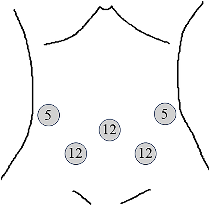
The patients’ median age was 62.5 years (range, 47–84 years), and 17 were men. The median BMI (body mass index) was 22.2 (range, 17.6–27.5) and Performance status (PS) was favorable (PS 0) in all patients. The median tumor size at preoperative diagnosis was 15 mm (range, 7–27 mm). The macroscopic tumor types were 0-I, 0-IIa, and 0-IIc, with submucosal tumors (SMT) noted in 2, 18, 8, and 2 cases, respectively. Tumors were located in the first, second, and third portions of the duodenum in 5, 20, and 5 cases, respectively. Regarding the horizontal localization of the tumor in the duodenum, 1, 12, 8, and 9 tumors were located at the papilla-ampulla side, the counterpart of the papilla-ampulla, the anterior wall, and the posterior wall, respectively (Table 1). Here, 7 and 23 patients underwent the anterior and transmesocolic approaches, respectively. Five cases out of seven were men for the anterior approach and twelve cases out of twenty-three were men for the transmesocolic approach. The median age was 70 years (range, 58–84 years) for the anterior approach and 62 years (range, 47–79) for the transmesocolic approach. The median BMI was 22.1 (range, 17.6–24.6) for the anterior approach and 22.4 (range, 19.7–27.5) for the transmesocolic approach. The macroscopic tumor types were 0-I, 0-IIa, 0-IIc, and SMT noted in 1, 3, 3, and 0 cases for the anterior approach and, 1, 15, 5, and 2 cases for the transmesocolic approach, respectively. The horizontal localization of the tumor in the duodenum, 0, 1, 4 and 2 tumors were located at the papilla-ampulla side, the counterpart of the papilla-ampulla, the anterior wall, and the posterior wall for the anterior approach and, 1, 11, 4, and 7 tumors for the transmesocolic approach, respectively. There were no statistical differences in these clinical features described above. The median tumor size was 12 mm (range, 7–20 mm) for the anterior approach and 20 mm (range, 8–27 mm) for the transmesocolic approach; a significant difference was observed between the two approaches (p = 0.02). Tumors were located in the first, second, and third portions of the duodenum in 5, 2, and 0 cases for the anterior approach, and 0, 18, and 5 cases for the transmesocolic approach, respectively; a significant difference was observed between the two approaches (p < 0.001). The median total operation time was 281 min (range, 168–401 min) for the anterior approach and 297 min (range, 182–535 min) for the transmesocolic approach (p = 0.76). The median ESD time was 47 min (range, 20–180 min) for the anterior approach and 75 min (range, 5–120 min) for the transmesocolic approach (p = 0.66). The median laparoscopic time was 135 min (range, 22–359 min) for the anterior approach and 138 min (range, 68–421 min) for the transmesocolic approach (p = 0.81). The median amount of bleeding was 10 mL (range, 0–126 mL) for the anterior approach and 0 mL (range, 0–1117 mL) for the transmesocolic approach (p = 0.50) (Table 2). During ESD, seromuscular layer injury occurred in four cases, and the right branch of the middle colic artery was injured by the high-frequency ESD knife in one case. All intraoperative complications were treated with laparoscopic suturing. No postoperative complications, such as delayed perforation or pancreatitis, were observed.
All patients underwent curative resection. Pathological analysis revealed 21 cases of adenoma, 7 of adenocarcinoma, 1 of gastrointestinal tumor, and 1 of neuroendocrine tumor (Table 1). All seven adenocarcinoma cases showed intramucosal invasion without vessel invasion or exposure at the resection edge. No disease recurrence was observed via endoscopy or CT surveillance during the median follow-up period of 35 months.
Five patients underwent additional surgical procedures during D-LECS (Table 2). Three patients were converted to laparoscopic full-thickness tumor resection; among them, one underwent duodenojejunal bypass due to a large defect in the duodenal wall. One patient underwent laparoscopic cholecystectomy, and another underwent ESD for a remnant gastric adenoma after subtotal esophagectomy. These additional procedures extended the total surgical time compared to the simple D-LECS procedure.
To compare the two different D-LECS approaches without surgical bias, 25 cases, excluding the five described above, were re-evaluated. The anterior and transmesocolic approaches were used in 4 and 21 cases, respectively. The patients’ median age was 65 years (range, 58–74 years) for the anterior approach and 62 years (range, 47–79 years) for the transmesocolic approach. The median BMI was 19.7 (range, 17.6–24.6) for the anterior approach and 21.8 (range, 19.7–27.5) for the transmesocolic approach. Tumors were located in the first, second, and third portions of the duodenum in 2, 2, and 0 cases for the anterior approach, and 0, 16, and 5 cases for the transmesocolic approach, respectively; a significant difference was observed between the two approaches (p < 0.01). The median tumor size was 15.5 mm (range, 8–20 mm) for the anterior approach and 20 mm (range, 9–27 mm) for the transmesocolic approach. There were no statistical differences in these clinical features described above. The median total operation time was 258 min (range, 168–401 min) for the anterior approach and 295 min (range, 182–416 min) for the transmesocolic approach. The median ESD time was 45 min (range, 20–100 min) for the anterior approach and 77 min (range, 45–120 min) for the transmesocolic approach. The median laparoscopic procedure time was 126 min (range, 66–359 min) for the anterior approach and 126 min (range, 68–233 min) for the transmesocolic approach. The median amount of bleeding was 20 mL (range, 5–32 mL) for the anterior approach and 0 mL (range, 0–75 mL) for the transmesocolic approach. No significant differences were observed in these factors between the two approaches (Table 3 and Supplemental Fig. 1). Body mass index tended to positively correlate with the total operation time, laparoscopic time, and the amount of bleeding (Supplemental Fig. 2).
