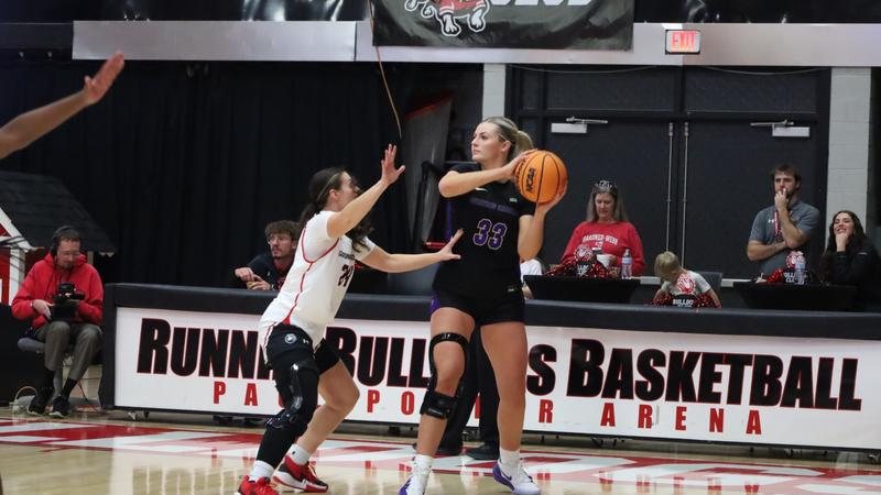
Comparative Analysis of Myocardial Protection Solutions in Pediatric E

Introduction

In recent years, advancements in medical technology have led to the diversification of surgical strategies for complex congenital heart disease in pediatric patients, particularly with respect to extracorporeal circulation procedures. Myocardial protection is a critical component of cardiopulmonary bypass surgery and serves as an important factor influencing the success of the procedure.1 The choice of an appropriate myocardial protection solution is essential for mitigating myocardial ischemia-reperfusion injury, reducing the incidence of postoperative cardiac arrhythmia, and optimizing patient outcomes.2

Cardiac arrhythmia is a frequently encountered complication following pediatric surgery with extracorporeal circulation for complex congenital heart defects. Serum sodium, the predominant extracellular cation, plays a key role in maintaining osmotic pressure, acid-base balance, and neuromuscular excitability.3 Serum sodium levels, which are crucial for osmotic homeostasis, acid-base balance, and cellular excitability, can be significantly affected by CPB-related hemodilution and electrolyte shifts.4,5 Factors such as extracorporeal circulation and hemodilution may induce fluctuations in serum sodium concentrations, potentially resulting in multi-organ dysfunction and adversely influencing postoperative recovery and clinical outcomes.6

Histidine-Tryptophan-Ketoglutarate (HTK) solution and St. Thomas cardioplegic solution are widely used myocardial protection solutions in clinical practice. HTK is classified as an intracellular-type cardioplegic solution, characterized by low sodium and calcium concentrations. It contains 15 mmol/L sodium, 10 mmol/L potassium, 4 mmol/L magnesium, 50 mmol/L chloride, 180 mmol/L histidine, 18 mmol/L histidine hydrochloride, 30 mmol/L mannitol, 2 mmol/L tryptophan, and 1 mmol/L α-ketoglutarate. In contrast, St. Thomas solution is an extracellular-type cardioplegia, formulated with 470 mL normal saline, 7.5 mL of 10% KCl, 2 mL of 25% MgSO₄, 1.5 mL of 5% CaCl2, 2 mL of 10% procaine, and 20 mL of 5% sodium bicarbonate. These differences in ionic composition and buffering capacity influence their mechanisms of myocardial protection, metabolic effects, and potential impact on electrolyte balance.

Despite their widespread use, there remains limited comparative data on how HTK and St. Thomas solutions affect postoperative cardiac rhythm stability and serum sodium levels in pediatric patients undergoing CPB. To assess the comparative efficacy of these two solutions in pediatric patients undergoing surgery with extracorporeal circulation for complex congenital heart disease, this retrospective study analyzed clinical data from 60 such patients. The impact of these cardioplegic agents on perioperative cardiac rhythm and serum sodium levels is presented in the following sections.

Materials and Methods

General Data

Following approval from the institutional ethics committee, a retrospective analysis was conducted on the clinical data of 60 pediatric patients diagnosed with complex congenital heart disease who underwent open-heart surgery with extracorporeal circulation between January 2022 and October 2024. Patients were categorized into a St. Thomas cardioplegic solution group (n = 30) and an HTK solution group (n = 30) based on the myocardial protection solution administered during surgery. Although this study was retrospective, written informed consent was obtained from patients’ legal guardians during preoperative discussions, which included information regarding potential future use of anonymized clinical data for research purposes.

Inclusion criteria were as follows: (1) Diagnosis of complex congenital heart disease confirmed through imaging examinations such as echocardiography and cardiovascular angiography;7 (2) Age between 1 month and 6 years; (3) Indication for corrective open-heart surgery with extracorporeal circulation; (4) Availability of complete clinical data; and (5) Provision of informed consent from guardians following explanation of the study’s purpose, methods, and potential risks.

Exclusion criteria were as follows: (1) History of systemic autoimmune disease; (2) Presence of severe preoperative cardiac arrhythmias unresponsive to medical or other interventions, potentially confounding the assessment of intraoperative cardiac rhythm; and (3) Preoperative use of vasoactive medications.

Methods

Patients in both groups underwent open-heart surgery with the use of extracorporeal circulation. Following routine induction of anesthesia, assisted ventilation was provided via a ventilator. Core temperature during intravenous anesthesia was maintained at approximately 28°C during cardioplegic arrest. Extracorporeal circulation was established using a priming solution composed of albumin (15 g), 5% sodium bicarbonate (15 mL), suspended red blood cells (1–2 units), Plasma-Lyte Solution (200 mL), and heparin (5–10 mg). Cannulation of the aorta, right atrial appendage, and superior and inferior vena cava was performed to initiate extracorporeal circulation. The use of 1–2 units of suspended red blood cells during cardiopulmonary bypass varied due to significant differences in preoperative hemoglobin levels among patients. The target hematocrit during cardiopulmonary bypass was maintained above 25%, and above 30% postoperatively. The cannula size was selected based on the patient’s body weight. Cardioplegia delivery was performed via the cardiopulmonary bypass pump system. During cardiopulmonary bypass, blood pressure was maintained at 30–40 mmHg, temperature at approximately 28°C, and blood gas management followed a pH-stat strategy. Cannulation of the superior and inferior vena cava was performed in all cases.

For myocardial protection, patients in the St. Thomas cardioplegic solution group received St. Thomas cardioplegic solution, with a perfusion duration of 1.5–2 minutes at a dosage of 20 mL/kg. When the duration of aortic cross-clamp exceeded 30 minutes, an additional perfusion was administered. Cardioplegia perfusion pressure was maintained within the range of 100 and 150 mmHg. The HTK solution group received HTK solution via a single perfusion, at a dosage of 50 mL/kg, over 5–8 minutes, with perfusion pressure maintained between 80–100 mmHg. Following myocardial protection solution perfusion, ice slush was uniformly applied in both groups to assist in cardiac arrest. St. Thomas cardioplegic solution is administered every 30 minutes, whereas HTK solution requires re-administration only every 2 hours. The lower perfusion pressure of HTK solution is possibly due to its unique properties. It typically requires 4–5 minutes to deliver the full volume, compared to about 2 minutes for standard cardioplegic solutions, resulting in a lower perfusion pressure.

The St. Thomas cardioplegic solution mainly consists of 470 mL of normal saline, 7.5 mL of 10% potassium chloride (KCl), 2 mL of 25% magnesium sulfate (MgSO₄), 1.5 mL of 5% calcium chloride (CaCl2), 2 mL of 10% procaine, and an appropriate amount of 5% sodium bicarbonate (NaHCO₃). The HTK solution contains the following components: 0.8766 grams of sodium chloride (NaCl), 0.6710 grams of potassium chloride (KCl), 0.1842 grams of potassium hydrogen 2-oxoglutarate, 0.8132 grams of magnesium chloride hexahydrate (MgCl2·6H2O), 3.7733 grams of histidine hydrochloride monohydrate, 27.9289 grams of histidine, 0.4085 grams of tryptophan, 5.4651 grams of mannitol, and 0.0022 grams of calcium chloride dihydrate (CaCl2·2H2O). HTK is characterized as an isotonic solution with low sodium and slightly elevated potassium concentrations, and it uses histidine as a buffering agent.

Once cardiac arrest was achieved, surgical correction of the cardiac malformation was performed by the same surgeon for all patients. Following the completion of the intracardiac repairs and rewarming, the aortic cross-clamp was removed to restore myocardial perfusion. In cases of ventricular fibrillation, electrical defibrillation was promptly performed. If reperfusion was delayed, spontaneous recovery of myocardial activity was awaited. After successful cardiac reperfusion, extracorporeal circulation was discontinued, and patients were subsequently transferred to the intensive care unit for further postoperative management. Electrolyte monitoring was conducted immediately upon return to the ICU, one hour postoperatively, and subsequently based on clinical condition.

A combination of conventional ultrafiltration and modified ultrafiltration techniques was employed. conventional ultrafiltration was performed during cardiopulmonary bypass primarily to manage fluid balance and reduce hemodilution, while modified ultrafiltration was initiated immediately after weaning from bypass to concentrate blood components and reduce inflammatory mediators.

Observation Indicators

Serum sodium levels were measured at multiple perioperative time points in both groups, including preoperatively, 30 minutes intraoperatively, 60 minutes intraoperatively, 12 hours postoperatively, and 48 hours postoperatively.

Changes in myocardial enzyme profile indicators were evaluated at 12 and 48 hours postoperatively in both groups. The biomarkers assessed included Creatine Kinase MB (CK-MB) and B-type Natriuretic Peptide (BNP).

The incidence of postoperative cardiac arrhythmia and the duration of cardiac reperfusion were recorded and compared between the two groups.

Statistical Methods

All statistical analyses were performed using IBM SPSS version 22.0. Continuous variables were assessed for normality using the Shapiro–Wilk test. Variables conforming to a normal distribution were presented as mean ± standard deviation (SD) and compared between groups using independent samples t-tests. For non-normally distributed data, results were expressed as median (interquartile range) and analyzed using the Mann–Whitney U-test. Categorical variables were expressed as frequencies and percentages [n (%)], and comparisons between groups were made using the Chi-square test (χ²-test) or Fisher’s exact test, as appropriate depending on expected cell counts. Repeated measures (eg, serum sodium levels at multiple time points) were analyzed using repeated-measures ANOVA, with Bonferroni correction applied for post-hoc comparisons where needed. A two-tailed p-value < 0.05 was considered statistically significant.

Results

The St. Thomas cardioplegic solution group comprised 17 male and 13 female patients, all diagnosed with tetralogy of Fallot. The age range was 0.55 to 5 years (mean ± SD: 3.40 ± 1.06 years). The weight range was 8.01 to 17.67 kg (mean ± SD: 13.02 ± 1.25 kg). The HTK solution group included 16 male and 14 female patients, also diagnosed with tetralogy of Fallot. Their ages ranged from 0.5 to 6 years (mean ± SD: 3.47 ± 1.01 years) and weights ranged from 8.23 to 17.59 kg (mean ± SD: 13.21 ± 1.21 kg). No statistically significant differences were observed between the groups in terms of baseline characteristics (p > 0.05).

Comparison of Perioperative Serum Sodium Levels Between Groups

As presented in Table 1, no statistically significant difference was observed in preoperative serum sodium concentrations between the HTK solution and the St. Thomas cardioplegic solution groups (138.02±2.33 vs 138.38±2.36, p > 0.05). However, at 30 and 60 minutes intraoperatively, serum sodium levels were significantly lower in the HTK solution group compared to the St. Thomas cardioplegic solution group (p < 0.05). No significant differences were noted in serum sodium levels between the two groups at 12 and 48 hours postoperatively (p > 0.05).

Table 1 Comparison of Serum Sodium Levels at Different Perioperative Time Points Between the Two Groups ( ± s, mmol/L)

Comparison of Postoperative Myocardial Enzyme Profile Indicators Between Groups

As presented in Table 2, no significant differences were observed in preoperative myocardial enzyme profile indicators between the HTK solution and St. Thomas cardioplegic solution groups (p > 0.05). However, at 12 and 48 hours postoperatively, CK-MB and BNP levels were significantly lower in the HTK solution group compared to the St. Thomas cardioplegic solution group (HTK 12 h: 32.86±10.09 vs 40.01±12.04 μg/L; HTK 48 h: 4.26±1.10 vs 6.99±1.12 μg/L; BNP 12 h: 5406.85±989.31 vs 9889.42±1011.75 ng/L; BNP 48 h: 4701.45±2551.30 vs 7745.36±6085.13 ng/L, all p < 0.05).

Table 2 Comparison of Myocardial Enzyme Markers at Various Postoperative Stages Between the Two Groups ( ±s)

Comparison of Postoperative Cardiac Arrhythmia Incidence and Cardiac Reperfusion Time Between Groups

As presented in Table 3, no significant difference was observed in the incidence of postoperative cardiac arrhythmia between the HTK solution and St. Thomas cardioplegic solution groups (p > 0.05). However, cardiac reperfusion time was significantly shorter in the HTK solution group compared to the St. Thomas cardioplegic solution group (p < 0.05).

Table 3 Comparison of Postoperative Cardiac Arrhythmia Incidence and Cardiac Reperfusion Duration Between the Two Groups

Discussion

Complex congenital heart disease represents a prevalent form of congenital cardiac anomalies in pediatric populations and is frequently managed through open-heart surgery involving extracorporeal circulation. Although this surgical approach effectively corrects structural abnormalities and enhances cardiac function, it also induces a series of pathophysiological changes, particularly myocardial ischemia-reperfusion injury. This condition contributes to myocardial cell damage and apoptosis, impairing both systolic and diastolic function, and increasing the risk of postoperative complications such as cardiac arrhythmia and low cardiac output syndrome.8 To mitigate ischemia-reperfusion injury, myocardial protection techniques play a key role, with the choice of cardioplegic solution constituting a critical element of these strategies. An optimal myocardial protection solution should provide adequate delivery of oxygen and nutrients during extracorporeal circulation, reduce myocardial metabolic demand, minimize energy consumption, preserve myocardial cell structure and function, and mitigate ischemia-reperfusion injury.9

St. Thomas cardioplegic solution is widely utilized in cardiac surgery and contains key electrolytes such as sodium, potassium, calcium, and magnesium. These components contribute to reduced myocardial oxygen consumption and support the preservation of myocardial cell integrity and function.10 Similarly, HTK solution is frequently used for myocardial protection. It functions by inhibiting ion channel activity at the cellular membrane, reducing intracellular calcium overload, lowering myocardial metabolic rates, and alleviating ischemia-reperfusion injury. Additionally, HTK solution possesses buffering properties that assist in maintaining acid-base balance and osmotic pressure, both of which are essential for intracellular stability.11,12

Findings from this study demonstrated that intraoperative serum sodium concentrations at 30 and 60 minutes were significantly lower in the HTK solution group compared to the St. Thomas cardioplegic solution group (p < 0.05), whereas no significant differences were observed between the groups at 12 and 48 hours postoperatively (p > 0.05). This trend may be attributed to the composition of HTK solution, which is characterized by low sodium, low potassium, and high magnesium content. The administration of HTK solution in relatively large volumes introduces a low-sodium load into circulation, resulting in a dilutional decrease in serum sodium levels. However, the implementation of postoperative sodium supplementation, tailored to each patient’s specific serum sodium concentrations, effectively restored sodium ion concentrations.13

Further analysis demonstrated that postoperative levels of CK-MB and BNP levels at 12 and 48 hours were significantly lower in the HTK solution group compared to the St. Thomas cardioplegic solution group (p < 0.05). Additionally, no significant difference was observed in the incidence of postoperative cardiac arrhythmia between the two groups (p > 0.05), while the cardiac reperfusion time was significantly shorter in the HTK solution group (p < 0.05). These findings indicate that, compared to St. Thomas cardioplegic solution, HTK solution is associated with improved myocardial enzyme profile indicators and shorter reperfusion times, without an increased risk of postoperative cardiac arrhythmia.

The observed effects of HTK solution can be attributed primarily to its histidine-tryptophan-α-ketoglutarate buffering system, which effectively maintains intracellular pH levels and mitigates acidosis-induced myocardial cell damage. Additionally, the high magnesium concentration in HTK solution stabilizes myocardial cell membranes, inhibits calcium influx, reduces myocardial excitability and metabolic activity, lowers myocardial energy consumption, and preserves myocardial cell integrity, thereby minimizing CK-MB release. Enhanced myocardial protection thereby reduces the extent of myocardial injury, leading to reduced BNP secretion, a biomarker indicative of myocardial function.14

Previous studies have demonstrated that the ionic composition of HTK solution—characterized by low sodium, low potassium, and high magnesium—closely resembles the intracellular environment, facilitating the maintenance of ionic equilibrium across cellular membranes.15 This ionic stability enhances the suppression of myocardial cell automaticity and excitability, stabilizes myocardial membrane potential, and reduces the incidence of postoperative cardiac arrhythmia. Moreover, due to its comprehensive myocardial protective properties, HTK solution minimizes ischemic damage to myocardial tissue. Consequently, myocardial cells are able to more rapidly regain structural and functional integrity following reperfusion, allowing for a quicker restoration of spontaneous cardiac activity and a subsequent reduction in cardiac reperfusion time.16

The limitations of this study should also be acknowledged. First, the sample size was relatively small. A larger cohort would enhance the statistical power of the findings and improve the generalizability of the results. Second, the number of observation time points and outcome indicators was limited. A more comprehensive follow-up schedule and a broader set of clinical and biochemical parameters are needed. Lastly, due to the retrospective nature of the study and the limitations of available clinical data, no additional outcome parameters were available for analysis.

Conclusion

In conclusion, our retrospective study contributes to the limited comparative data on myocardial protection strategies in pediatric cardiac surgery by directly evaluating the intraoperative and early postoperative effects of HTK and St. Thomas cardioplegic solutions. Our findings demonstrate that HTK solution, while associated with transient intraoperative serum sodium reduction, provides superior myocardial protection, as evidenced by significantly reduced cardiac reperfusion time and lower levels of CK-MB and BNP at 12 and 48 hours postoperatively, without increasing the risk of arrhythmias. These results suggest that HTK may be better suited for the physiological characteristics of pediatric patients undergoing extracorporeal circulation. This study adds to the current body of knowledge by offering evidence from a single-surgeon cohort, thereby reducing inter-operator variability. Future prospective, multicenter studies with larger sample sizes and extended follow-up are recommended to validate these findings and explore long-term cardiac and neurological outcomes associated with different cardioplegic strategies in this vulnerable population.

Abbreviations

HTK, Histidine-Tryptophan-Ketoglutarate solution; CKMB, Creatine Kinase MB; BNP, B-type Natriuretic Peptide.

Data Sharing Statement

All data generated or analysed during this study are included in this article. Further enquiries can be directed to the corresponding author.

Ethics Approval and Consent to Participate

The study was conducted in accordance with the Declaration of Helsinki (as was revised in 2013). The study was approved by Ethics Committee of theShanxi Children’s Hospital. The written, informed consent was obtained from all the patients’ parents or legal guardians.

Acknowledgments

We are particularly grateful to all the people who have given us help on our article.

Funding

No external funding was received to conduct this study.

Disclosure

The authors declare no conflicts of interest in this work.

References

1. Chiari P, Fellahi JL. Myocardial protection in cardiac surgery: a comprehensive review of current therapies and future cardioprotective strategies. Front Med. 2024;11:1424188. doi:10.3389/fmed.2024.1424188

2. Kelleher ST, McMahon CJ, James A. Necrotizing Enterocolitis in Children with Congenital Heart Disease: a Literature Review. Pediatr Cardiol. 2021;42(8):1688–1699. doi:10.1007/s00246-021-02691-1

3. Chen QK, Cheng ZY, Li ZY, et al. Comparison of Myocardial Protection Effects of HTK Solution and del Nido Solution in Cardiac Valve Surgery. Chin J Cardiovasc Res. 2023;21(1):30–35.

4. Bernal A, Zafra MA, Simón MJ, Mahía J. Sodium Homeostasis, a Balance Necessary for Life. Nutrients. 2023;15(2):395. doi:10.3390/nu15020395

5. Sagar N, Lohiya S. A Comprehensive Review of Chloride Management in Critically Ill Patients. Cureus. 2024;16(3):e55625. doi:10.7759/cureus.55625

6. Sachdeva R, Armstrong AK, Arnaout R, et al. Novel Techniques in Imaging Congenital Heart Disease: JACC Scientific Statement. J Am Coll Cardiol. 2024;83(1):63–81. doi:10.1016/j.jacc.2023.10.025

7. Sun GC, Li SJ. Chinese Expert Consensus on Surgical Treatment of Congenital Heart Disease (Part V): reoperation. Chin J Thorac Cardiovasc Surg Clin. 2020;27(6):609–613.

8. Li C, Yan YC, Chen HT, et al. Propensity Score Matching Study of Myocardial Protection Effects of HTK and del Nido Solutions in Neonatal Arterial Switch Operation. Chin J Thorac Cardiovasc Surg Clin. 2023;30(9):1301–1307.

9. Flores S, Riley CM, Sassalos P, et al. ALCAPA in Children with Complex Congenital Heart Disease: a Multicenter Study. Pediatr Cardiol. 2025;46(3):610–620. doi:10.1007/s00246-024-03473-1

10. Hitakshi Modi P, Jani S, M O, et al. Abstract 4142966: bullying Behavior in Children with Congenital Heart Disease: key Findings From The National Survey of Children’s Health. Circulation. 2024;150(Sup 1):3. doi:10.1161/circ.150.suppl_1.4142966

11. Lopez-Barreda R, Schaigorodsky L, Rodríguez-Pinto C, et al. Barriers to healthcare access for children with congenital heart disease in eight Latin American countries. Paediatr Anaesth. 2024;34(9):893–905. doi:10.1111/pan.14880

12. Mao D, Fu L, Zhang W. Risk Factors and Nomogram Model of Postoperative Delirium in Children with Congenital Heart Disease: a Single-Center Prospective Study. Pediatr Cardiol. 2024;45(1):68–80. doi:10.1007/s00246-023-03297-5

13. Foronda G, Ferreira ADMV, PDCGC R, et al. Implementation of a High-Risk Outpatient Clinic for Children with Complex Congenital Heart Disease in a Reference Service in Brazil. Congenital Heart Disease. 2023;18(6):649–656. doi:10.32604/chd.2023.027987

14. Said SM, Essa Y. Right Axillary Thoracotomy Should Be the Standard of Care for Repair of Non-Complex Congenital Heart Defects in Infants and Children. Congenital Heart Disease. 2024;19(4):407–417. doi:10.32604/chd.2024.055636

15. Wang LF, Zhou ZQ, Qi HW, et al. Comparison of Clinical Effects of HTK Solution and Warm Blood Perfusion on Myocardial Protection in Adult Valve Replacement Surgery. Chin J Evid-Based Cardiovasc Med. 2020;12(11):1341–1343.

16. Dai WS, Lin WH, Lin SH, Chen Q, Cao H. Postoperative health-related quality of life in children with congenital heart disease: a short-term follow-up study. J Cardiothorac Surg. 2023;18(1):17. doi:10.1186/s13019-023-02110-x

Continue Reading