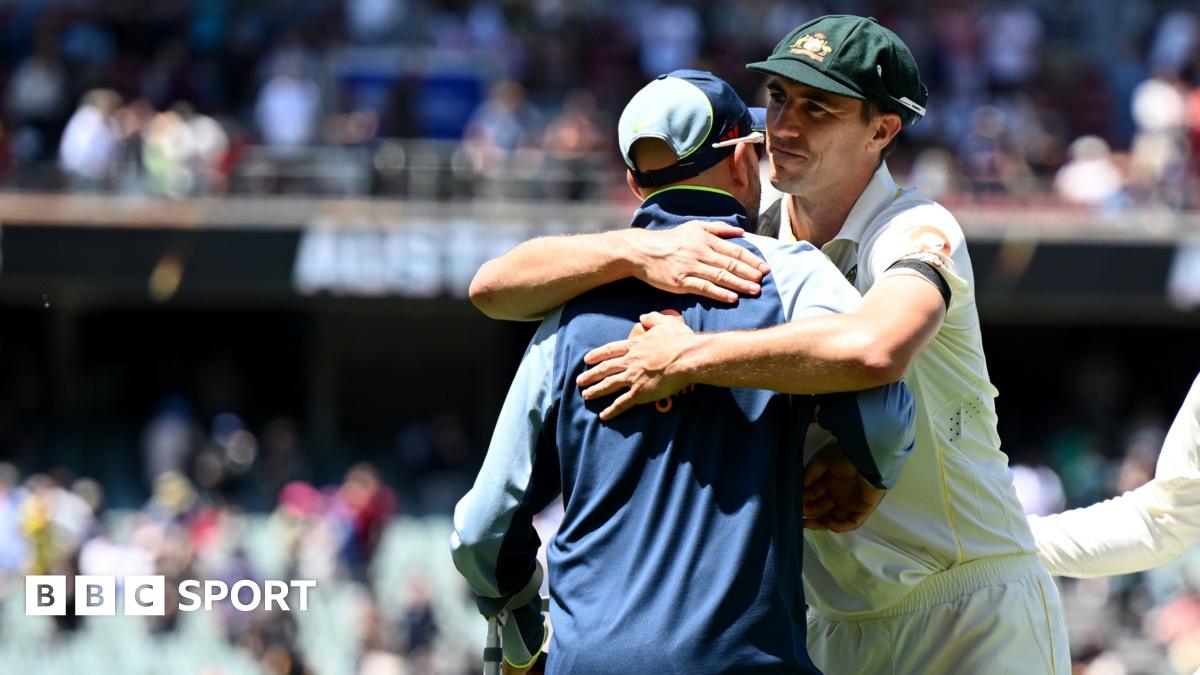
Preparation of SDS-EDTA-treated chromatography paper strips
Paper strips were prepared as previously described [4]. Briefly, Whatman grade 17 Chr pure cellulose chromatography paper with a thickness of 0.92 mm, a high absorbency of 870 g water/m2 and a linear flow rate of 190 mm/30 min was used (Cytiva Whatman, Kent, UK). The chromatography paper was cut into strips of 80 mm x 4 mm, while wearing disposable gloves to prevent contamination. The strips were immersed for 2 min in a solution containing 2% (w/v) sodium dodecyl sulfate (SDS), 10 mM ethylenediaminetetraacetic acid (EDTA), and 60 mM Tris–HCl [tris(hydroxymethyl) aminomethane hydrochloride]. Following immersion, the strips were air-dried overnight at room temperature (20–25 °C).
Cells
Human embryonic lung cells (HEL-299, CCL-137 ATCC) were grown in Dulbecco’s Modified Eagle’s Medium (DMEM, Thermo Fisher Scientific) supplemented with 10% foetal calf serum (FCS), 2 mM l-glutamine, 0.1 mM non-essential amino acids, 1 mM sodium pyruvate, and 10 mM HEPES, at 37 °C in a 5% CO2 humidified atmosphere.
Viruses
The following viral strains were used: MPXV (GenBank accession number ON622712.1), successfully isolated from genital lesion swabs from a Belgian patient, HSV-1 (Kos strain, ATCC VR-1493), HSV-2 (G strain, ATCC VR-3393), and VZV (Oka strain, ATCC VR-1832). Viral stocks were prepared and titrated in HEL cells: MPXV (2.5 × 10⁷ PFU/mL), HSV-1 (2 × 10⁶ PFU/mL), HSV-2 (3 × 10⁵ PFU/mL), and VZV (4.5 × 10⁴ PFU/mL).
All MPXV-related work was conducted in the high-containment BSL3 facilities of the KU Leuven Rega Institute under the official permit with reference number AMV/02062020/S88219.2020/0221, and according to the institutional guidelines. VZV and HSV work was conducted in BSL2 facilities at the Rega Institute following biosafety guidelines.
Sample dilutions, loading on the chromatography paper strips and incubation
All viral stocks (MPXV, VZV, HSV-1 and HSV-2) were serially diluted 1:100, 1:1.000, 1:10.000 and 1:100.000. The SDS-EDTA-treated chromatography paper strips were immersed for approximately 1 min (until the paper was saturated) into the different dilutions of the vital stocks. The strips were allowed to air dry overnight at room temperature. After complete drying, each strip was kept in a separate 50 mL Eppendorf tube and stored under different temperature conditions, -20 °C, 4 °C, room temperature (22 °C), and 37 °C. Storage durations included 1, 7, 14, 60, and 120 days. Negative controls for SDS-EDTA strips (without application of virus) were used for MPXV in each time point.
Viral inactivation tests
To assess potential viral survival on SDS-EDTA-treated chromatography paper strips, viral clearance studies were conducted using cell-based infectivity assays.
A 1:10 dilution of each virus (MXPV, VZV, HSV-1, and HSV-2) was applied to both SDS-EDTA-treated and untreated chromatography paper strips. The strips were then placed in sterile tubes containing 3 mL Universal Transport Medium (UTM), vigorously mixed and filtered through a 0.45 μm membrane. Subsequently, 0.1 mL of this UTM filtrate was inoculated onto HEL cells (for MPXV and VZV) or E1SM cells (for HSV-1 and HSV-2). Cell cultures were monitored daily for cytopathic effects over a 14-day period. Viral titers were quantified via serial 10-fold dilutions of the samples in 96-well microtiter plates pre-seeded with HEL or E1SM cells.
Viral DNA extraction
After each storage time interval at the different temperatures, the DNA extraction was performed on the virus-laden strips. The SDS-EDTA strips were cut in half and put into an Eppendorf tube with 1000 µl of RNA-free water. After a 10-minute incubation, the SDS-EDTA strips were squeezed thoroughly with the pipette tip to release absorbed viral material. A 400 µL aliquot of the resulting supernatant was used for viral nucleic acid extraction using the MagMAX™ Viral/Pathogen Nucleic Acid Isolation Kit, automated on a on Kingfisher Flex-96 purification system (ThermoFisher Scientific, Europe). Negative controls for DNA extraction step were used for virus at each time point.
Real-time polymerase chain reaction (qPCR)
Primer and probe sequences are provided in Supplementary Data (Table 1). MPXV primers and probe [7] and HSV-1/HSV-2 primers and probes [8] were published previously. qPCR amplification was conducted on a QuantStudio 7 Flex Real Time PCR system (Applied Biosystems, ThermoFisher). To amplify the targets of interest, a mix for each virus was made using 5 µL TaqMan™ Fast Virus 1-step Master Mix (Applied Biosystems) with 1 µL of each forward and reverse primers (stock concentration 20 µM) and 0.5 µL probe (stock concentration 10 µM). Supplemented with 7.5 µL RNase free water to a total of 15 µL. A total of 5 µL viral DNA was added to the reaction mixes. Thermal cycling conditions were 20 s at 95 °C, followed by 45 cycles of 3 s at 95 °C and 30 s at 60 °C. Analysis was done using QuantStudio Real-Time PCR software (Applied Biosystems, ThermoFisher).