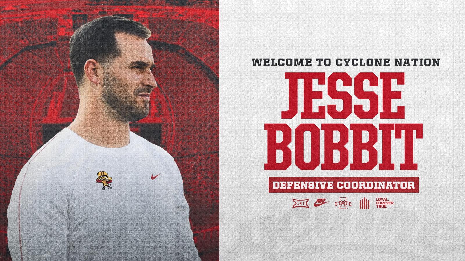
Introduction
Klebsiella pneumoniae (KP) is a highly adaptable opportunistic pathogen and a major contributor to global mortality associated with antimicrobial resistance.1 Taxonomically, it is a member of the family Enterobacteriaceae and genus Klebsiella. It is a Gram-negative, facultatively anaerobic bacillus characterized by the presence of a prominent capsule, frequent occurrence of pili, and the absence of both spores and flagella.2 Based on virulence and pathogenic features, KP strains are broadly classified into two distinct types: classical KP (cKP) and hypervirulent KP (HvKP). Compared to cKP, HvKP exhibit potent virulence and are capable of causing community-acquired infections in otherwise healthy individuals. These infections involve multiple organ systems and can manifest as pneumonia, liver abscesses, endophthalmitis, and meningitis.3 According to systematic reviews, KP is a leading etiological agent of Gram-negative meningitis and bacteremia in low-and middle-income countries.4 Infections caused by HvKP are typically acute in onset, characterized by rapid progression to disseminated systemic infections, and associated with significant morbidity and poor prognostic outcomes. According to the 2017 Infectious Diseases Society of America (IDSA) recommendations, intraventricular drainage and intrathecal antibiotic administration may be implemented for meningitis patients with severe central nervous system (CNS) infections.5 In this case, the primary genetic markers of the hvKP strain were associated with rmpA and siderophore systems.
Case Presentation
The patient was a 63-year-old female with a four-year history of diabetes mellitus. She had been receiving drug treatment and was able to maintain a relatively normal lifestyle, although glycemic control remained poor. Before acute deterioration, for 72 hours, the patient developed prodromal symptoms including fatigue and anorexia. She presented to the emergency department with a 24-hour history of progressive delirium, altered mental status, and tympanic fever, and was subsequently hospitalized for urgent evaluation and treatment. Initial laboratory tests showed leukocytosis, with a white blood cell (WBC) count of 20.81×10⁹ /L (normal range: 4.0–9.5×10⁹ /L) and neutrophil predominance at 90.6% (normal range: 40.0–75.0%). Blood glucose was significantly elevated at 24.13 mmol/L (normal range: 4.1–5.9 mmol/L), and C-reactive protein (CRP) level was significantly elevated at 328.25 mg/L (normal range: ≤10 mg/L). Blood gas analysis indicated a pH of 7.49 (normal range: 7.35–7.45), PaO2 of 83 mmHg (normal range: 80–100 mmHg), PCO2 of 19 mmHg (normal range: 35–45 mmHg), HCO3− of 13.2 mmol/L (normal range: 21–27 mmol/L), and substantially elevated levels of lactic acid at 2.9 mmol/L (normal range: 0.9–1.7 mmol/L). Procalcitonin (PCT) was significantly elevated at 23.15 ng/mL (normal range: 0–0.05 ng/mL). Brain MRI scans revealed multiple intracranial infectious lesions accompanied by pneumocephalus (Figure 1A). Chest CT scans revealed bilateral lung inflammation, multiple pulmonary abscesses, and small bilateral pleural effusions (Figure 1B and C). Abdominal CT revealed cavitary changes in the right hepatic lobe (Figure 1D).
Figure 1 Imaging findings suggest multiple infectious lesions. (A) Brain MRI scan: multiple intracranial infectious lesions accompanied by pneumocephalus. Arrow points to the high signal areas in the temporal lobes. (B) Chest CT: multiple areas of consolidation in the right lung, with an arrow indicating one such consolidation. (C) Chest CT: mediastinal window shows consolidation with cavity in the right lung. Arrow indicates the cavity. (D) Abdominal CT: liver Abscess and arrow points to the liver lesion.
|
The patient was diagnosed with sepsis and admitted to the intensive care unit (ICU) for further treatment. Empiric antibiotic therapy was initiated with intravenous meropenem (2.0 g every 8 hours, administered for over 3 hours) in combination with linezolid (600 mg every 12 hours). Shortly thereafter, the patient exhibited progressive neurological deterioration, characterized by deepening coma, bilateral anisocoria, and respiratory distress. Mechanical ventilation was employed to manage dyspnea. Subsequently, a lumbar puncture was performed to access the cerebrospinal fluid (CSF), which appeared purulent and turbid (Figure 2D1), with an opening pressure of 375 mmH2O (equivalent to approximately 3.68 kPa). Biochemical analysis of the CSF revealed the following abnormalities: elevated glucose 1.11 mmol/L, increased levels of chlorine at 119.3 mmol/L, elevated levels of protein at 10.36 mg/L, and significantly elevated nucleated cell count of 85000 × 106/L (Table 1).
![]() |
Table 1 Susceptibility Test Result of the Bronchoalveolar Lavage Fluid and Cerebrospinal Fluid Culture Was Obtained on October 14, 2022 (Day 2)
|
![]() |
Figure 2 Cerebrospinal fluid (CSF) samples demonstrating varying degrees of appearance. (D1) Appearance of CSF on day 1. (D2–D5) The appearance of CSF following CSF exchange therapy and intrathecal injection was gradually cleared from day 2 to day 5.
|
On the second day of hospitalization, the patient demonstrated a deteriorated condition. The glasgow coma scale (GCS) score decreased from 7 to 4. Due to the high viscosity of the CSF, drainage was ineffective, prompting the placement of a lumbar cisternal drainage catheter via subarachnoid puncture to facilitate CSF replacement therapy. Daily CSF exchange therapy was initiated, involving the removal of 10 mL of CSF followed by the infusion of an equal volume of normal saline, for a total of approximately 30 mL per day. To further reduce inflammatory response and enhance antibiotic concentration, intrathecal injection of 3 mg dexamethasone combined with 50 mg amikacin was conducted following each CSF exchange. Concurrent intravenous anti-infective therapy was continued with meropenem (2.0 g every 8 hours) and linezolid (600 mg every 12 hours). This combined regimen of CSF exchange and intrathecal injections was continued for five consecutive days, CSF, during which the CSF was gradually cleared (Figure 2D1–D5).
On the fourth day after admission, KP was isolated from blood, bronchoalveolar lavage fluid (BALF), and CSF cultures (Table 2), with a positive string test indicative of a hypervirulent phenotype (Figure 3A and B). Whole-genome sequencing (WGS) further confirmed the hypervirulent phenotype of the KP strain, multilocus sequence typing (MLST) analysis confirmed that the isolate belongs to sequence type 65 (ST65-K1), identifying key virulence determinants including the rmpA transcriptional activator, aerobactin synthesis gene cluster (iucABCD), salmochelin siderophore system (iroBCDE), enterobactin biosynthesis genes (entABCDEFS), iron uptake-related genes (fepABCDG), type 3 fimbrial genes (mrkABCDFHIJ), type 1 fimbrial genes (fimABCDEFGHIK), and K1 capsular serotype-associated loci. Collectively, the clinical presentation and laboratory findings were consistent with the diagnosis of a disseminated infection due to HvKP (Table S1).
![]() |
Table 2 Laboratory Data in Cerebrospinal Fluid Laboratory During Treatment
|
![]() |
Figure 3 Klebsiella pneumoniae (KP) identification. (A) Shows a petri dish containing a bacterial culture on a blood agar plate. An arrow points to a specific colony morphology. (B) Blood agar plate showing positive string test for the KP isolated from the patient.
|
By the eighth day of hospitalization, the patient exhibited improvement in neurological functions, presenting with lethargy but able to open her eyes spontaneously. The motor function of the upper extremities was partially preserved, with the ability to perform simple command-based movements. However, due to persistent severe pulmonary infection, a tracheostomy was performed (Figure 4A and B). Ongoing treatment included intravenous antimicrobial therapy, which was de-escalated to ceftriaxone (2 g IV once daily) combined with moxifloxacin (0.4 g IV once daily), based on antimicrobial susceptibility testing. Supportive care comprised albumin infusion, parenteral nutrition, and symptomatic management.
![]() |
Figure 4 Repeat chest CT on day 8 reveals bilateral cavitary infectious lung lesions. (A) Demonstrating right lower lobe consolidation (arrow). (B) Showing cavitation within the consolidated area and progression of the infection (arrow).
|
Following comprehensive treatment, the patient exhibited marked improvement in consciousness level, including regained spontaneous eye-opening and command-following movements of the upper extremities. Successful weaning from invasive mechanical ventilation was achieved on day 11 of hospitalization. Repeat brain MRI on day 11 demonstrated complete resolution of the previously identified purulent meningoencephalitis lesions (Figure 5A and B), while concurrent thoracic and abdominal CT scans revealed significant therapeutic response with >80% reduction in pulmonary and hepatic abscesses volumes (Figure 5C and D). A multidisciplinary team implemented a stepwise decannulation protocol on day 20, resulting in the successful removal of the tracheostomy tube and the initiation of structured neurorehabilitation. The patient was discharged on hospital day 33 after successful clinical cure. At discharge, final neurological examination revealed a GCS score of 15, preserved language function, and full muscle strength (grade V) in all four limbs, without residual neurological deficits. At the six-month follow-up, the patient could independently perform all activities of daily living.
![]() |
Figure 5 Repeat imaging involving brain MRI, chest and abdominal CT scans on day 11. (A and B) Brain MRI shows that intracranial infection lesions have been cleared. (C) Chest CT shows that the right lung abscess has significantly reduced in size (arrow indicates lesion). (D) Abdominal CT shows that the liver abscess has significantly reduced in size (arrow indicates lesion).
|
Discussion
Etiologically, HvKP is primarily associated with community-acquired infections. It is significantly prevalent among Asian, Pacific Islanders, and Hispanic populations. While HvKP typically affects individuals with diabetes or a compromised immune system, it has also been shown to affect healthy individuals. Typically, the disease is invasive, with liver abscesses representing the most prevalent initial presentation, followed by metastatic infections such as endophthalmitis, pulmonary abscesses, meningitis, splenic abscesses, and necrotizing fasciitis. These infections progress rapidly, resulting in exacerbated neurological functioning and suboptimal prognostic outcomes.6 Additionally, studies have reported cases of post-traumatic systemic invasive infections due to HvKP.7 Studies indicate that diabetes is an independent risk factor for systemic HvKP infections, a phenomenon potentially attributable to the reduced immunity under hyperglycemic conditions.8
For instance, the patient in this case report had pre-existing diabetes, resulting in rapidly progressing disseminated infections involving the brain, lungs, and liver following HvKP infection. Key virulence factors for KP include capsular serotypes (K1 and K2), hypermucoviscous phenotype, virulence plasmids, lipopolysaccharides, iron acquisition systems, and fimbriae.9,10 Hypermucoviscous KP strains expressing K1 and K2 capsular serotypes are strongly associated with treatment-resistant liver abscesses and recurrent invasive infections. HvKP exhibits enhanced iron acquisition capabilities, capsule production, and biofilm formation compared to cKP. These pathological mechanisms contribute to its enhanced invasiveness.11 The ST65-K1 hypervirulent Klebsiella pneumoniae (hvKP) strain described in this study carries key virulence determinants, including the capsular hypermucoviscosity regulator rmpA and siderophore gene clusters. The rmpA gene drives excessive capsular polysaccharide expression, conferring a hypermucoid phenotype that promotes invasive dissemination. Concurrently, the siderophore clusters enable high-affinity ferric iron (Fe³+) scavenging, breaching host nutritional immunity in iron-restricted organs such as the liver, thereby confirming the diagnosis of the HvKP infection. Research has shown that HvKP-related mortality increases significantly with invasion of multiple organs (such as liver, brain, lungs), with reported mortality rates of 45% for KP meningitis.12,13 Empirical antibiotic regimens for KP infections recommend cephalosporins combined with aminoglycosides. For intracranial infections, third-generation cephalosporins—such as ceftriaxone or cefotaxime—with high CSF penetration capability are recommended as first-line treatment options. Additionally, carbapenem agents—such as imipenem and meropenem—are recommended in cases where extended-spectrum β-lactamase (ESBL) production is suspected.14 Meropenem is recommended as the first-line therapy for severe infections involving multiple sites,15 particularly in critically ill diabetic patients undergoing intensive care.16 For HvKP, antibiotics with strong tissue penetration are highly recommended, coupled with abscess drainage in cases where the procedure is clinically feasible. In this case, percutaneous drainage was not conducted given the small size (3 cm) of the liver abscess size.17 HvKP-related intracranial infections progress rapidly, characterized by significantly high mortality rates, and an undefined optimal treatment duration. Studies indicate that survival and favorable neurological outcomes are associated with initial GCS scores ≤7 at the time of antibiotic initiation.18 Aztreonam, aminoglycosides, and carbapenems are the primary therapeutic options for KP meningitis, although they exhibit varying efficacy. According to the 2023 statistics from the CHINET China Bacterial Resistance Surveillance Network (www.chinets.com), KP ranks as the second most common pathogen among clinical isolates, accounting for 14.22% of cases. Notably, carbapenem resistance rates have exceeded 20%. Key resistance mechanisms identified include ESBLs, carbapenemases such as KPC, and metallo-β-lactamases, including the NDM.19 HvKP-induced community-acquired meningitis with septic shock is associated with a high hospitalization rate, with a 28-day mortality.20 In this case, intrathecal amikacin and dexamethasone were administered based on the results from the susceptibility test. Intrathecal administration of amikacin enables direct attainment of therapeutic concentrations in the CSF, thereby enhancing antimicrobial efficacy.21 Additionally, findings from meta-analyses indicate that intrathecal dexamethasone exhibits significant efficacy in reducing capillary permeability, inflammation, cerebral edema, and intracranial pressure, while enhancing antibiotic activity.22
The patient in this case presented with an acute-onset, a history of diabetes mellitus, and disseminated invasive infection. The disease progressed rapidly, resulting in impaired consciousness and respiratory failure. Based on clinical manifestations and genomic sequencing, the diagnosis was confirmed to be HvKP invasive syndrome complicated by severe intracranial infection. Due to the high viscosity of the CSF, preventing drainage through conventional methods, an integrated approach involving CSF exchange and intrathecal injections with amikacin and dexamethasone was used to treat the patient. This approach was therapeutically efficacious, resulting in improved clinical outcomes and subsequent discharge of the patient in stable condition.
Conclusion
Efficacious treatment of HvKP-related intracranial infections necessitates early recognition, potent antibiotic therapy with high tissue penetration ability, and quick CSF drainage. In this case, CSF exchange, combined with intrathecal injection with amikacin and dexamethasone, significantly improved clinical outcomes. This case represents the first reported application of this integrated regimen for HvKP meningitis, highlighting its significant therapeutic potential for broader clinical utility. However, this case provides an example but does not establish a generalizable standard of care, and that further studies or reports are needed to validate safety and outcomes.
Abbreviations
KP, Klebsiella pneumoniae; cKP, classical KP; HvKP, Hypervirulent KP; IDSA, Infectious Diseases Society of America; CNS, central nervous system; WBC, white blood cell; CRP, C-reactive protein; PCT, Procalcitonin; ICU, intensive care unit; CSF, cerebrospinal fluid; GCS, glasgow coma scale; BALF, bronchoalveolar lavage fluid; WGS, whole-genome sequencing; MLST, multilocus sequence typing; ESBL, extended-spectrum β-lactamase.
Data Sharing Statement
Data on the case clinical information, informed consent form, and images are available for review from the corresponding author upon request.
Ethical Approval
The publication of de-identified case details was expressly authorized under the original study approval by theAffiliated Lu’an Hospital of Anhui Medical University Institutional Review Board (Approval No. 2025LLKS-KY-042).
Consent for Publication
Written informed consent was obtained from patient and her families for the publication of case details and images. The complete signed consent form is archived at the Institutional Review Board (IRB) of Affiliated Lu’an Hospital of Anhui Medical University under approval number (Approval No. 2025LLKS-KY-042).
Acknowledgments
The authors thank the patient’s family for their consent to participatein this study as well as the medical, nursing, radiologist, and laboratory staff who were involved in the patient’s care.
Funding
The authors declare that financial support was received for the research in publication of this article. This research is supported by the Lu’an City Science and Technology Bureau Research Project (No.2024lakj013).
Disclosure
The authors report no conflicts of interest in this work.
References
1. Murray CJL, Ikuta KS, Sharara F, et al. Global burden of bacterial antimicrobial resistance in 2019: a systematic analysis. Lancet. 2022;399(10325):629–655. doi:10.1016/s0140-6736(21)02724-0
2. Bengoechea JA, Sa Pessoa J. Klebsiella pneumoniaeinfection biology: living to counteract host defences. FEMS Microbiol Rev. 2018;43(2):123–144. doi:10.1093/femsre/fuy043
3. Zhu J, Wang T, Chen L, Du H. Virulence factors in hypervirulent Klebsiella pneumoniae. Front Microbiol. 2021;12. doi:10.3389/fmicb.2021.642484
4. Hallmaier-Wacker LK, Andrews A, Nsonwu O, et al. Incidence and aetiology of infant gram-negative bacteraemia and meningitis: systematic review and meta-analysis. Arch Dischildhood. 2022;107(11):988–994. doi:10.1136/archdischild-2022-324047
5. Tunkel AR, Hasbun R, Bhimraj A, et al. 2017 infectious diseases Society of America’s Clinical Practice guidelines for healthcare-associated ventriculitis and meningitis*. Clinl Infect Dis. 2017;64(6):e34–e65. doi:10.1093/cid/ciw861
6. Namikawa H, Oinuma KI, Yamada K, Kaneko Y, Kakeya H, Shuto T. Predictors of hypervirulent Klebsiella pneumoniae infections: a systematic review and meta-analysis. J Hosp Infect. 2023;134:153–160. doi:10.1016/j.jhin.2023.02.005
7. Chen H-Q, Mo Z-H, Wei W-X. Case report: trauma-induced Klebsiella pneumoniae invasive syndrome presenting with liver abscess, lung abscess, endophthalmitis, and purulent meningitis. Front Med. 2025;11. doi:10.3389/fmed.2024.1513831
8. Li L, Yuan Z, Chen D, Xie X, Zhang B. Clinical and microbiological characteristics of invasive and hypervirulent Klebsiella pneumoniae infections in a teaching hospital in China. Infect Drug Resist. 2020;Volume 13:4395–4403. doi:10.2147/idr.S282982
9. Dong N, Yang X, Chan EW-C, Zhang R, Chen S. Klebsiella species: taxonomy, hypervirulence and multidrug resistance. eBioMedicine. 2022;79. doi:10.1016/j.ebiom.2022.103998
10. Wyres KL, Lam MMC, Holt KE. Population genomics of Klebsiella pneumoniae. Nat Rev Microbiol. 2020;18(6):344–359. doi:10.1038/s41579-019-0315-1
11. Fang CT, Lai SY, Yi WC, Hsueh PR, Liu KL, Chang SC. Klebsiella pneumoniae genotype K1: an emerging pathogen that causes septic ocular or central nervous system complications from pyogenic liver abscess. Clinl Infect Dis. 2007;45(3):284–293. doi:10.1086/519262
12. Yang X, Wang Y, Zhao S, et al. Clinical characteristics and prognosis of Klebsiella pneumoniae meningitis in adults. Heliyon. 2024;10(7). doi:10.1016/j.heliyon.2024.e28010
13. Sun R, Zhang H, Xu Y, Zhu H, Yu X, Xu J. Klebsiella pneumoniae-related invasive liver abscess syndrome complicated by purulent meningitis: a review of the literature and description of three cases. BMC Infect Dis. 2021;21(1). doi:10.1186/s12879-020-05702-3
14. Siu LK, Yeh K-M, Lin J-C, Fung C-P, Chang F-Y. Klebsiella pneumoniae liver abscess: a new invasive syndrome. Lancet Infect Dis. 2012;12(11):881–887. doi:10.1016/s1473-3099(12)70205-0
15. Committe ADAPP. 16. Diabetes care in the hospital: standards of medical care in diabetes—2022. Diabetes Care. 2022;45(Supplement_1):S244–S253. doi:10.2337/dc22-S016
16. Zerem E, Hadzic A. Sonographically guided percutaneous catheter drainage versus needle aspiration in the management of pyogenic liver abscess. Am J Roentgenol. 2007;189(3):W138–W142. doi:10.2214/ajr.07.2173
17. Russo TA, Marr CM. Hypervirulent Klebsiella pneumoniae. Clin Microbiol Rev. 2019;32(3). doi:10.1128/cmr.00001-19
18. Fang CT. Klebsiella pneumoniae meningitis: timing of antimicrobial therapy and prognosis. Qjm. 2000;93(1):45–53. doi:10.1093/qjmed/93.1.45
19. Han X, Yao J, He J, et al. Clinical and laboratory insights into the threat of hypervirulent Klebsiella pneumoniae. Int J Antimicrob Agents. 2024;64(3):107275. doi:10.1016/j.ijantimicag.2024.107275
20. Jung J, Park K-H, Park SY, et al. Comparison of the clinical characteristics and outcomes of Klebsiella pneumoniae and Streptococcus pneumoniae meningitis. Diagn Microbiol Infect Dis. 2015;82(1):87–91. doi:10.1016/j.diagmicrobio.2015.02.006
21. Jones RN, Sader HS, Beach ML. Contemporary in vitro spectrum of activity summary for antimicrobial agents tested against 18 569 strains non-fermentative gram-negative bacilli isolated in the SENTRY antimicrobial surveillance program (1997–2001). Int J Antimicrob Agents. 2003;22(6):551–556. doi:10.1016/s0924-8579(03)00245-0
22. Gao Y, Su J, Ma Y, et al. Efficacy and safety of intrathecal dexamethasone combined with isoniazid in the treatment of tuberculous meningitis: a meta-analysis. BMC Neurol. 2024;24(1). doi:10.1186/s12883-024-03701-4