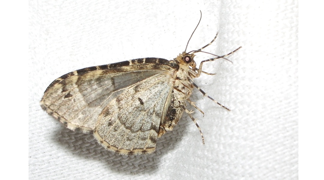
It is estimated that one-third of the 50 million people worldwide with epilepsy are resistant to anti-seizure medications. These patients, having drug-resistant epilepsy, have limited treatment options beyond surgery to control their seizures. Even surgical interventions become difficult in many of these patients due to challenges in pinpointing the anatomical source of their seizures, such as the seizures originating from multiple regions of the brain. Deep brain stimulation (DBS), a treatment that involves an implanted device that delivers an electrical current directly to areas of the brain, has emerged as a promising alternative, offering partial seizure control for patients who are not eligible for resective surgery.
In DBS, electrical impulses are delivered to parts of the brain, which are the epicenter of seizures, using precisely implanted electrodes. Electrical stimulation of the anterior nucleus of the thalamus is already approved for epilepsy in Europe and Canada. The attention is now turning towards another thalamic target, the centromedian nucleus (CM). Scientists estimate that targeting the CM, with its extensive cortical and subcortical connections, could be an effective avenue for treating general and frontal lobe seizures, including those associated with conditions like Lennox–Gastaut syndrome.
However, targeting the CM is not easy. It is small in size, located deep, and is near other thalamic nuclei, making it hard to pinpoint using standard imaging techniques. This raises the risk of faulty electrode placement, leading to poor surgical outcomes. As such, these limitations hinder CM-DBS from being widely adopted as a treatment.
In a recent review article, a team of researchers from the University Hospital La Princesa, Madrid, led by Dr. Cristina Virgina Torres Díaz, in collaboration with the University Medical Center of the Johannes Gutenberg University Mainz, reviewed advanced approaches for improving the accuracy of targeting the CM during DBS. These methods include high-resolution magnetic resonance imaging (MRI) techniques, intraoperative microelectrode recordings (MER), and diffusion tensor imaging (DTI) tractography. Together, these tools offer a multimodal approach towards seizure control and better outcomes. The article was published in Volume 1, Issue 2 of the journal Brain Network Disorders and made available online on March 9, 2025.
“Our primary goal was to reduce targeting errors and expand the clinical viability of CM-DBS,” says Dr. Torres Díaz, corresponding author of the study. “By integrating advanced imaging and neurophysiology, we can more confidently localize the CM, especially in patients with complex anatomy or structural abnormalities.”
The paper starts by discussing an MRI sequence known as magnetization-prepared 2 rapid acquisition gradient echo (MP2RAGE), used for high-resolution brain imaging. MP2RAGE enhances the contrast between the CM and surrounding thalamic structures, facilitating clearer anatomical differentiation. Together with 3D brain atlases and image gradient analysis, MP2RAGE allows more accurate visualization of the CM. Other techniques, such as quantitative susceptibility mapping (QSM) and edge-enhancing gradient echo with multi-image co-registration and averaging (EDGE-MICRA), have also additional potential for improving CM delineation.
Next, the authors examined the role of intraoperative MER in improving DBS localization. In MER, microelectrodes are used to record electrical activity from deep brain structures and help differentiate between neighboring tissues based on neural firing patterns. MER data from some studies show that the CM exhibits distinctive ‘tonic activity’ and ‘lower spike rates’ compared to adjacent nuclei (e.g., the ventral lateral nucleus). This neurophysiological signature can help guide electrode placement during DBS surgery.
The review also explored the application of DTI tractography, which can help identify relevant brain pathways and improve stimulation by targeting specific circuits. Studies that used tractography showed that the optimal stimulation areas were closely linked to fiber tracts connecting the CM to the brainstem, cerebellum, sensorimotor cortex, and supplementary motor area. Patients whose electrodes were optimally aligned with these pathways experienced significant reductions (50% or more) in seizure frequency.
“Through the review of our own patient series, we found that patients who responded most favorably to CM-DBS had strong structural and functional links between the stimulation site and specific brain networks involved in motor regulation and arousal,” Dr. Torres Díaz explained. “This highlights the importance of targeting not just a nucleus, but the circuits it controls.”
This review serves as a comprehensive roadmap for implementing CM-DBS in patients with drug-resistant epilepsy. By combining imaging modalities, electrophysiological mapping, and connectivity analysis, a team of surgeons can accurately implant electrodes and even accommodate for differences in brain structure and seizure network. This tailored approach has the potential to improve outcomes while minimizing surgical risks.
“As diagnostic tools advance and improve our understanding of brain networks, CM-DBS could offer life-changing results for patients once deemed untreatable,” concludes Dr. Torres Díaz. “Precision targeting is not just a technical achievement; it is a path to renewed hope for people with the most challenging forms of epilepsy.”
Source:
Brain Network Disorders Editorial Office
Journal reference:
Bazarra Castro, G. J., et al. (2025). Targeting the centromedian nucleus of the thalamus for epilepsy. Brain Network Disorders. doi.org/10.1016/j.bnd.2024.11.002