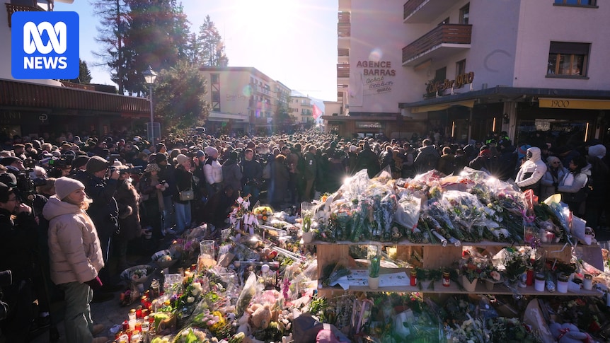
Severe community acquired pneumonia with secondary organizing pneumoni

Introduction

Pseudomonas aeruginosa (P. aeruginosa) is a common Gram-negative bacterium in clinical practice, which is characterized by its ability to colonize, adapt through mutation, and develop resistance to multiple antibiotics. Although the overall isolation rate of P. aeruginosa has decreased in recent years,1 it remains a major pathogen in hospital-acquired pneumonia and ventilator-associated pneumonia (VAP). Surveillance data indicate that it accounts for approximately 20–30% of VAP episodes, particularly in intensive care units, and is associated with high mortality and prolonged hospitalization.2,3 Although primarily a nosocomial pathogen, its role in community-acquired pneumonia is increasingly recognized. A 12-year surveillance data on community-acquired pneumonia in China found that the detection rate of P. aeruginosa in severe community acquired pneumonia (SCAP) was second only to Klebsiella, accounting for about 10.39%. The study has shown that co-infection with P. aeruginosa is an important factor in the progression of SCAP in adult aged 18 years and above.4 These observations highlight that, despite its relatively low prevalence in the community, P. aeruginosa infections warrant close attention due to their potential for severe and rapidly progressive disease.

Beyond pulmonary disease, P. aeruginosa is also capable of causing disseminated infections, particularly in immunocompromised individuals. Endophthalmitis and skin infections have all been reported, often carrying a poor prognosis.5,6 The emergence of highly virulent strains, characterized by enhanced type III secretion system (T3SS) activity and exotoxin production, further contributes to necrotizing pneumonia and systemic involvement.7 The bacterium employs various virulence factors to enhance its pathogenicity, leading to severe pulmonary damage and systemic inflammatory responses.

In addition, although uncommon, there are documented cases in which P. aeruginosa pneumonia was followed by the development of bronchiolitis obliterans organizing pneumonia (BOOP)—a form of organizing pneumonia confirmed by lung biopsy. This suggests that, beyond acute pulmonary injury, P. aeruginosa may instigate aberrant reparative responses in certain patients.8

We report a case of severe community-acquired pneumonia with secondary organizing pneumonia, endophthalmitis, and ecthyma gangrenosum caused by a disseminated infection with highly virulent P. aeruginosa. This case illustrates the pathogen’s ability to spread systemically and cause severe pulmonary and extrapulmonary complications, emphasizing the importance of early recognition and appropriate management strategies in both community and hospital settings.

Case Presentation

The patient was a 61-year-old female who presented at a local hospital with a “persistent high fever” on May 24, 2024. The examination results showed that white blood cell counts and C-reactive protein were significantly increased, and Chest Computed Tomography (CT) showed bilateral lung infection. Before admission, the patient received empirical anti-infective therapy at the local hospital, including Ampicillin-Sulbactam, Azithromycin, Moxifloxacin, and Piperacillin-Tazobactam. Despite these treatments, the patient still had persistent high fever, pain in the left eye, blurred vision, and gradually developed drowsiness. Over the past year, she underwent long-term chemotherapy for a “Malignant Tumor of the right breast (Lateral, Invasive Cancer, Grade III, T2N0cM0, StageIIa, Triple Negative)” and was fitted with a Port Infusion System four months ago. Personal history and family history are not abnormal.

Vital signs on admission were as follows (05/27/2024): Temperature 37.5°C, Heart Rate 116 beats/min, Respiratory Rate 32 beats/min, Blood Pressure 125/85 mmHg. The patient presented with drowsiness, delayed speech, hyperemia in the left conjunctiva, yellowish-white secretions within the conjunctiva sac, patchy bleeding in the left temporal and inferior conjunctiva, and turbidity of the left cornea. Bright red erosive erythema and obvious tenderness were present on the lips, with several white hyperplastic erosive plaques on the back of the tongue, and several white blisters scattered in the mucosa of the upper palate. The skin of the whole body can be seen scattered red papules of different sizes, and some surrounding areas are flushed, blistered and ruptured (Figure 1A–C). The breath sounds in the lungs are coarse and moist rales can be audible. The cardiac examination revealed no abnormalities.

Figure 1 (A) The bright red erosive erythema could be seen on the lips, and several white hyperplastic erosive plaques could be seen on the back of the tongue. (B) Scattered red papules of different sizes could be seen on the skin of the whole body (marked by red arrow). (C) Red halosis, blisters and ulcers could be seen around some parts.

Laboratory tests after admission showed: PH 7.5, Oxygen partial pressure 79.6mmHg, oxygen saturation 97.2%, oxygenation index 241mmHg (oxygen flow rate 3L/min), white blood cell count 39.41×10⁹/L, neutrophil count 37.66×10⁹/L, C-reactive protein 272.4mg/L, hemoglobin 102g/L, platelet count 51×109/L, procalcitonin 11.45ng/mL, interleukin-6 62.7pg/mL, D-dimer 4.18mg/L. Liver and kidney function tests and respiratory virus assays were negative. Antinuclear antibody profile, antineutrophil cytoplasmic antibodies, anti-O antibodies, rheumatoid factor, lupus anticoagulant were negative. Chest CT revealed multiple lobe pneumonia with bilateral pleural effusion (Figure 2A). Abdominal and craniocerebral CT showed no abnormalities. Upon admission to our hospital, anti-infective treatment was given (Omadacycline 0.1g intravenous qd, double the initial dose + Piperacillin-tazobactam 4.5g intravenous q6h) and Methylprednisolone (30mg, once daily) for anti-inflammatory therapy. After three days of anti-infection treatment, the patient’s body temperature remained high, consciousness cleared, oral mucosa and rash improved, but the vision in the left eye did not recover and was accompanied by unbearable pain, with inflammatory indicators showing improvement.

Figure 2 (A) On admission, chest CT showed multiple lung consolidation and pleural effusion, mainly in the left lung. (B) After 1 week of hormone therapy, the lesion was better than before absorption. (C) After 3 months of hormone therapy, chest CT showed that the lesion was significantly improved than before.

Sputum culture and blood culture (draw from both arms-aerobic) results showed P. aeruginosa(05/30/2024). The antibiotic sensitivity results of the blood culture indicate that P. aeruginosa is sensitive to all antimicrobial agents. The results of catheter tip and intraductal blood culture at infusion port were negative. Based on the drug sensitivity results, the treatment regimen was adjusted to include Amikacin 1g once daily and Meropenem 1g every 8 hours for anti-infection purposes. After four days, the patient continued to experience persistent high fever. A subsequent chest CT scan revealed that the inflammation had deteriorated. A bronchoscopy showed a large amount of white sputum retention, mucosal hyperemia and edema (06/05/2024). Metagenomics next-generation sequencing of alveolar lavage fluid revealed that the number of unique reads of P. aeruginosa were 652 and the relative abundance was 42.58% (06/07/2024). The bronchoalveolar lavage fluid culture’s drug sensitivity results indicate P. aeruginosa with intermediate sensitivity to ticarcillin-clavulanic acid and sensitivity to other antibiotics (06/08/2024).

Considering the patient’s high fever and the possibility of endophthalmitis, the ophthalmologist recommends performing enucleation of the ocular contents (06/06/2024). Preoperative physical examination showed no visual acuity in the left eye, limited eye movement, highly hyperemia and edema of the eyelid and conjunctiva, edema of the corneal stroma, yellowish white flocculent exudation in the anterior chamber, pupil dilated and fixed, light reflection disappeared (Figure 3A). A large amount of yellow pus overflow after opening the cornea of the left eye during the operation (Figure 3B). Upon removal of the left eye’s contents post-surgery, yellow purulent material was observed (Figure 3C). The postoperative pathology of the eyeball (06/08/2024) showed that a large number of neutrophil infiltrates were observed around the tissue, presenting suppurative inflammatory changes and abscess formation (Figure 4A). The growth of P. aeruginosa with sensitive to all antimicrobial agents was observed in the eye pus medium (06/09/2024) (Figure 5).

Figure 3 (A) Left eye vision, limited eye movement, moderately elevated intraocular pressure, eyelid and bulbar conjunctival hyperemia and edema, corneal stroma edema, yellow and white flocculent exudation in the anterior chamber, pupil dilated and fixed, light reflection disappeared. (B) A lot of yellow pus overflow after opening the cornea of the left eye during the operation. (C) The contents of the left eye removed after surgery: yellow purulent material was observed.

Figure 4 (A) The postoperative pathology of the eyeball showed that a large number of neutrophil infiltration was observed around the tissue, presenting suppurative inflammatory changes and abscess formation (HE×40). (B) Large tissue necrosis was observed in the puncture tissue of the left lung, and the alveolar cavity was filled with fibrinoid exudate and granulation tissue (HE×40).

Figure 5 The growth of Pseudomonas aeruginosa in chocolate medium of eye pus.

To detect resistance and virulence factors of P. aeruginosa, the third-generation whole-genome sequencing of the eyeball pus strain revealed a complete chromosome and no plasmid was detected. The total genome size was 6.14 MB, consistent with the range of P. aeruginosa. Multisite Sequence Typing identified the isolate as sequence type 1207. Additionally, we detected several virulence factors, including the T3SS effectors exoS, exoT, and exoY. The complete genome sequences of have been deposited in GenBank under the accession number PRJNA1294486. Virulence genes were identified via VFDB (https://www.mgc.ac.cn/VFs/), the result was provided in Supplementary Table 1. Antimicrobial resistance genes were identified using AMRFinderPlus v3.11.1 with database version 2024–10-22.1, the result was provided in Supplementary Table 2.

Following the patient’s fever persisted, and chest CT examination revealed that the inflammation had progressed further. After communicating with the patient’s family, an ultrasound-guided lung puncture biopsy was performed (06/11/2024). Post-operative pathology revealed extensive tissue necrosis in the puncture tissue of the left lung, with the alveolar cavity filled with fibrinoid exudate and granulation tissue (Figure 4B). The pathologist diagnosed it as organizing pneumonia (OP).

After multidisciplinary consultation, the patient was prescribed Cefoperazone sulbactam (3.0g intravenous q8h) for anti-infection and methylprednisolone (40mg intravenous qd) for anti-inflammatory therapy. The following day, the patient’s temperature had normalized. A week later, the chest CT examination revealed that the inflammation had improved (Figure 2B). The left eye recovered well post-surgery, and the skin rash gradually subsided (Figure 6). Following discharge, the patient continued the oral administration of prednisone tablets, initially at a dosage of 8 tablets per day, which was gradually decreased after two weeks. A reexamination of chest CT after three months revealed significant improvement in absorption (Figure 2C). The patient is currently undergoing long-term follow-up.

Figure 6 The skin rash gradually subsided.

Discussion

The patient initially presented with consolidation on chest CT and received empirical antibiotics (omadacycline plus piperacillin-tazobactam) without improvement, raising suspicion for OP. Early lung biopsy was recommended but declined. Initial methylprednisolone therapy (60 mg/day) failed to reduce fever. Targeted antibiotics against P. aeruginosa (meropenem plus amikacin) led to gradual clinical improvement and normalization of inflammatory markers, though low-grade fever persisted. A subsequent lung biopsy confirmed SOP, and reintroduction of methylprednisolone (40 mg/day) promptly normalized body temperature, with chest imaging showing lesion resolution within a week. Although bacterial infections are less commonly reported than viral causes, case reports indicate that P. aeruginosa pneumonia can trigger excessive pulmonary inflammation leading to SOP, such as BOOP.8 This case highlights the potential association of SOP with P. aeruginosa infection and underscores the importance of considering SOP in pneumonia patients not responding to standard therapy, particularly when P. aeruginosa infection is suspected.

In this case, the interval between symptom onset and the diagnosis of OP was 19 days, which is consistent with the previous report indicating that SOP usually develops 2–3 weeks after the initial infection, particularly following viral pneumonia.9 Other studies have also shown that OP commonly appears in the subacute stage after infection, further supporting this clinical timeline.10,11 The lack of corticosteroid response at the early stage in our patient was likely because OP had not yet developed, as corticosteroids are generally effective once OP is established.12 Nevertheless, relapse may occur and the optimal dosing strategy is still debated.13 Larger studies are still required to clarify the typical onset time of SOP and to identify factors influencing treatment outcomes.Clinicians should also be aware that in pneumonia patients who do not respond to targeted antimicrobial therapy, the possibility of secondary organizing pneumonia should be carefully considered. Our case highlights that P. aeruginosa bloodstream infection may progress to systemic dissemination with multiple organ involvement. This pathogen is known to cause a wide spectrum of infections, ranging from localized forms such as green nail syndrome, otitis externa, and hot-tub folliculitis to severe systemic complications including ecthyma gangrenosum and endogenous endophthalmitis.5,6 Ecthyma gangrenosum, a cutaneous lesion strongly associated with P. aeruginosa bacteremia, has been described predominantly in immunocompromised patients and is often linked to poor prognosis.14,15 Endophthalmitis caused by P. aeruginosa has also been reported; while some cases were related to hematogenous spread from distant foci, others resulted from direct inoculation, such as contaminated eye drops, yet both underscore the organism’s potential for fulminant ocular destruction.16 In contrast, postoperative cases related to urinary tract infection have also been documented, highlighting the organism’s ability to invade through non-hematogenous routes such as surgical contamination.17 Furthermore, severe community-acquired pneumonia due to highly virulent P. aeruginosa has been documented in otherwise healthy individuals, with rapid progression and high fatality despite appropriate antimicrobial therapy.18 Taken together, these reports indicate that P. aeruginosa can cause diverse invasive syndromes beyond its classical opportunistic infections, reinforcing the importance of early recognition and aggressive management. Studies have shown that the colonization and infection of hypervirulent Klebsiella pneumoniae are closely related to its specific virulence factors, such as K1/K2 serotype, rmpA gene and siderophore, etc.19 These factors collectively lead to disseminated infections, including suppurative liver abscess, osteomyelitis, and endophthalmitis. P. aeruginosa is also rich in virulence factors and can evade the immune system’s attack and antibacterial drugs. It also possesses a range of endogenous and acquired antimicrobial resistance mechanisms, including reduced outer membrane permeability, efflux pumps, target modification, etc.3 It has been reported that the risk of acute exacerbation in patients with positive exoU/pldA bronchoectasis is 6.8 times higher than that in patients with negative bronchoectasis, and the heterogeneity of P. aeruginosa virulence genes affects acute exacerbation of bronchoectasis.20 In this case, the antibiotic sensitivity results of blood culture indicate that P. aeruginosa is sensitive to all antimicrobial agents. We conducted third-generation whole-genome sequencing on the eyeball pus strain and detected the presence of T3SS effectors (exoS, exoT, and exoY). T3SS is one of the most important virulence factors in the arsenal of P. aeruginosa, which can help inject toxic proteins into host cells and are also closely associated to the virulence of P. aeruginosa.21 There is reason to believe that the patient is suffering from a systemic disseminated infection caused by high-virulence P. aeruginosa. Clinicians should recognize that high-virulence P. aeruginosa can cause rapidly progressing, systemic infections, highlighting the need for early identification and prompt, targeted treatment.

Our research team will plan to construct a mouse model of systemic disseminated infection caused by P. aeruginosa and further explore whether there are high virulence factors or new virulence factors in P. aeruginosa, leading to systemic disseminated infection, which is intended to provide a new perspective for the treatment of P. aeruginosa infection.

Conclusion

This case represents the first report of SCAP with disseminated highly virulent P. aeruginosa infection complicated by SOP, endophthalmitis, and ecthyma gangrenosum. It underscores the ability of P. aeruginosa to cause severe community-onset disseminated disease and the importance of recognizing secondary organizing pneumonia as a potential sequela to ensure timely management.

Abbreviations

P. aeruginosa, Pseudomonas aeruginosa; VAP, ventilator-associated pneumonia; SCAP, Severe community acquired pneumonia; BOOP, bronchiolitis obliterans organizing pneumonia; CT, Computed Tomography; T3SS, Type III secretory system; OP, organizing pneumonia; SOP, secondary organizing pneumonia.

Data Sharing Statement

The data supporting this study’s the findings are available from the corresponding author (Xuedong Liu) upon reasonable request.

Ethics Approval and Consent to Participate

Written informed consent was obtained from the individual for the publication of any potentially identifiable images or data included in this article. This research was carried out according to the principles of the Declaration of Helsinki and was approved by the Ethics Committees of the Qingdao Municipal Hospital (2025-KY-010). Qingdao Municipal Hospital has approved the release of the case details.

Acknowledgments

We acknowledge the patient and her family who gave informed consent for this publication.

Author Contributions

All authors made a significant contribution to the work reported, whether that is in the conception, study design, execution, acquisition of data, analysis and interpretation, or in all these areas; took part in drafting, revising or critically reviewing the article; gave final approval of the version to be published; have agreed on the journal to which the article has been submitted; and agree to be accountable for all aspects of the work.

Funding

This work was supported by the National Clinical Key Specialty Construction Project (Department of Respiratory Medicine) (grant number GJZDZK-002) and Qingdao Science and Technology Wellness Promotion Demonstration Program [grant number 24-1-8-smjk-13-nsh].

Disclosure

The authors report no conflicts of interest in this work.

References

1. Lyu J, Chen H, Bao J, et al. Clinical distribution and drug resistance of pseudomonas aeruginosa in Guangzhou, China from 2017 to 2021. J Clin Med. 2023;12(3):1189. doi:10.3390/jcm12031189

2. Kalanuria AA, Ziai W, Mirski M. Ventilator-associated pneumonia in the ICU. Crit Care. 2014;18(2):208. doi:10.1186/cc13775

3. Horcajada JP, Montero M, Oliver A, et al. Epidemiology and treatment of multidrug-resistant and extensively drug-resistant pseudomonas aeruginosa infections. Clin Microbiol Rev. 2019;32(4):e00031–19. doi:10.1128/CMR.00031-19

4. Liu YN, Zhang YF, Xu Q, et al. Infection and co-infection patterns of community-acquired pneumonia in patients of different ages in China from 2009 to 2020: a national surveillance study. Lancet Microbe. 2023;4(5):e330–e339. doi:10.1016/S2666-5247(23)00031-9

5. Gargouri L, Maaloul I, Kamoun T, et al. Ecthyma gangrenosum: a manifestation of community-acquired pseudomonas aeruginosa septicemia in three infants. Arch Pediatr. 2015;22(6):616–620. doi:10.1016/j.arcped.2015.03.006

6. Spernovasilis N, Psichogiou M, Poulakou G. Skin manifestations of pseudomonas aeruginosa infections. Curr Opin Infect Dis. 2021;34(2):72–79.

7. Hauser AR. The type III secretion system of pseudomonas aeruginosa: infection by injection. Nat Rev Microbiol. 2009;7(9):654–665.

8. Akbar DH, Alamoudi OS. Bronchiolitis obliterans organizing pneumonia associated with pseudomonas aeruginosa infection. Saudi Med J. 2000;21(11):1081–1084.

9. Asai N, Yokoi T, Nishiyama N, et al. Secondary organizing pneumonia following viral pneumonia caused by severe influenza B: a case report and literature reviews. BMC Infect Dis. 2017;17(1):572. doi:10.1186/s12879-017-2677-1

10. Radzikowska E, Fijolek J. Update on cryptogenic organizing pneumonia. Front Med. 2023;10:1146782. doi:10.3389/fmed.2023.1146782

11. Limkul L, Tovichien P. Secondary organizing pneumonia after infection. World J Clin Cases. 2024;12(36):6877–6882. doi:10.12998/wjcc.v12.i36.6877

12. Cendon L, Rafecas Codern A, de la Rosa D, et al. Systematic review of systemic corticosteroids for treatment of organizing pneumonia. Open Respir Arch. 2022;4(4):100211.

13. Drakopanagiotakis F, Paschalaki K, Abu-Hijleh M, et al. Cryptogenic and secondary organizing pneumonia: clinical presentation, radiographic findings, treatment response, and prognosis. Chest. 2011;139(4):893–900. doi:10.1378/chest.10-0883

14. Wiedemann GM, Schneider J, Verbeek M, et al. An eschar-like souvenir from a journey to Colombia: ecthyma gangrenosum as a differential diagnosis of tropical diseases in immunocompromised patients – a case report. BMC Infect Dis. 2021;21(1):344. doi:10.1186/s12879-021-05998-9

15. Hadano Y, Yoshida-Sakai N, Imamura Y, et al. Acute myeloid leukaemia presenting with ecthyma gangrenosum as the first manifestation: a case report. Medicine. 2021;100(18):e25867. doi:10.1097/MD.0000000000025867

16. Wang T, Jain S, Glidai Y, et al. Extensively drug-resistant pseudomonas aeruginosa panophthalmitis from contaminated artificial tears. IDCases. 2023;33:e01839. doi:10.1016/j.idcr.2023.e01839

17. Chin M, Khan I. Postoperative pseudomonas aeruginosa endophthalmitis associated with asymptomatic bacteriuria. BMJ Case Rep. 2022;15(4):e246985. doi:10.1136/bcr-2021-246985

18. Barp N, Marcacci M, Biagioni E, et al. A fatal case of pseudomonas aeruginosa community-acquired pneumonia in an immunocompetent patient: clinical and molecular characterization and literature review. Microorganisms. 2023;11(5):1112. doi:10.3390/microorganisms11051112

19. Lee CR, Lee JH, Park KS, et al. Antimicrobial resistance of hypervirulent Klebsiella pneumoniae: epidemiology, hypervirulence-associated determinants, and resistance mechanisms. Front Cell Infect Microbiol. 2017;7:483. doi:10.3389/fcimb.2017.00483

20. Luo RG, Miao XY, Luo LL, et al. Presence of pldA and exoU in mucoid pseudomonas aeruginosa is associated with high risk of exacerbations in non-cystic fibrosis bronchiectasis patients. Clin Microbiol Infect. 2019;25(5):601–606. doi:10.1016/j.cmi.2018.07.008

21. Veetilvalappil VV, Manuel A, Aranjani JM, et al. Pathogenic arsenal of pseudomonas aeruginosa: an update on virulence factors. Future Microbiol. 2022;17(6):465–481. doi:10.2217/fmb-2021-0158

Continue Reading