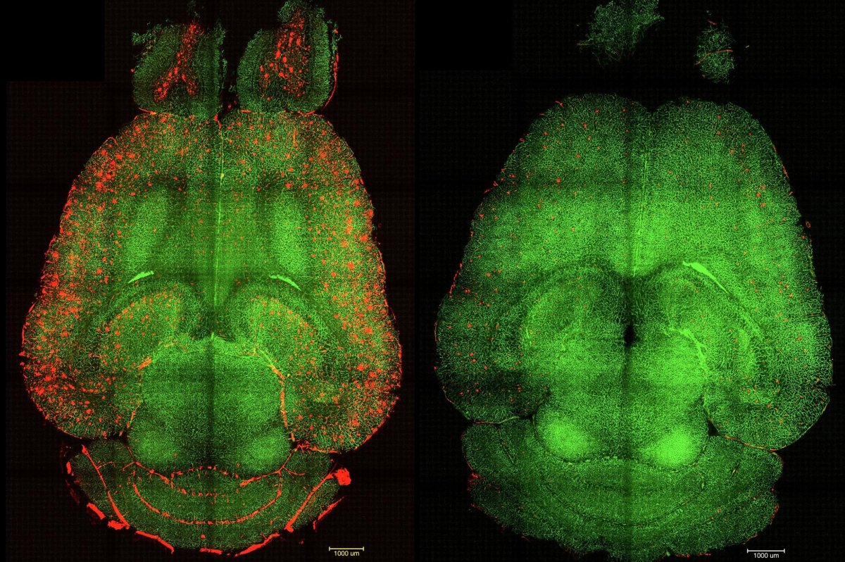
1 of 2 | Before-and-after fluorescence microscope images of a mouse brain show red-colored build-ups of toxic amyloid beta plaque (L) and the same brain 12 hours after being treated with nanoparticles. Spanish and Chinese scientists say their…

1 of 2 | Before-and-after fluorescence microscope images of a mouse brain show red-colored build-ups of toxic amyloid beta plaque (L) and the same brain 12 hours after being treated with nanoparticles. Spanish and Chinese scientists say their…