The 2R Artificiality/Adobe Stock
NEWS BRIEF
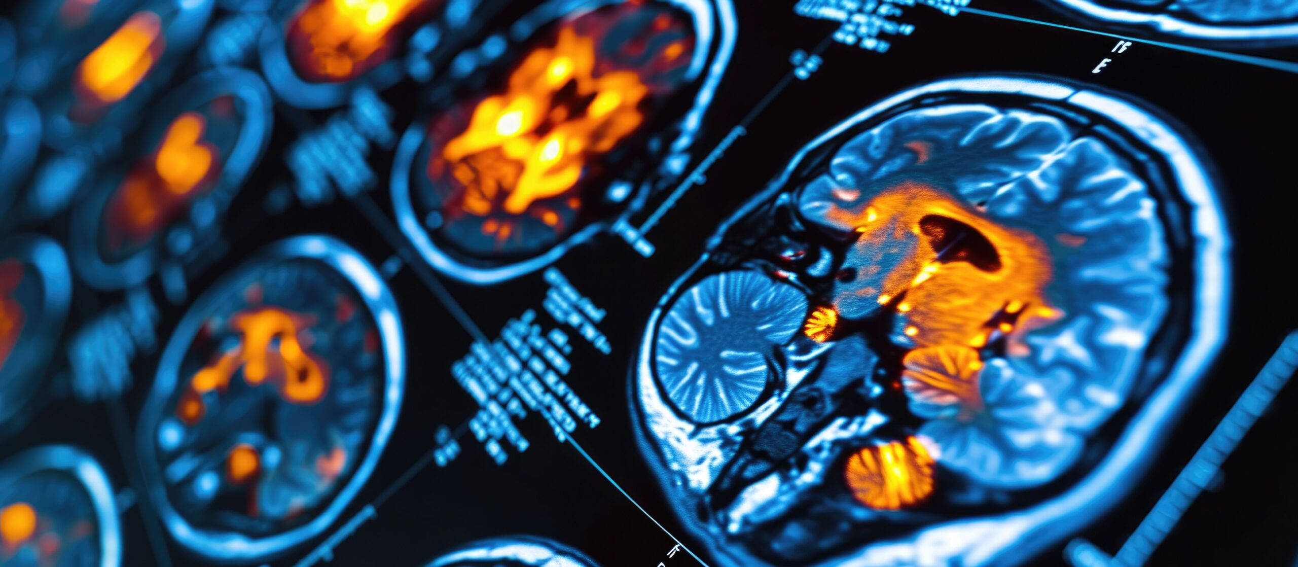
Using new, ultra high field structural imaging in 7 Tesla (7T) MRI technology, a recent study found that cortical thickness in the parahippocampus (PHC) region is correlated with major depressive disorder (MDD) and traits of neuroticism.1 Patients with MDD had significantly lower cortical thickness in the PHC region, and further analysis showed association between neuroticism and PHC thickness. Nießen et al concluded that PHC thickness may serve as a biomarker for depression and provide a neurobiological basis for neuroticism.
The study compared a group of 43 participants with MDD and a control group of 43 (20 females and 23 males in each group) participants. The MDD group met diagnostic criteria by both the IDC-10 and DSM-5. Ages ranged from 18 to 61, with a mean of 31.4. There was no significant difference for age and sex between groups. Some participants in the MDD group had other diagnoses along with MDD: 8 had a form of personality disorder, 3 had posttraumatic stress disorder, 2 had alcohol abuse issues, 1 had social phobia, 1 had somatoform disorder, 1 had trichotillomania, and 2 had previous psychotic symptoms.
In investigating potential neuromorphological correlates of MDD and the association between PHC morphology and neuroticism, the study authors used 7T MRI scanning to obtain clearer, more detailed images to examine PHC cortical thickness. Using the NEO Five Factor Inventory scale (also known as the Big Five personality inventory), authors also compared scores on the neuroticism item with PHC thickness. Because of their use of a 7T MRI scanner, this study was able to obtain clearer spatial resolution and tissue contrast of the PHC than previous studies using more standard 1.5 or 3T imaging. Cortical thickness estimate measurements are also more accurate with 7T scans.2,3
Investigators found that patients with MDD had significantly lower PHC thickness compared with the control group (P=0.002), though this significance was only found for the left hemisphere. Further analysis of the link between neuroticism and PHC thickness revealed a significant association between neuroticism and cortical thickness in both hemispheres (left: P=0.012, right: P=0.08). Participants in the MDD group had significantly higher neuroticism scores than the control group. The investigators posited that reduced PHC thickness may be indicative of a disconnect between the hippocampal formation and cortical regions. The region is associated with memory processing, emotion, and at rest mind wandering; effects of reduced cortical thickness in the PHC may lead to bias in associate memory processing, negative affect, and depressive rumination.4,5 Such structural alterations in the PHC may then play a role in perpetuating the dysfunctional cognitive processes seen in MDD and neurotic personality profiles.
This study was also the first to employ 7T MRI technology to investigate parahippocampus morphology in MDD cases. The documentation of structural alteration to the PHC provides further evidence that these changes can indicate emotional dysregulation and perhaps represent early manifestation or predispositions to MDD. Evidence of neurobiological changes related to MDD and neuroticism may provide a pathophysiological foundation of MDD, investigators posited. The study authors also note that their results “underscore the importance of multimodal assessments in MDD, potentially contributing to the foundation of individualized clinical decision-making and paving the way towards precision psychiatry.”
References
1. Nießen D, Rajkumar R, Akkoc Altinok DC, et al. 7-Tesla ultra-high field MRI of the parahippocampal cortex reveals evidence of common neurobiological mechanisms of major depressive disorder and neurotic personality traits. Transl Psychiatry 2025;15:227.
2. Scholtens LH, de Reus MA, van den Heuvel MP. Linking contemporary high resolution magnetic resonance imaging to the von Economo legacy: A study on the comparison of MRI cortical thickness and histological measurements of cortical structure. Hum Brain Mapp. 2015;36(8):3038-3046.
3. Okada T, Fujimoto K, Fushimi Y, et al. Neuroimaging at 7 Tesla: a pictorial narrative review. Quant Imaging Med Surg. 2022;12(6):3406-3435.
4. Aminoff EM, Kveraga K, Bar M. The role of the parahippocampal cortex in cognition. Trends Cogn Sci. 2013;17(8):379-390.
5. Li M, Lu S, Zhong N. The parahippocampal cortex mediates contextual associative memory: evidence from an fMRI study. Biomed Res Int. 2016;9860604
