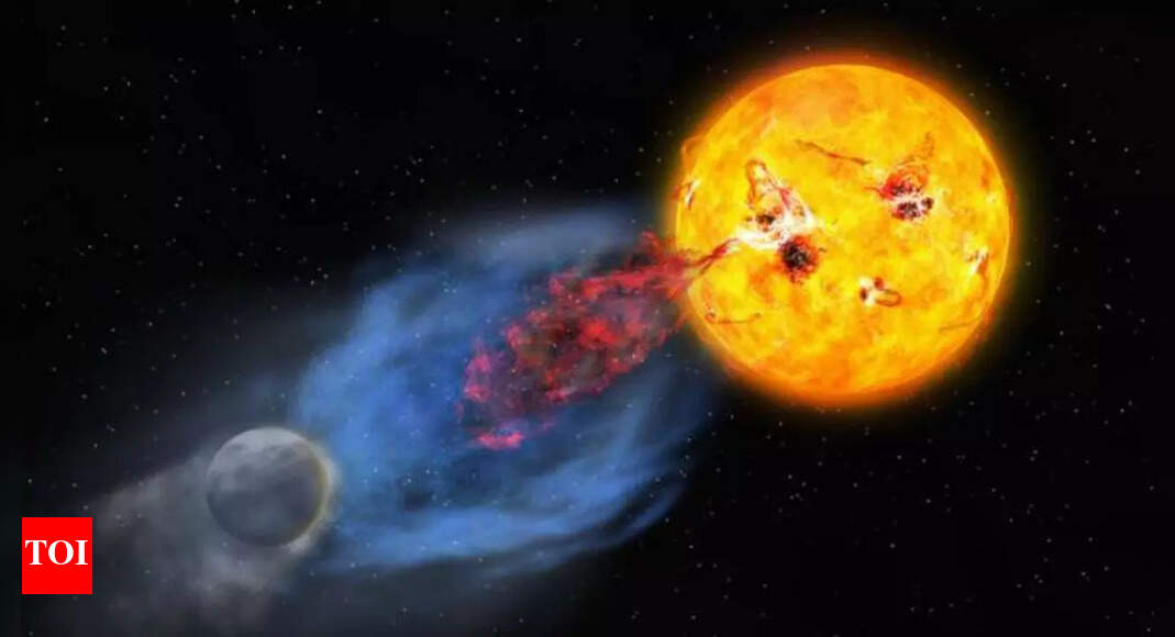
Association Between Atherogenic Index of Plasma and Endometriosis: Evi

Introduction

Approximately 10–15% of women of reproductive age are diagnosed with endometriosis,1 a common gynecological ailment. It manifests with endometrial-like tissue outside the uterus, often inducing chronic pelvic pain, infertility, and various significant symptoms that heavily impact the quality of life.2 The origin of endometriosis is attributed to genetic, hormonal, and immunological factors; however, it remains not fully elucidated. Notably, recent studies have identified abnormal lipid profiles in patients with endometriosis.3–6 Abnormal lipid metabolism may indirectly influence the growth and function of endometrial tissue by modulating inflammatory responses and immune function.7–9

The Atherogenic Index of Plasma (AIP) is calculated as the logarithm of the ratio of plasma triglycerides (TG) to high-density lipoprotein cholesterol (HDL-C).10 It is an emerging biomarker used to assess lipid metabolism disorders, reflecting the balance between atherogenic and anti-atherogenic lipoproteins.11 A high AIP level points to an increased risk of cardiovascular disease (CVD), stemming from the high prevalence of small, dense low-density lipoprotein (LDL) particles. These particles are particularly prone to oxidation and arterial infiltration, thereby contributing to atherosclerosis.12 Thus, AIP was initially used to predict atherosclerosis risk.11 Mechanistically, elevated AIP levels indicate increased small, dense LDL particles that are prone to oxidation.13 Oxidized LDL can stimulate inflammatory cytokine release (eg, IL-6, TNF-α),14 promote oxidative stress, and facilitate endometrial cell adhesion and invasion—key processes in endometriosis development.15

While AIP has been extensively studied in the context of cardiovascular risk, its relationship with endometriosis remains unclear. Investigating whether elevated AIP levels contribute to an increased prevalence of endometriosis in women is the primary focus of this study. Specifically, the study aims to: (1) compare AIP levels between women with and without endometriosis, and (2) assess the impact of elevated AIP on the likelihood of having endometriosis. To address this, we used data from the National Health and Nutrition Examination Survey (NHANES), a nationally representative survey of the US population with standardized health, dietary, and laboratory data. Its comprehensive lipid measurements and robust sampling design make it well-suited for investigating metabolic factors in gynecological conditions. Clinically, identifying an association between elevated AIP and endometriosis could encourage healthcare providers to include lipid profile evaluations in the routine management of women with endometriosis. This would enable early detection and intervention for lipid abnormalities, potentially mitigating the cardiovascular risks associated with both conditions.

Methods

Study Participants in NHANES

Initiated in 1999, the NHANES targets the evaluation of health and nutritional conditions in US adults and children. Conducted biennially, it accumulates detailed data from physical examinations, interviews, and laboratory tests. This data plays a crucial role in researching public health matters, establishing health policies, and detecting health trends and risk factors. Our study utilized the dataset covering the years 1999 to 2006. To enhance the accuracy and reliability of the results, we included only those participants who fulfilled the predetermined criteria. We omitted individuals with incomplete data on endometriosis, AIP, and related covariates. After these omissions, 1817 participants remained eligible for analysis. Figure 1 illustrates the inclusion and exclusion process of this study, with the number and percentage of exclusions indicated at each stage.

Figure 1 Flow chart of eligible participants’ selection.

Endometriosis

A diagnosis of endometriosis for women aged 20–54 was determined via a reproductive health questionnaire, which asked, “Has a doctor or another health professional ever informed you about a diagnosis of endometriosis?”

Measurement of the AIP

AIP was considered the exposure variable in this study and was defined as log10 [TG (mmol/L)/HDL-C (mmol/L)].10 The data for TG and HDL-C was procured from the “Laboratory Data” area of NHANES. Visit the NHANES website for more detailed laboratory test information.

Covariates Assessment

Our study’s covariates cover extensive social and health dimensions including racial identity, age, age at menarche, educational level, marital status, family income-to-poverty ratio (PIR), body mass index (BMI), smoking patterns, alcohol usage, hypertension, diabetes, fertility status, moderate to vigorous recreational physical activities, and female hormone use. Specifically, socioeconomic factors (education, marital status, PIR) may influence health behaviors and healthcare access, while reproductive and lifestyle factors directly impact hormonal balance and inflammatory processes relevant to endometriosis. The grouping of age at menarche, derived from a reproductive health questionnaire, consisted of three categories: under 12 years, 12 to 13 years, and 14 years or more. We queried participants about their smoking status with the question, “Have you smoked at least 100 cigarettes in your lifetime?” and measured alcohol consumption by asking, “In the past 12 months, have you consumed 4 to 5 or more drinks per day?” Histories of diabetes and hypertension were acquired from self-reports in our health survey questionnaire.

Statistical Analysis

Our analyses adhered to NHANES guidelines, incorporating fasting subsample weights for enhanced accuracy. Categorical variables underwent comparisons via the chi-square test, and continuous variables were assessed with the t-test. Means and standard errors illustrated continuous data, while categorical data were represented by percentages. For robustness testing, AIP was segmented into quartiles, with the first quartile as the reference. We constructed three multivariable logistic regression models to probe the associations with endometriosis. The initial model was devoid of adjustments. The subsequent model took into account age and race. The final model was extensively adjusted for various covariates such as age, race, educational background, BMI, age at menarche, marital status, PIR, smoking and alcohol habits, hypertension, diabetes, fertility status, moderate to vigorous recreational physical activities, and female hormone use. We have detailed the outcomes as odds ratios (ORs) with 95% confidence intervals (CIs). To elucidate the relationship between AIP and the risk of endometriosis, we employed restricted cubic splines (RCS), making adjustments for the confounders mentioned in model three. We conducted subgroup analyses and interaction tests for characteristics including age, age at menarche, smoking status, alcohol intake, fertility status, moderate to vigorous recreational physical activities, and female hormone use. The efficacy of various lipid indicators was analyzed using the XGBoost algorithm. All statistical work was performed using R and RStudio (version 4.3.2), with P<0.05 as the criterion for statistical significance.

Results

Characteristics of the Participants

Table 1 provides a summary of the primary characteristics and weighted estimates for 1817 study participants. Among these, 146 had endometriosis, while 1671 did not. Statistically significant differences (P<0.05) were observed between endometriosis and non-endometriosis groups in demographic characteristics (age, racial distribution), lifestyle factors (smoking status), female hormone use, and lipid profile parameters (total cholesterol, AIP levels). Data indicated that endometriosis-afflicted individuals tended to be older, with mean ages of 40.78 years compared to 39.18 years for controls. They were more frequently smokers of over 100 cigarettes (60.2% vs 43.5%). Additionally, their total cholesterol (TC) (206.54 mg/dL vs 196.65 mg/dL) and AIP levels (0.38 vs 0.28) were consistently higher.

Table 1 Baseline Characteristics of the Study Population Stratified by Endometriosis

Weighted Logistic Regression and Subgroup Analyses

To probe the relationship between AIP and endometriosis, we implemented weighted univariate and multivariate logistic regression analyses. Table 2 details that, upon adjusting for all relevant covariates, there was a 157.8% increase in the likelihood of endometriosis for every unit increase in AIP (OR=2.578, CI: 1.232–5.394, P=0.013). AIP was categorized into quartiles from its original continuous form. In the model with full adjustments, the frequency of endometriosis in the highest AIP quartile was 1.762 times that of the lowest quartile (OR=1.762, 95% CI: 1.056–3.103, P=0.047). Further, restricted cubic spline analysis (Figure 2) demonstrated a linear association between AIP and endometriosis after considering all covariates (P=0.042 for overall, P=0.879 for non-linearity).

Table 2 Association Between AIP and Endometriosis

Figure 2 Restricted cubic spline plot of the association between AIP and endometriosis. Model was adjusted for age, race, educational background, BMI, age at menarche, marital status, PIR, smoking and alcohol habits, hypertension, diabetes, fertility status, moderate to vigorous recreational physical activities, and female hormone use.

Details of Subgroup Analysis

As shown in Figure 3, when the association between AIP and endometriosis was observed within subgroups stratified by age, age at menarche, smoking status, alcohol intake, fertility status, moderate to vigorous recreational physical activities, and female hormone use, no interaction effects were observed for these covariates (P>0.05). This indicates that the positive correlation between AIP and endometriosis remains strong and consistent across all subgroups.

Figure 3 The relationship between AIP and endometriosis according to different subgroups.

Results of the XGBoost Model

To determine the relative importance of various lipid metrics in endometriosis, researchers employed machine learning modeling using the XGBoost algorithm (Figure 4). The machine learning results indicate that AIP is the most critical lipid metric. This is followed by TC, LDL-C, HDL-C, and TG. The model’s predictive performance was evaluated using 5-fold cross-validation. The lowest test logloss obtained during cross-validation was 0.365, indicating a relatively good agreement between predicted probabilities and actual outcomes and suggesting a low risk of overfitting. Furthermore, subgroup validation across different age groups demonstrated that AIP consistently remained the top-ranking lipid metric (Supplementary Figure 1).

Figure 4 Results of the XGBoost algorithm.

Abbreviations: AIP, atherogenic index of plasma; HDL-C, high-density lipoprotein cholesterol; LDL-C, low-density lipoprotein cholesterol; TG, triglycerides; TC, total cholesterol.

Discussion

Our records indicate that this is the first cross-sectional analysis dedicated to examining the link between AIP and the prevalence of endometriosis. Our findings highlight a positive correlation between AIP and endometriosis prevalence, even after adjusting for other covariates in multivariable regression analysis. Additionally, subgroup analysis results indicate that this positive association remains consistent across different population subgroups. Therefore, these findings provide preliminary clinical evidence of the association between AIP and endometriosis.

Various studies underscore the connection between lipid anomalies and endometriosis. He et al utilized Mendelian randomization to demonstrate significant associations between elevated TG, reduced HDL-C, and particularly pelvic peritoneal endometriosis.16 Furthermore, Li et al observed that endometriosis increases the risk of metabolic syndrome and raises TG levels.4 Similarly, Nahar et al documented significantly higher levels of TC, LDL-C, and TG, and lower HDL-C in women with endometriosis than in healthy controls.5 A study with a sample size of 120 individuals reported elevated levels of TC, TG, LDL-C, and HDL-C in women with endometriosis.17

Higher TG and LDL-C levels result in the formation of small, dense LDL particles. These particles are very susceptible to oxidation.13 Oxidized LDL particles are known to activate endothelial cells and macrophages, which then release pro-inflammatory cytokines like interleukin-6 (IL-6) and tumor necrosis factor-alpha (TNF-α).14 These cytokines significantly contribute to the inflammatory processes associated with endometriosis.18,19 Another mechanism by which lipid abnormalities may affect endometriosis is oxidative stress. Dyslipidemia, particularly with high LDL-C, leads to enhanced production of reactive oxygen species (ROS).20 When there is an imbalance between ROS production and antioxidant defenses, oxidative stress develops, causing cellular and tissue damage. In endometriosis, oxidative stress is a significant factor in the inflammatory environment.21 Oxidized lipids can enhance the adhesion of endometrial cells to the peritoneal surface and promote their invasion, which are critical steps in the establishment and progression of endometriotic lesions.15

Recent metabolomic studies have provided important evidence of lipid metabolic dysregulation in endometriosis. Using advanced techniques such as nuclear magnetic resonance (NMR) and liquid chromatography–mass spectrometry (LC-MS), researchers have identified a range of characteristic lipid alterations in patients with endometriosis.22 Notably, multiple studies have consistently reported significantly elevated levels of phospholipids, sphingolipids, and phosphatidic acid,23,24 which are closely associated with inflammatory responses and oxidative stress. These metabolomic findings support the hypothesis that AIP, as a marker of lipid imbalance, may reflect underlying metabolic disturbances in endometriosis.

The observed relationship between AIP and endometriosis has significant implications for the cardiovascular health of women with endometriosis. Elevated AIP levels indicate adverse lipid profiles characterized by high TG and low HDL-C, which are established risk factors for CVD.25 This dysregulation in lipid metabolism not only exacerbates the inflammatory environment but also contributes to the pathogenesis of atherosclerosis. Endometriosis correlates with heightened cardiovascular risk, encompassing coronary heart disease and stroke. Mendelian randomization research highlights a marked increase in stroke risk, advocating for recognition of endometriosis as a multifaceted systemic disease rather than merely a reproductive disorder.26 A large cohort study conducted by Blom et al revealed that hospitalization rates for CVD and secondary CVD events are higher among women with endometriosis. This condition may be linked to inflammatory dysregulation, enhanced oxidative stress, and negative lipid profiles.27 Okoli et al completed a systematic review and meta-analysis that substantiated a substantial link between endometriosis and increased risks of major cardiovascular events, ischemic heart disease, and cerebrovascular accidents.28 It is clinically vital to comprehend how AIP, endometriosis, and cardiovascular disease interact. Elevated AIP in women with endometriosis can serve as a useful biomarker for identifying those at high risk for cardiovascular complications. This underscores the necessity for comprehensive cardiovascular risk assessments in women with endometriosis, including regular monitoring of lipid and inflammatory markers. Early intervention strategies focusing on lifestyle modifications, anti-inflammatory treatments, and lipid-lowering therapies can mitigate these risks and improve overall health outcomes for women with endometriosis.

Furthermore, the observed association between AIP and endometriosis is statistically significant in women under the age of 40 but not in women over 40. This suggests that age-related differences in metabolic and hormonal environments may play a role. In younger women, hormonal fluctuations are more pronounced, which may exacerbate the metabolic and inflammatory processes associated with endometriosis.29 Additionally, the chronic nature of endometriosis means that by the age of 40, many women may have already undergone significant pathological changes, making it more challenging to detect the impact of AIP at this stage. To develop targeted prevention and treatment strategies for endometriosis, understanding age-specific dynamics is crucial.

This research thoroughly examines the specific influence of AIP on endometriosis risk, underscoring the essential role of lipid metabolism disorders in evaluating this risk. However, attention must be given to several limitations. Most critically, as a cross-sectional study, we cannot determine the causal direction between AIP and endometriosis. This fundamental limitation necessitates cautious interpretation of our findings. Second, although we adjusted for many covariates, unmeasured confounding may still exist. In particular, dietary habits—such as fat and cholesterol intake—are important factors influencing both lipid metabolism and endometriosis risk, but were not accounted for in our analysis. Third, the diagnosis of endometriosis in this study was based on self-reported physician diagnosis, which may lead to misclassification bias. Future studies should validate these findings using clinically confirmed cases of endometriosis. Fourth, the exclusion of participants with incomplete data may have led to selection bias; however, this concern is partially alleviated by the extensive adjustment for relevant covariates. Finally, the study population was limited to women in the United States, which restricts the generalizability of the results to other regions and populations. Therefore, future longitudinal prospective studies and comprehensive data collection are essential to confirm the association between AIP and the risk of endometriosis.

Conclusions

Our findings highlight a positive correlation between AIP and the risk of endometriosis among US females. These results provide preliminary evidence that lipid metabolism indicators may have potential as biomarkers for endometriosis risk assessment; however, further longitudinal and clinical studies are needed to validate AIP as a reliable risk assessment tool for endometriosis.

Data Sharing Statement

The data used in this study are publicly available and can be retrieved from the NHANES website: (https://www.cdc.gov/nchs/nhanes/).

Ethics Approval and Informed Consent

The study protocol (Protocol Number: Protocol #2005–06, Protocol #98-12) was approved by the NCHS Research Ethics Review Board (ERB) and all participants provided written informed consent prior to participation.

Based on Item 1 and Item 2 of Article 32 of the Measures for Ethical Review of Life Science and Medical Research Involving Human Subjects, dated February 18, 2023, China, our study is exempt from additional ethical approval.

The relevant legislation details are as follows:

Article 32: Research involving human data or biological samples, where no harm is caused to individuals and no sensitive personal information or commercial interests are involved, may be exempt from ethical review. This is to reduce unnecessary burdens on researchers and to facilitate the progress of life science and medical research involving humans.

(1) Research using publicly available data that has been legally obtained, or data generated through observation without interference with public behavior.

(2) Research using anonymized data.

Acknowledgments

We would like to thank the NHANES database for providing the data source for this study.

Author Contributions

All authors made a significant contribution to the work reported, whether that is in the conception, study design, execution, acquisition of data, analysis and interpretation, or in all these areas; took part in drafting, revising or critically reviewing the article; gave final approval of the version to be published; have agreed on the journal to which the article has been submitted; and agree to be accountable for all aspects of the work.

Disclosure

The authors declare no competing interests in this work.

References

1. Smolarz B, Szyllo K, Romanowicz H. Endometriosis: epidemiology, classification, pathogenesis, treatment and genetics (Review of Literature). Int J Mol Sci. 2021;22(19):10554. doi:10.3390/ijms221910554

2. Parasar P, Ozcan P, Terry KL. Endometriosis: epidemiology, diagnosis and clinical management. Curr Obstet Gynecol Rep. 2017;6(1):34–41. doi:10.1007/s13669-017-0187-1

3. Mu F, Rich-Edwards J, Rimm EB, Spiegelman D, Forman JP, Missmer SA. Association between endometriosis and hypercholesterolemia or hypertension. Hypertension. 2017;70(1):59–65. doi:10.1161/HYPERTENSIONAHA.117.09056

4. Li B, Zhang Y, Zhang L, Zhang L. Association between endometriosis and metabolic syndrome: a cross-sectional study based on the national health and nutrition examination survey data. Gynecol Endocrinol. 2023;39(1):2254844. doi:10.1080/09513590.2023.2254844

5. Nahar K, Khanam NN, Chowdhury AA, Khan NJ, Mohamed Z. Association of dyslipidemia with endometriosis: a case control study. Mymensingh Med J. 2023;32(1):118–124.

6. Zheng R, Du X, Lei Y. Correlations between endometriosis, lipid profile, and estrogen levels. Medicine. 2023;102(29):e34348. doi:10.1097/MD.0000000000034348

7. Andersen CJ. Lipid metabolism in inflammation and immune function. Nutrients. 2022;14(7):1414. doi:10.3390/nu14071414

8. Jackson LW, Schisterman EF, Dey-Rao R, Browne R, Armstrong D. Oxidative stress and endometriosis. Hum Reprod. 2005;20(7):2014–2020. doi:10.1093/humrep/dei001

9. Santanam N, Murphy AA, Parthasarathy S. Macrophages, oxidation, and endometriosis. Ann N Y Acad Sci. 2002;955:183–98; discussion 19–200, 396–406. doi:10.1111/j.1749-6632.2002.tb02779.x

10. Dobiasova M, Frohlich J. The plasma parameter log (TG/HDL-C) as an atherogenic index: correlation with lipoprotein particle size and esterification rate in apoB-lipoprotein-depleted plasma (FER(HDL)). Clin Biochem. 2001;34(7):583–588. doi:10.1016/S0009-9120(01)00263-6

11. Fernandez-Macias JC, Ochoa-Martinez AC, Varela-Silva JA, Perez-Maldonado IN. Atherogenic index of plasma: novel predictive biomarker for cardiovascular illnesses. Arch Med Res. 2019;50(5):285–294. doi:10.1016/j.arcmed.2019.08.009

12. Chapman MJ, Ginsberg HN, Amarenco P, et al. Triglyceride-rich lipoproteins and high-density lipoprotein cholesterol in patients at high risk of cardiovascular disease: evidence and guidance for management. Eur Heart J. 2011;32(11):1345–1361. doi:10.1093/eurheartj/ehr112

13. Krauss RM. Small dense low-density lipoprotein particles: clinically relevant? Curr Opin Lipidol. 2022;33(3):160–166. doi:10.1097/MOL.0000000000000824

14. Castro CA, Buzinari TC, Lino RLB, et al. Profile of IL-6 and TNF in foam cell formation: an improved method using fluorescein isothiocyanate (FITC) probe. Arq Bras Cardiol. 2022;119(4):533–541. doi:10.36660/abc.20210682

15. Polak G, Barczynski B, Kwasniewski W, et al. Low-density lipoproteins oxidation and endometriosis. Mediators Inflamm. 2013;2013:624540. doi:10.1155/2013/624540

16. He X, Xie S, Liu Y. The association of circulating lipoprotein lipids and apolipoproteins with risk of endometriosis: a Mendelian randomization study. Postgrad Med J. 2024;100(1186):578–583. doi:10.1093/postmj/qgae011

17. Melo AS, Rosa-e-Silva JC, Rosa-e-Silva AC, Poli-Neto OB, Ferriani RA, Vieira CS. Unfavorable lipid profile in women with endometriosis. Fertil Steril. 2010;93(7):2433–2436. doi:10.1016/j.fertnstert.2009.08.043

18. Guo F, He Y, Fan Y, et al. G-CSF and IL-6 may be involved in formation of endometriosis lesions by increasing the expression of angiogenic factors in neutrophils. Mol Hum Reprod. 2021;27(11). doi:10.1093/molehr/gaab064

19. Abutorabi R, Baradaran A, Sadat Mostafavi F, Zarrin Y, Mardanian F. Evaluation of tumor necrosis factor alpha polymorphism frequencies in endometriosis. Int J Fertil Steril. 2015;9(3):329–337.

20. O’Donnell RW, Johnson DK, Ziegler LM, DiMattina AJ, Stone RI, Holland JA. Endothelial NADPH oxidase: mechanism of activation by low-density lipoprotein. Endothelium. 2003;10(6):291–297. doi:10.1080/10623320390272280

21. Van Langendonckt A, Casanas-Roux F, Donnez J. Oxidative stress and peritoneal endometriosis. Fertil Steril. 2002;77(5):861–870. doi:10.1016/S0015-0282(02)02959-X

22. Angioni S, Saponara S, Succu AG, Sigilli M, Scicchitano F, D’Alterio MN. Metabolomic Characteristics in Endometriosis Patients. In: Genazzani AR, Nisolle M, Petraglia F, Taylor RN, editors. Endometriosis Pathogenesis, Clinical Impact and Management: Volume 9: Frontiers in Gynecological Endocrinology. Cham: Springer International Publishing; 2021:9–17.

23. Angioni S, Saponara S, Vitale SG. Metabolomics analysis in endometriosis patients: is it a step toward the future? Gynecol Endocrinol. 2023;39(1):2227276. doi:10.1080/09513590.2023.2227276

24. Adamyan L, Pivazyan L, Zarova E, et al. Metabolomic biomarkers of endometriosis: a systematic review. J Endometr Uterine Disord. 2024;7:100077. doi:10.1016/j.jeud.2024.100077

25. Caselli C, De Caterina R, Smit JM, et al. Triglycerides and low HDL cholesterol predict coronary heart disease risk in patients with stable angina. Sci Rep. 2021;11(1):20714. doi:10.1038/s41598-021-00020-3

26. Zheng M, Zheng S. Endometriosis Increases the Risk of Stroke: a Mendelian Randomization Study. Stroke. 2023;54(2):e30–e3. doi:10.1161/STROKEAHA.122.041163

27. Blom JN, Velez MP, McClintock C, et al. Endometriosis and cardiovascular disease: a population-based cohort study. CMAJ Open. 2023;11(2):E227–E36. doi:10.9778/cmajo.20220144

28. Okoli U, Charoenngam N, Ponvilawan B, Jaroenlapnopparat A, Mettler S, Obiejesie O. Endometriosis and risk of cardiovascular disease: systematic review and meta-analysis. J Women’s Health. 2023;32(12):1328–1339. doi:10.1089/jwh.2023.0091

29. Pluchino N, Freschi L, Wenger JM, Streuli I. Innovations in classical hormonal targets for endometriosis. Expert Rev Clin Pharmacol. 2016;9(2):317–327. doi:10.1586/17512433.2016.1129895

Continue Reading