By analyzing CT images with 3D software, small liver tumors can be successfully treated using ablation. This has been demonstrated by research conducted by Radboudumc.
Liver tumors up to three centimeters in size can be treated in various ways. In consultation with the patient, the treating physician decides whether to perform surgery or ablation, a method in which the tumor is punctured with a thin needle and heated so that it breaks down. The latter method is less invasive; it takes less time, there is less chance of complications, and the patient recovers more quickly.
Higher chance of cancer recurrence
However, the chance of cancer recurrence was found to be relatively high with ablation compared to surgery. “By looking at the procedure and technical possibilities together, we have arrived at a new process,” says oncological surgeon Martijn Stommel.
3D analysis during treatment
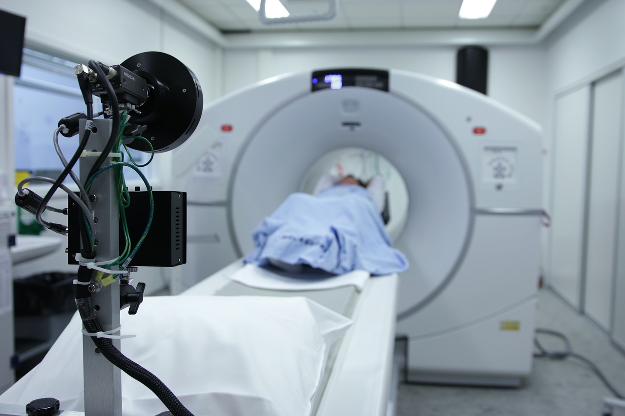
Part of this new process is a 3D analysis. The ablation does not take place in an operating room, but in a room with a CT scanner. Immediately after treatment, while the patient is still under anesthesia, a CT scan is made.
Previously, doctors assessed the CT scan visually, but this is now done using advanced 3D software. This provides a much more accurate image of the tumor. By superimposing images from before and after the operation, it is immediately clear whether the entire tumor has been treated.
More effective treatment
The results are promising. Thanks to the analysis with the 3D software, the number of recurrent liver tumors within two years after ablation has fallen sharply, from 33% to less than 10%.
The 3D analysis is a great example of the smart use of technology that directly benefits patients. Less invasive procedures are better for patients, but they also mean lower costs and less strain on the healthcare system.
AI as a digital weapon against skin cancer
Each week in our EverydAI column, we highlight an application of AI that makes your life easier. Even if you’re not an AI expert. Today: AI and skin cancer.