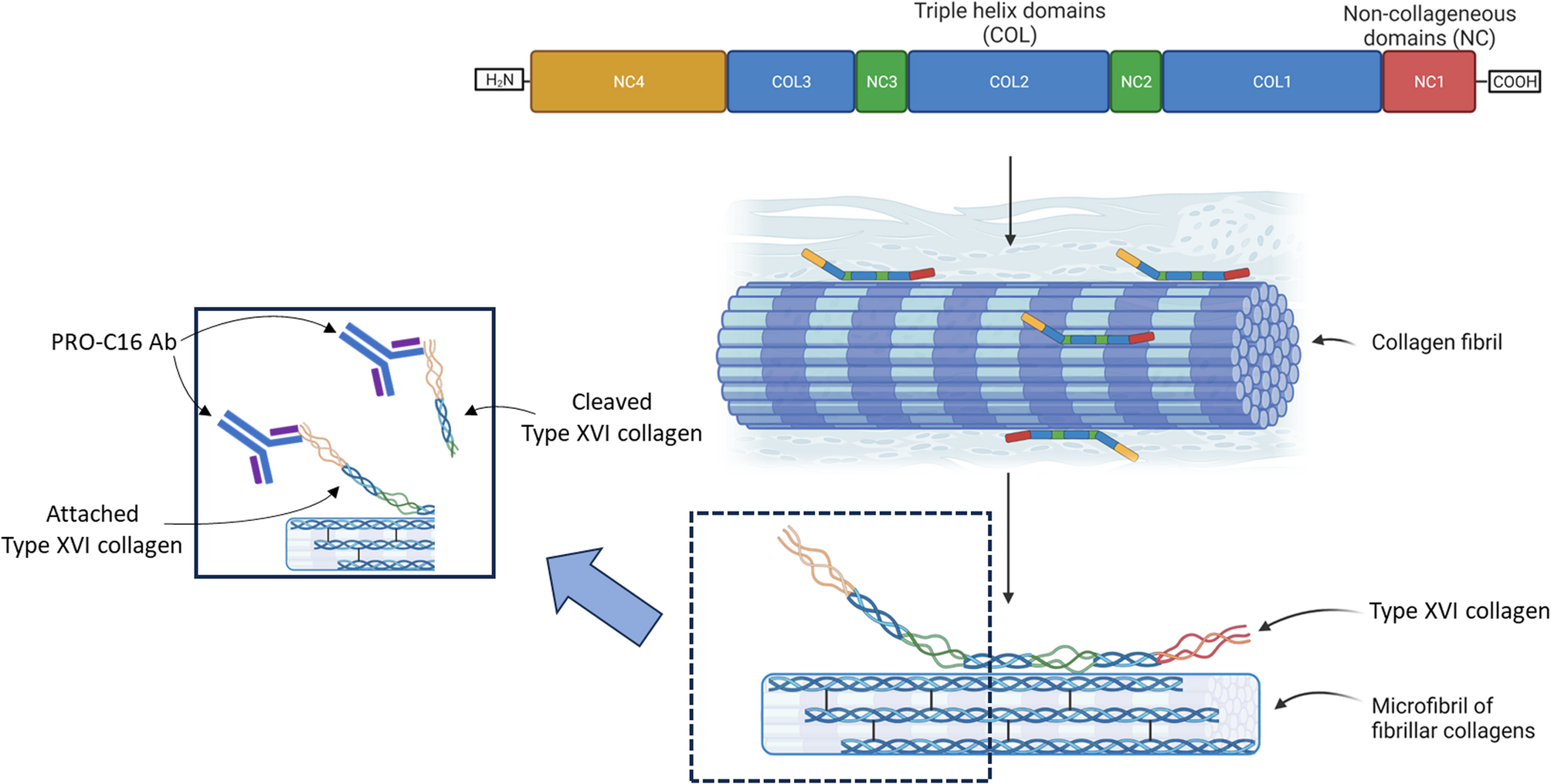
Based on two independent patient cohorts, the results presented herein provide evidence that PRO-C16 could be a clinically relevant biomarker for identifying CD patients with fibrostenotic strictures and have utility for clinical development in patients treated with novel anti-fibrotic agents. This assumption was also supported by results obtained in the chronic DSS colitis model, where PRO-C16 was also demonstrated to be elevated in the DSS rats compared to healthy rats (vehicle control). Furthermore, these results are in line with a previous report by Jensen et al., demonstrating that PRO-C16 is elevated in patients with UC [24]., where, despite the rarity of fibrostenotic strictures, there is active ECM remodeling resulting in intestinal fibrosis [6, 26,27,28]. In addition, PRO-C16 was also demonstrated to be elevated in patients with primary sclerosing cholangitis [29]. This indicates that PRO-C16 is not a diagnostic biomarker but rather reflects an active fibrotic process. Recently, Bourgonje et al. demonstrated that CD patients with stricturing disease had significantly less degradation of type I, III, and IV collagens, which fits with the overall notion that the tissue balance in stricturing CD patients has increased accumulation of collagen and reduced collagen fibrolysis [30].
The results obtained in this study are in line with the current biology and knowledge on the biology of type XVI collagen and its possible implication in tissue fibrosis [5]. Overexpression of type XVI collagen is believed to promote and support chronic inflammation, thus contributing to fibrogenesis [6, 21]. As type XVI collagen belongs to the FACIT collagen family, it is conceivable that other FACIT collagens, e.g. type IX, XII, XIV, XIX, XX, XI, and XXII collagens, could also be implicated and promoting intestinal fibrogenesis, due to their association with fibrillar collagens such as type I, III, and V collagens whose excessive deposition is the hallmark of tissue fibrosis [21,22,23], which is in line with the elevated serum levels of PRO-C16 in fibrostenotic CD patients observed in this study.
We also observed differences between the two cohorts. The B3 group from Cohort 1 has a sample size of n = 13, whereas Cohort 2’s B3 group has a sample size of n = 3. CD patients with the B3 are often presented with concomitant intestinal fibrosis [31]. In this study, B3 patients demonstrated low levels of PRO-C16. We also did not observe any statistical differences between the disease location. Since fibrostenotic strictures are often located in the terminal ileum [3]. Given PRO-C16’s stronger association with this phenotype, it is likely that PRO-C16 also would be more associated with ileal disease. Even though the PRO-C16 data is encouraging, this warrants validation in future prospective studies are essential for further evaluating the PRO-C16, e.g., as a biomarker for fibrostenotic strictures and to monitor intestinal fibrosis development. If successful, then PRO-C16 could have the potential to also be applied as a pharmacodynamic biomarker for anti-fibrotic treatments in CD.
PRO-C16 was elevated in the DSS colitis model compared to the control rats, which could indicate that the fibrogenesis was successfully induced, as increased collagen deposition and fibrogenesis were observed in the submucosa and mucosa. Furthermore, it seemed that the PRO-C16 serum level peak followed the same degree of fibrosis that could be observed from the Masson trichrome staining. Especially after the second cycle of DSS at day 21 and after the fourth cycle of DSS at day 49, severe fibrogenesis was present in the mucosa and submucosa space of the DSS rats. PRO-C16 serum levels were also lower in the DSS rats at cycle 3 at day 35, which also seemed to follow the pattern observed from the Masson trichrome staining, revealing only mild to moderate fibrogenesis in the DSS rats (Fig. 6) compared to the control rats. However, while we don’t know the exact course of the variability of PRO-C16 measurements after the DSS cycles, we believe it could indicate that continuous tissue destruction and remodeling will increase the fibrogenesis and intestinal fibrosis, which is reflected by the elevated levels of PRO-C16 after the second cycle. The drop in PRO-C16 levels after cycle 3 could be an indication of a change in the phenotype with less fibrosis but increased tissue destruction, where we see that the PRO-C16 is elevated again after cycle 4.
Two independent cohorts were included, where PRO-C16 serum levels were proven to be significantly elevated in CD patients with fibrostenotic strictures, and the elevated PRO-C16 serum levels in the chronic rat DSS model suggest that PRO-C16 could be related to intestinal fibrogenesis. While this strengthens the overall robustness of the study, there are also several limitations. Even though MRE is not preferred for consecutive evaluation of fibrostenotic stricture development, it would still be relevant to evaluate PRO-C16 serum levels and their association with the MRE findings, which is also one of the limitations of the Montreal classification of the B2 phenotype as it lacks objective fibrosis quantification, which can also result in unrecognized intestinal fibrosis development in the B1 group. The absence of histology fibrosis assessment is also a limitation of this study. However, as it is impracticable and impossible to assess the entire intestinal segments of the ileum and colon, future studies focusing on PRO-C16 should also include CT-imaging methodologies in addition to the histological assessment. The two human cohorts included were cross-sectional studies and some discrepancies were observed e.g., cohort 1 demonstrated higher disease activity (CDAI) and a greater sample size of CD patients with the B3 phenotype compared to cohort 2, and CRP and fecal calprotectin was only available for cohort 1, which makes a direct comparison difficult. For future validation studies, it would be relevant to test PRO-C16 in prospective studies and more homogeneous studies for the potential to monitor intestinal fibrosis development. The sample size for the two cohorts included is also low, and together with the lack of strong association of PRO-C16 with inflammation, it would also be relevant to evaluate PRO-C16 in patients with CD who have a quiescent inflammatory disease but with a progressing stricturing phenotype in a longitudinal study. The DSS model reflects especially colonic inflammation and fibrosis, where the chronic model is more relevant for studying fibrogenesis [32]. However, the DSS model does not replicate transmural fibrosis and stricture formation and therefore does not 100% represent human CD patients with stricturing disease [33]. Furthermore, for the chronic DSS study included in this paper we did not have the opportunity to assess the intestinal fibrotic grading, however as with the Masson Trichrome staining’s we confirm that we successfully induced intestinal fibrosis which corresponds to the serum measurements and is also in line with what we observed in the human patients CD. Given PRO-C16’s association with intestinal fibrosis, based on the murine DSS model, it would also be relevant to evaluate how PRO-C16 relates to the risk of recurrence in a post-operative CD patient population, and treatment response would provide additional clarity in this patient population with unmet clinical needs.
Since disease location and creeping fat are relevant factors for intestinal fibrosis [34]. Future studies should also aim for a bigger sample size where meaningful stratification is based on disease location and the presence of creeping fat. Furthermore, NOD2 homozygosity has proven to be a strong predictor of intestinal stenosis in CD, independent of the IL23R genotype; as such, it would be relevant to investigate PRO-C16 association with NOD2 and IL23R genotypes [31]. Finally, the PRO-C16 marker is not specific for CD, and for future studies, non-IBD controls should be included to investigate how PRO-C16 is regulated in other diseases. However, in the context of CD and intestinal fibrosis, PRO-C16 could be a relevant marker.
