Aniket Bera, director of the Ideas Lab at Purdue University, has worked on two very different projects in the AI space – the restoration of a fragment of film from 1899, believed to be the oldest surviving footage of India, and an earlier…
Author: admin
-

‘Pure euphoric escapism’: why Adventureland is my feelgood movie | Jesse Eisenberg
While casting his knockout quasi-biopic The Social Network, film-maker David Fincher must’ve really dug how eventual Mark Zuckerberg portrayer Jesse Eisenberg handled being dumped on screen. A year before the award-lassoing Facebook drama,…
Continue Reading
-
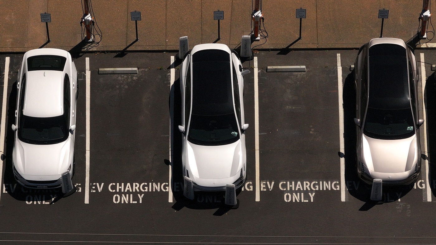
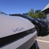
2025 was a roller coaster year for EVs : NPR
Electric cars sit parked at a charging station in Corte Madera, Calif., in May 2025.
Justin Sullivan/Getty Images North America
hide captiontoggle caption
Justin Sullivan/Getty Images North America
The electric vehicle industry has taken a pummeling this year. The Trump administration, as expected, reversed a whole suite of federal policies that promoted or encouraged EVs.
California’s ability to require the sale of EVs: gone. Federal rules about emissions and fuel economy — being rewritten. Federal penalties for car companies that sell too many gas guzzlers: zeroed out. The $7,500 federal tax credit? Kaput.
Meanwhile, automakers delayed or canceled a host of unprofitable EV plans.
The all-electric Ram 1500 REV was canceled before a single one was built. The all-electric Ford Lightning was discontinued despite some glowing reviews. (Both pickups will be replaced with extended-range electric vehicles, which come with both a big battery and a backup gas tank.)
The buzzy Volkswagen Buzz is still available in other countries, but no longer in the U.S. The GM Brightdrop van is no more. The list goes on.
As for sales? “It’s a roller-coaster ride,” says Stephanie Valdez Streaty, who monitors EVs for the data and services company Cox Automotive.
Sales spiked in August and September, during the last weeks that the federal tax credit was available, as buyers rushed to take advantage of the expiring opportunity. Cox estimated EVs hit an all-time high of 11.6% of the new vehicle market in September. Then sales crashed by 50% in October.
But here’s a twist.
“Among U.S. shoppers who are in [the] market for new vehicles, the interest in electric vehicles actually ticked up a bit after the tax credit went away,” says Brent Gruber, who runs the EV practice at consumer insights company J.D. Power.
It’s the EV story you might not have heard this year: Despite the political and product planning whiplash, consumer appetite for EVs has been on a very smooth ride.
Overall, about 25% of new car shoppers are very interested in buying an EV, according to J.D. Power surveys. And with minor fluctuations, “it’s held pretty consistent,” Gruber says, despite what he calls the “turbulence” of this year.
“There’s still a tremendous amount of interest,” he says. “And from an EV owner perspective, we continue to see high levels of satisfaction once people do get into those products.” In fact, EV owners are 94% likely to repurchase another EV for their next vehicle, he says.
BJ Birtwell runs the Electrify Expo, a traveling festival dedicated to EVs. He says EVs have suffered from being politicized, with a lot of right-of-center Americans rejecting them out of hand.
“There’s still a cloud of skepticism around EVs across some parts of the country,” he says. But put a skeptic behind the wheel of a new EV, he says, “and I’ll tell you what I see: Smiles for miles.” Test drives reveal the cars are fun to drive, he says, and a little research can show that charging at home is easier and cheaper than they thought.
An American slowdown
Still, while Americans remain interested in EVs, it’s undeniable that battery-powered vehicles are taking off more slowly than industry execs expected a few years ago. That’s not just because of the policy reversal; it’s also because of market realities. For example, while charging might be easy at home, it’s a hassle for apartment dwellers who don’t have that option. Meanwhile, vehicle prices — a challenge for the entire auto market — are even higher for EVs. Lower fuel and maintenance costs can’t always overcome that up-front sticker shock, even for people who are hypothetically interested in buying.
This slowdown will have global consequences for the environment and for human beings: It locks in higher carbon emissions and air pollution for years to come.
The legacy automakers, of course, have lost billions of dollars on the EV designs they’ve canceled or postponed. But the delay hurts more than just the big-name auto brands. A whole network of suppliers sell parts to the automakers, and they also bear the burden when plans change.
Ken O’Trakoun of RPM Partners works with auto suppliers in distress. “The whiplash,” he says, “between demand going up and demand receding, it has impacted a number of suppliers.” They made investments in factories to supply automakers for vehicles that either aren’t being made, or are being made at much lower volumes. “It’s pretty disruptive.”
The “ripple effect” from those suppliers “creates impacts on jobs,” Valdez-Streaty notes.
Automakers, too, have laid off or reassigned employees away from battery plants and EV production lines as part of their adjusted timelines.
A clear global trend
But while automakers are slowing their EV plans down significantly, they’re not giving up on them, either.
Partly that’s because of the enduring consumer interest; as long as there’s a market, the automakers want to serve it. And partly that’s because the automakers are all global companies. They want to be able to sell to the rest of the world, too.
“On a global scale, internal combustion engine cars already peaked back, like, eight or nine years ago,” says Huiling Zhou, U.S. EV analyst for the research group BloombergNEF.

About one in four cars sold worldwide this year was electric, Zhou says — driven by China’s remarkably fast embrace of those vehicles. And China, increasingly, is exporting cars around the world.
That means that the global market for cars that run on gas or diesel is shrinking, while the market for battery-powered cars is expanding — and China is dominating it.
If automakers want to compete around the world, they simply can’t afford to get off the EV roller coaster.
Continue Reading
-

PureHealth introduces pilot phase of Nada, an AI-based tool to assist doctors in documenting medical notes during patient visits
PureHealth has launched the pilot phase of Nada, an AI-powered service that functions as a secure digital assistant, supporting physicians by capturing and organising clinical notes from medical conversations in real time.
The pilot…
Continue Reading
-

Big travel day ahead for those heading home from holidays after winter snow storm flight delays and cancellations
NEW YORK (WABC) — Today marks another of the season’s biggest travel days as folks head home after the holidays.
The good news is that today we’re dealing with rain instead of snow.
The flight board isn’t currently showing delays or cancellations…
Continue Reading
-

Prince Harry torn between Meghan, royal family after King Charles warning
Prince Harry torn between Meghan, royal family after King Charles warning Prince Harry, who had hoped to reconcile with the royal family and bring his two children back into the fold, has received a strict warning…
Continue Reading
-

Why space debris demands global action
Earth’s orbit is getting crowded — and dangerously so. Over the last several decades, thousands of satellites, spent rocket stages, fragments from explosions and collisions, and even tiny shavings of paint have amassed around our…
Continue Reading
-
Interior Minister visits Shaheen Chowk Underpass in Islamabad – RADIO PAKISTAN
- Interior Minister visits Shaheen Chowk Underpass in Islamabad RADIO PAKISTAN
- CDA chief inspects work on Shaheen Chowk underpass project Daily Times
- Shaheen Chowk Underpass to open for traffic by Dec 31 The Nation (Pakistan )
- Shaheen Chowk…
Continue Reading
-
Over one million laptops distributed among students: Muqam – RADIO PAKISTAN
- Over one million laptops distributed among students: Muqam RADIO PAKISTAN
- Youth empowerment cornerstone of govt policy, says Muqam Dawn
- Pakistan taught India unforgettable lesson in May war: PM Shehbaz Geo News
- Federal Minister for Kashmir and…
Continue Reading
-
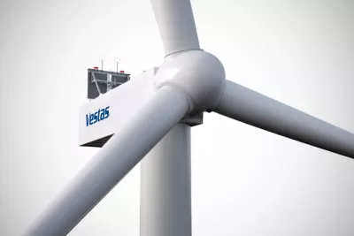
Vestas secures 390 MW offshore order in South Korea
Press Release:
News release from Vestas Asia Pacific
Seoul, 29 December 2025Vestas has received a 390 MW order for the Shinan-Ui offshore wind project in Jeollanam Province, South Korea. The project is developed by Shinan-Ui Offshore Wind, a consortium consisting of Hanwha Ocean, SK Eternix, KOMIPO (Korea Midland Power Co., Ltd.), Future Energy Fund, and Hyundai Engineering & Construction. The project marks Vestas’ first offshore wind order in South Korea, underscoring our commitment to advancing renewable energy globally and supporting South Korea’s clean energy transition.
The order includes 26 V236-15.0 MW offshore wind turbines as well as a 20-year service agreement to support reliable and optimised asset performance.
Purvin Patel, President of Vestas Asia Pacific, commented, “We are proud to partner with Hanwha Ocean on this milestone project in South Korea and introduce our industry-leading V236-15.0 MW offshore wind turbines to South Korean waters. These turbines, featuring world-class technology, are already being deployed in their inaugural European projects, with production ramping up to enable large-scale global deployment. Vestas brings decades of offshore experience and together with our customers, we are committed to driving the large-scale deployment of offshore wind in South Korea and delivering clean, secure and homegrown energy for the country.”
Jong Hyun Son, Head of Eco Energy & Industrial EPC Division, Hanwha Ocean, commented, “Hanwha Ocean will take a leading role in the Korean offshore wind industry through the successful execution of the Shinan-Ui Offshore Wind Project. The Shinan-Ui Project represents Korea’s first utility-scale offshore wind development to fully incorporate global standards, including the latest-generation offshore wind turbines, wind turbine installation vessels, offshore transformer stations, and onshore grid connection systems. We are pleased to partner with Vestas for the supply of cutting-edge wind turbine technology and value the strong local supply chain supporting foundations, cables, electrical components, and transportation and installation works.”
Turbine deliveries are scheduled to commence in 2027, with commercial operations of the wind farm expected to begin in 2028.
Vestas’ flagship offshore wind turbine, the V236-15.0 MW is built on proven, world-class technology and received its type certification in 2023, ensuring safety and quality. Since its launch, Vestas has secured more than 9 GW of firm orders globally, demonstrating its strong competitiveness in the offshore wind market.
For more information, please contact:
Megumi Sakuma
Marketing & Communications Manager
Mail: mgskm@vestas.com
Tel: +81 90 6723 5325About Vestas
Vestas is the energy industry’s global partner on sustainable energy solutions. We design, manufacture, install, and service onshore and offshore wind turbines across the globe, and with more than 197 GW of wind turbines in 88 countries, we have installed more wind power than anyone else. Through our industry-leading smart data capabilities and unparalleled more than 159 GW of wind turbines under service, we use data to interpret, forecast, and exploit wind resources and deliver best-in-class wind power solutions. Together with our customers, Vestas’ more than 37,000 employees are bringing the world sustainable energy solutions to power a bright future.For updated Vestas photographs and videos, please visit our media images page on: https://www.vestas.com/en/media/images.
We invite you to learn more about Vestas by visiting our website at www.vestas.com and following us on our social media channels:
Continue Reading