Nine-time Grand Prix winner Oscar Piastri has won Australian sport’s highest honour, the Don Award, with the Formula One star calling it a “massive privilege”.
The 24-year-old has spring-boarded into the elite this year, igniting national…

Nine-time Grand Prix winner Oscar Piastri has won Australian sport’s highest honour, the Don Award, with the Formula One star calling it a “massive privilege”.
The 24-year-old has spring-boarded into the elite this year, igniting national…
Nine-time Grand Prix winner Oscar Piastri has won Australian sport’s highest honour, the Don Award, with the Formula One star calling it a “massive privilege”.
The 24-year-old has spring-boarded into the elite this year, igniting national…
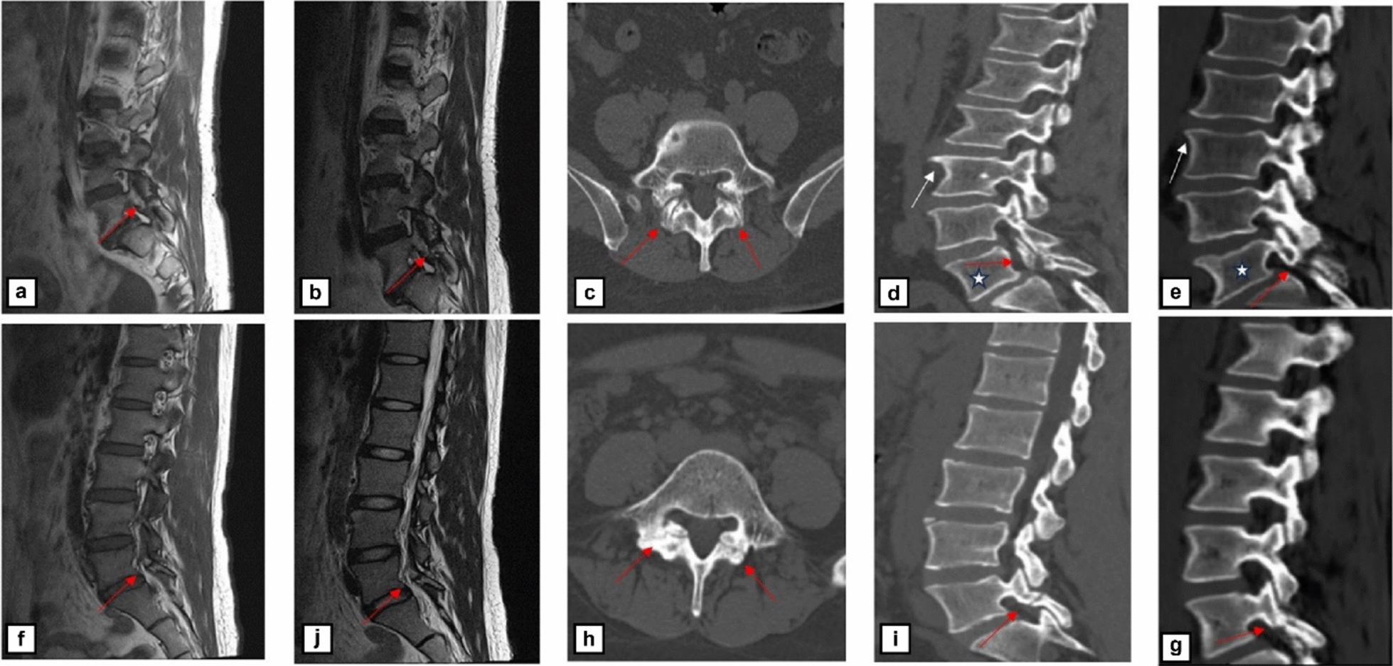
Lumbar spondylolysis—a cortical interruption of the pars interarticularis—is a common cause of low back pain, particularly in adolescents and athletes, and confident diagnosis hinges on clear depiction of the cortical breach [13]. Conventional CT remains the reference for osseous assessment but entails ionizing radiation, whereas routine T1- and T2-weighted MRI avoids radiation yet often fails to delineate subtle cortical discontinuities. Deep learning now enables MRI-based sCT that reproduces CT-like bone contrast from routine MR sequences and may bridge this gap; however, evidence specific to lumbar pars defects remains limited [14]. Therefore, we aimed to determine whether sCT generated from 1.5 T lumbar MRI can reliably depict lumbar spondylolysis underlying spondylolysis, using conventional CT as the reference standard and benchmarking against standard MRI in a retrospective case series [15].
Our study demonstrated that this deep learning-based sCT, derived from conventional lumbar MRI, provides diagnostic performance equivalent to that of traditional CT for identifying lumbar spondylolysis, significantly surpassing the capabilities of standard MRI sequences. The core value of sCT lies in its precise reproduction of cortical structures as seen on a CT bone window, whereas conventional T1W and T2W sequences are prone to missing non-displaced or occult fractures due to the inherent signal suppression of cortical bone [16]. In our study, the deep learning model was trained with a cycle‑consistent adversarial objective on anatomically aligned MRI–CT volumes; alignment was used to promote structural correspondence, without paired L1 supervision. This approach enhanced the visualization of subtle cortical breaches on sCT; quantitative HU agreement with CT was not evaluated [17]. In contrast to prior research that has primarily focused on large joints or other anatomical regions, we present one of the earliest clinical case series, to our knowledge, applying MRI-derived sCT specifically to lumbar spondylolysis [18,19,20].
The practical feasibility of sCT for diagnosing lumbar spondylolysis lesions in the lumbar arch was confirmed in a series of cases. Among all three patients with lumbar spondylolysis, the diagnostic performance of sCT and conventional CT assessments showed marked consistency. Nevertheless, conventional MRI sequences faced challenges in clearly delineating the left-sided lumbar spondylolysis in one patient, highlighting the superiority of sCT over conventional MRI. Both sCT and conventional CT accurately identified the cortical disruption of the pars interarticularis. More importantly, the two modalities demonstrated high concordance in depicting the anatomical morphology of the lumbar spondylolysis. Specifically, they provided equivalent imaging information regarding crucial diagnostic details such as the course of the fracture line, the sharpness of cortical margins, and the presence of surrounding osteosclerosis. The use of sCT obviates the need for ionizing radiation and concurrently provides a pathway for identifying spondylolisthesis, osteophytic sclerosis, and related features within a single scan. Our investigation, which identified vertebral osteophytes and sclerosis in the bilateral L5 lumbar spondylolysis of a 60-year-old patient using sCT, hints at its prospective applicability in diagnosing a wide array of musculoskeletal pathologies. This innovative clinical model promises to yield dual benefits for both patients and practitioners by mitigating radiation exposure and streamlining the clinical workflow.
In comparison with the latest international literature, numerous studies have already confirmed that sCT demonstrates high concordance with conventional CT for morphologic assessment in large joints like the hip and the cervical spine, emphasizing its global applicability in evaluating osseous structures [21,22,23]. For instance, Morbée et al. [21] and Florkow et al. [22] both reported that sCT-based depiction of hip bone structures could completely substitute for conventional CT, offering a radiation-free option suitable for large-scale screening in adolescents and young adults. van der Kolk et al. [23] further established that sCT meets the non-inferiority standard for visualizing cortical bone in the cervical spine. Our study, however, focuses on diagnosing lumbar spondylolysis, which is frequently missed on conventional MRI. We are the first to validate the utility of sCT in the diagnostic dimension of microfractures using real-world clinical data, thereby closing the loop in the application chain from the assessment of large bone structures to the visualization of micro-architectural bone injury. This highlights our study’s innovation and contribution to the field. Notably, Abel et al. [24] pioneered the use of sCT for preoperative geometric measurements of the lumbar spine, confirming its near-perfect agreement with CT for surgical planning parameters and providing a solid foundation for promoting sCT as a “single-scan, comprehensive-assessment” tool. Our work further underscores that the unique value of sCT in diagnosing microfractures has not been thoroughly explored in existing literature, a new frontier expanded by our research.
Mechanistically, deep learning-empowered sCT effectively overcomes the physical limitations of MRI for cortical bone imaging, achieving CT-like bone contrast for morphological assessment [25]. Our sample data indicate that the discrepancies between sCT and CT in the visualization of cortical fissures and in diagnostic concordance are minimal. This “one-stop-shop,” radiation-free, holistic assessment provides a safe and efficient diagnostic pathway for high-risk cohorts such as adolescents and athletes [26]. On a clinical application level, it can also significantly reduce the healthcare costs and procedural complexities associated with repetitive, multimodal imaging.
We acknowledge several limitations in this study. First, this proof-of-concept was established using a relatively small and specific cohort of adults with low back pain. The generalizability of our model’s performance to pediatric populations, asymptomatic individuals, or patients with varying degrees of skeletal maturity warrants validation in larger, multicenter, prospective trials. Second, our data were acquired on a 1.5 T MRI scanner. Although our model demonstrated high performance under this constraint, future research should investigate whether the use of higher field-strength magnets (e.g., 3 T) and more advanced MRI sequences could further enhance the resolution and fidelity of sCT images. Finally, this study focused on diagnostic accuracy and did not include longitudinal follow-up. Future research must correlate sCT findings with clinical outcomes, such as fracture healing rates, progression to spondylolisthesis, and response to conservative or surgical management.
In conclusion, this study provides compelling evidence that MRI-based synthetic CT is a high-precision, radiation-free modality for the diagnosis of lumbar spondylolysis. By overcoming the intrinsic limitations of conventional MRI for cortical bone assessment, this technology offers a robust alternative to conventional CT, thereby enhancing diagnostic confidence and patient safety. The future of musculoskeletal imaging will likely involve greater integration of such AI-driven, multimodal analyses, and our work establishes a validated application that can be readily translated into clinical practice, paving the way for broader investigations into other subtle skeletal pathologies.
Shchetynska-Marinova T, Amendt K, Sadick M, Keese M, Sigl M (2021) Aortitis – An Interdisciplinary Challenge. In Vivo 35:41–52
Google Scholar
Töpel I, Zorger N, Steinbauer M (2016)…

Original story from Chiba University
An intranasal HPV vaccine has exhibited encouraging results for tackling cervical cancer in a mouse model.
Cervical cancer, which affects the reproductive tract, is one of the most common cancers…
Garmin could be exploring a major design shift for its future adventure watches, with a newly granted patent…

The proposed axis offers a fresh perspective on migraine pathophysiology. Considering the clinical heterogeneity of migraine, do the authors anticipate that the lactylation–immune axis may be particularly relevant to certain clinical subtypes…

If you’re in the market for a new laptop, but you’re holding out for Black Friday deals to pick one up (smart move, by the way), you may be surprised as to what you can find before the date arrives. Companies are putting early Black Friday deals…

– Advertisement –
ISLAMABAD, Nov 18 (APP):The National Disaster Management Authority in partnership with Al-Khidmat Foundation on Tuesday sent its 25th aid consignment to Gaza a 100-ton shipment that raises total relief assistance to 2,427 tons…