A commission to protect Maine hospitals this week issued a report with more than a dozen recommendations.
They include codifying into state law that hospitals must give 120 days notice before closing a…
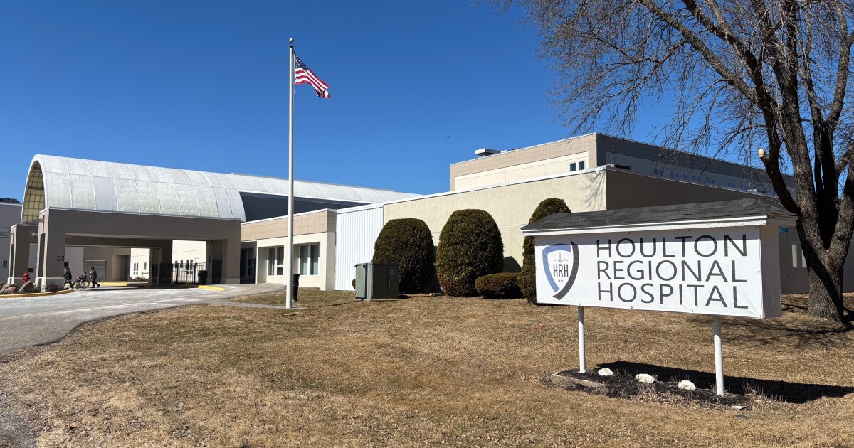
A commission to protect Maine hospitals this week issued a report with more than a dozen recommendations.
They include codifying into state law that hospitals must give 120 days notice before closing a…

Healthy women with elevated lipoprotein(a) levels are at greater risk of cardiovascular events over…
Westrick, E. R. & Ward, W. T. Adolescent idiopathic scoliosis: 5-Year to 20-Year Evidence-based surgical results. J. Pediatr. Orthop. 31, S61–S68. https://doi.org/10.1097/BPO.0b013e3181fd87d5 (2011).
Nam, Y., Choi, K., Jang, J., Kim, K. & Lee, G. Curve progression in adolescent idiopathic scoliosis with Cobb angles between 40 and 50 degrees at the late stage of skeletal growth: a minimum 5-year follow-up study. J. Clin. Med. 14 (15), 5272 (2025).
Miyanji, F. et al. A detailed analysis of the Lenke classification in surgical decision-making for adolescent idiopathic scoliosis. Spine Deform. 8 (1), 41–48 (2020).
Ilharreborde, B. et al. Hybrid constructs for tridimensional correction of the thoracic spine in adolescent idiopathic scoliosis: A comparative analysis of universal clamps versus hooks. Spine 35, 306–314. https://doi.org/10.1097/BRS.0b013e3181b7c7c4 (2010).
Cheung, K. M. C. & Luk, K. D. K. Prediction of correction of scoliosis with use of the fulcrum bending Radiograph*. J. Bone Joint Surg. 79, 1144–1150. https://doi.org/10.2106/00004623-199708000-00005 (1997).
Vedantam, R., Lenke, L. G., Bridwell, K. H. & Linville, D. L. Comparison of push-prone and lateral-bending radiographs for predicting postoperative coronal alignment in thoracolumbar and lumbar scoliotic curves. Spine 25, 76. https://doi.org/10.1097/00007632-200001010-00014 (2000).
Klepps, S. J., Lenke, L. G., Bridwell, K. H., Bassett, G. S. & Whorton, J. Prospective comparison of flexibility radiographs in adolescent idiopathic scoliosis. Spine 26, E74–E79. https://doi.org/10.1097/00007632-200103010-00002 (2001).
Hamzaoglu, A. et al. Assessment of curve flexibility in adolescent idiopathic. Scoliosis: Spine. 30, 1637–1642. https://doi.org/10.1097/01.brs.0000170580.92177.d2 (2005).
Lenke classification system of adolescent. Idiopathic scoliosis: treatment recommendations. Str. Course Lect 54, 537–542 (2005).
O’Brien, M. F., Kuklo, T. R. & Blanke, K. M. Spinal Deformity Study Group Radiographic Measurement Manual (Medtronic Sofamor Danek, 2005).
Watanabe, K. et al. Intraoperative spinal cord monitoring during intraoperative halo-femoral traction in scoliosis surgery. Spine 32, E849–E852. https://doi.org/10.1097/BRS.0b013e318074da84 (2007).
Faul, F., Erdfelder, E., Lang, A. G. & Buchner, A. GPower 3: a flexible statistical power analysis program for the social, behavioral, and biomedical sciences. Behav. Res. Methods. 39 (2), 175–191. https://doi.org/10.3758/BF03193146 (2007).
Faro, F. D., Marks, M. C., Pawelek, J. & Newton, P. O. Evaluation of a functional position for lateral radiograph acquisition in adolescent idiopathic scoliosis. Spine 29, 2284–2289. https://doi.org/10.1097/01.brs.0000142224.46796.a7 (2004).
Ilharreborde, B. et al. Angle measurement reproducibility using EOSThree-Dimensional reconstructions in adolescent idiopathic scoliosis treated by posterior instrumentation. Spine 36, E1306–E1313. https://doi.org/10.1097/BRS.0b013e3182293548 (2011).
Vidal, C., Ilharreborde, B., Azoulay, R., Sebag, G. & Mazda, K. Reliability of cervical lordosis and global sagittal spinal balance measurements in adolescent idiopathic scoliosis. Eur. Spine J. 22, 1362–1367. https://doi.org/10.1007/s00586-013-2752-2 (2013).
Potter, B. K. et al. Reliability of End, Neutral, and stable vertebrae identification in adolescent idiopathic scoliosis. Spine. 30, 1658–1663. https://doi.org/10.1097/01.brs.0000170290.05381.9a (2005).
Julien-Marsollier, F. et al. Benefits of a spine team for the surgical management of paediatric scoliosis. Orthop. Traumatology: Surg. Res. 103976. https://doi.org/10.1016/j.otsr.2024.103976 (2024).
Harrington, P. R. Technical details in relation to the successful use of instrumentation in scoliosis. Orthop Clin. North. Am (1972).
Moe, J. H. Methods of correction and surgical techniques in scoliosis. Orthop Clin. North. Am (1972).
Burton, D. C., Asher, M. A. & Lai, S-M. The selection of fusion levels using torsional correction techniques in the surgical treatment of idiopathic scoliosis. Spine 24, 1728. https://doi.org/10.1097/00007632-199908150-00015 (1999).
Ilharreborde, B., Sebag, G., Skalli, W. & Mazda, K. Adolescent idiopathic scoliosis treated with posteromedial translation: radiologic evaluation with a 3D low-dose system. Eur. Spine J. 22, 2382–2391. https://doi.org/10.1007/s00586-013-2776-7 (2013).
Reames, D. L. et al. Complications in the surgical treatment of 19,360 cases of pediatric scoliosis: A review of the scoliosis research society morbidity and mortality database. Spine 36, 1484–1491. https://doi.org/10.1097/BRS.0b013e3181f3a326 (2011).
Yagi, M., Takemitsu, M. & Machida, M. Chest cage angle difference and rotation of main thoracic curve are independent risk factors of postoperative shoulder imbalance in surgically treated patients with adolescent idiopathic scoliosis. Spine 38, E1209–E1215. https://doi.org/10.1097/BRS.0b013e31829e0309 (2013).
Sabharwal, S., Apazidis, A., Zhao, C., Hullinger, H. & Vives, M. Comparison of intraoperative supine and postoperative standing radiographs after posterior instrumentation for adolescent idiopathic scoliosis. J. Pediatr. Orthop. B. 20, 389–396. https://doi.org/10.1097/BPB.0b013e328347c2bc (2011).
Learch, T. J., Massie, J. B., Pathria, M. N., Ahlgren, B. A. & Garfin, S. R. Assessment of pedicle screw placement utilizing conventional radiography and computed tomography: A proposed systematic approach to improve accuracy of interpretation. Spine 29, 767–773. https://doi.org/10.1097/01.BRS.0000112071.69448.A1 (2004).
Cheh, G. et al. The reliability of preoperative supine radiographs to predict the amount of curve flexibility in adolescent idiopathic scoliosis. Spine. 32, 2668–2672. https://doi.org/10.1097/BRS.0b013e31815a5269 (2007).
Swany, L. M., Larson, A. N., Buyuk, A. F. & Milbrandt, T. A. Comparison of slot-scanning standing, supine, and fulcrum radiographs for assessment of curve flexibility in adolescent idiopathic scoliosis: a pilot study. Spine Deform. 9, 1355–1362. https://doi.org/10.1007/s43390-021-00349-9 (2021).
Ramchandran, S. et al. Impact of supine radiographs to assess curve flexibility in the treatment of adolescent idiopathic scoliosis. Glob. Spine J. 12, 1731–1735. https://doi.org/10.1177/2192568220988271 (2022).
Han, S-M. et al. Spinal sagittal alignment and postoperative adding-on in patients with adolescent idiopathic scoliosis after surgery. Orthop. Traumatol. 108, 103352. https://doi.org/10.1016/j.otsr.2022.103352 (2022).
Vidal, C., Ilharreborde, B., Queinnec, S. & Mazda, K. Role of intraoperative radiographs in the surgical treatment of adolescent idiopathic scoliosis. J. Pediatr. Orthop. 36, 178–186. https://doi.org/10.1097/BPO.0000000000000428 (2016).
Rodrigues, L. M. R. et al. Comparison between different radiographic methods for evaluating the flexibility of scoliosis curves. Acta Ortop. Bras. 22, 78–81. https://doi.org/10.1590/1413-78522014220200844 (2014).
Lenke, L. G. et al. Adolescent idiopathic scoliosis: a new classification to determine extent of spinal arthrodesis. J. Bone Joint Surg. Am. (2001).
Thawrani, D., Agabegi, S. S., Eismann, E., Martin, R. & Sturm, P. F. Accuracy and reliability of drawing central sacral vertical line on scoliosis radiographs in clinical practice. Spine Deformity. 1, 16–20. https://doi.org/10.1016/j.jspd.2012.10.003 (2013).
Uneri, A. et al. Intraoperative evaluation of device placement in spine surgery using known-component 3D–2D image registration. Phys. Med. Biol. 62, 3330–3351. https://doi.org/10.1088/1361-6560/aa62c5 (2017).
Jeantet, R-E., Simon, A-L., Happiette, A. & Ilharreborde, B. Bivertebral pedicle-supralaminar autostable claw for proximal fixation of magnetic growing rods in early-onset scoliosis. Orthop. Traumatol. 109, 103634. https://doi.org/10.1016/j.otsr.2023.103634 (2023).
Luhmann, S. J., Lenke, L. G., Bridwell, K. H. & Schootman, M. Revision surgery after primary spine fusion for idiopathic scoliosis. Spine 34, 2191–2197. https://doi.org/10.1097/BRS.0b013e3181b3515a (2009).
Jones, M. et al. A united Kingdom single centre review of the impact of extended waiting times in Early-Onset scoliosis: the effect of a delay to surgical treatment of greater than 12 months. Spine Deformity. 5, 446–447. https://doi.org/10.1016/j.jspd.2017.09.018 (2017).
Ohrt-Nissen, S., Luk, K. D. K., Samartzis, D. & Cheung, J. P. Y. Selection of the lowest instrumented vertebra in main thoracic adolescent idiopathic scoliosis: is it safe to fuse shorter than the last touched vertebra? Eur. Spine J. 29, 2018–2024. https://doi.org/10.1007/s00586-020-06398-4 (2020).
Compagnon, R. et al. Side bending radiographs and lowest instrumented vertebra in adolescent idiopathic scoliosis: A French quality-of-care study. Orthop. Traumatol. 108, 103350. https://doi.org/10.1016/j.otsr.2022.103350 (2022).
Iida, T. et al. Performance of forward roll maneuvers following corrective spinal fusion for idiopathic scoliosis patients. Orthop. Traumatol. 107, 103034. https://doi.org/10.1016/j.otsr.2021.103034 (2021).
Dang, N. R., Moreau, M. J., Hill, D. L., Mahood, J. K. & Raso, J. Intra-observer reproducibility and interobserver reliability of the radiographic parameters in the spinal deformity study groups AIS radiographic measurement Manual. Spine 30, 1064–1069. https://doi.org/10.1097/01.brs.0000160840.51621.6b (2005).
Ogon, M. et al. Interobserver and intraobserver reliability of lenke’s new scoliosis classification system. Spine 27, 858–862. https://doi.org/10.1097/00007632-200204150-00014 (2002).
Kuklo, T. R., Potter, B. K., Schroeder, T. M. & O’Brien, M. F. Comparison of manual and digital measurements in adolescent idiopathic scoliosis. Spine. 31, 1240–1246. https://doi.org/10.1097/01.brs.0000217774.13433.a7 (2006).
Mok, J. M. et al. Comparison of observer variation in conventional and three digital radiographic methods used in the evaluation of patients with adolescent idiopathic scoliosis. Spine 33, 681–686. https://doi.org/10.1097/BRS.0b013e318166aa8d (2008).
Liu, R. W. et al. Comparison of supine Bending, Push-Prone, and traction under general anesthesia radiographs in predicting curve flexibility and postoperative correction in adolescent idiopathic scoliosis. Spine. 35, 416–422. https://doi.org/10.1097/BRS.0b013e3181b3564a (2010).
Jeandel, C. et al. Enhanced recovery following posterior spinal fusion for adolescent idiopathic scoliosis: A medical and economic study in a French private nonprofit pediatric hospital. Orthop. Traumatol. 109, 103626. https://doi.org/10.1016/j.otsr.2023.103626 (2023).
Langlais, T. et al. Sagittal plane assessment of manual concave rod bending for posterior correction in adolescents with idiopathic thoracic scoliosis (Lenke 1 and 3). Orthop. Traumatol. 109, 103654. https://doi.org/10.1016/j.otsr.2023.103654 (2023).
Baldairon, F. et al. Analysis of factors associated with sagittal alignment deterioration after correction of degenerative scoliosis by in situ contouring. Orthop. Traumatol. 107, 103023. https://doi.org/10.1016/j.otsr.2021.103023 (2021).

Jan. 9 (UPI) — A man convicted of encouraging Muslims to fight overseas after the Sept. 11, 2001, terrorist attacks engaged in lawful free speech, a federal appellate court ruled Friday.
The 4th U.S. Circuit Court of Appeals’ three-judge panel…

Friday, 09 January 2026
| Alert Summary | |
|---|---|
| Category 1: | For Action |
| Alert Notification: | 2026.01 (Update 3) |
| Product Identification: | SMA Advanced Follow-On Milk; pack size: 800g |
| Batch Code | 51890742F2; expiry date: Jul-27 |
Message:
Further to FSAI food alert 2026.01, FSAI food alert 2026.01 (Update 1) and FSAI food alert 2026.01 (Update 2), the correct expiry date for the above batch of SMA Advanced Follow-On Milk is July-27.
Recall notices will be displayed at point-of-sale.
Questions and answers.
Nestlé is advising its customers that have purchased any of these batches to contact them:
Via its online form, sharing a photo of the product and the batch code: www.nestle.co.uk/en-gb/getintouch
By calling its careline on 1800 931 832.
Nature Of Danger:
Cereulide toxin is produced by the bacterium Bacillus cereus. The toxin may be pre-formed in a food and is extremely heat resistant. Consumption of foods containing cereulide toxin can lead to nausea and severe vomiting. Symptoms can appear within five hours. The duration of illness is usually 6 to 24 hours.
Action Required:
Manufacturers, wholesalers, distributors, caterers & retailers:
Retailers are requested to remove the implicated batches from sale and display recall notices at point-of-sale.
Wholesalers/distributors are requested to contact their affected customers and recall the implicated batches and provide a point-of-sale recall notice to their retailer customers.
Consumers:
Parents, guardians and caregivers are advised not to feed the implicated batches to infants or young children.
GAME NOTES | Game 15 — at Western Illinois | PDF
By Thomas Corhern, TTU Athletics Media Relations
MACOMB, Ill. – As the Tennessee Tech women’s basketball team enters Saturday’s contest, the Golden Eagles are in a little bit of unfamiliar…

Anorexia nervosa (or AN) doesn’t just result in fat loss. It can also result in a 20-30% loss of skeletal muscle strength and size, which is critical to longevity and the ability to do basic activities like grocery shopping or picking up…