Lorde. A$AP Rocky. JENNIE. Baby Keem. KATSEYE. That’s just a taste of who’s performing at the 2026 Governors Ball, and this year, Spotify is introducing a brand-new way to navigate…


Lorde. A$AP Rocky. JENNIE. Baby Keem. KATSEYE. That’s just a taste of who’s performing at the 2026 Governors Ball, and this year, Spotify is introducing a brand-new way to navigate…

Biofuels, such as rapeseed, are not an ideal alternative to non-fossil carbon.Credit: Krisztian Bocsi/Bloomberg/Getty
There’s a relatively new word doing the rounds in sustainability research and policy: defossilization. Beyond expert circles, it isn’t necessarily obvious that phasing out fossil fuels does not mean phasing out carbon. Under net-zero scenarios, carbon-based fuels are still needed, to provide power, for example, and for aviation. Carbon, currently often derived from fossil hydrocarbons, is also integral to everyday consumer products such as soaps and detergents, as well as medicines, fertilizers and plastics.
Worldwide, demand for ‘embedded’ carbon — that found in chemicals — is expected to double by 2050, according to the nova-Institute, a green-energy research institute in Hürth, Germany (see go.nature.com/4jpx6qi). But this carbon cannot come from the usual sources, such as coal, natural gas and oil. These must remain in the ground, and this is where defossilization comes in.
Chemistry can make plastics sustainable – but isn’t the whole solution
Defossilization means finding sustainable ways to make carbon-based chemicals. Alternative sources of carbon include the atmosphere and plants, as well as carbon in existing biological or industrial waste, such as used plastics or agricultural residue. In some cases, these chemicals will eventually return carbon dioxide to the atmosphere through burning or biodegradation. In principle, this will occur as part of a circular process, rather than one that has added greenhouse gases.
The subject of defossilization is of increasing research interest — as it needs to be — despite signs that some governments, including a number in Europe and that of the United States, are backsliding on their climate commitments. In this two-part Editorial, we describe some of the challenges faced by researchers, in both academia and industry, that scientists and policymakers need to solve to enable defossilization to happen on the scale required. In this first instalment, we focus on Europe. In the next, we explore advances under way in China.
Biomass from crops is a key source of non-fossil carbon, and one that can be obtained at scale. One driver of large-scale production is the European Union’s biofuels strategy. This mandates that transport fuels include biomass-derived products. Examples include biodiesel, which can be made from oils such as sunflower and palm, and bioethanol, which is synthesized from crops such as maize (corn) and wheat. But clearing existing cropland or converting uncultivated land to grow biofuels can’t be the alternative of choice, not least because of the attendant risk to biodiversity and soil health, and the demand it puts on water resources. There’s also some evidence that, by encouraging farmers to convert land previously used to grow food crops, the directive has pushed up food prices.

Reinvent oil refineries for a net-zero future
The extraction of carbon from lignocellulose — tough plant matter — in crop waste is an alternative with potential that remains mostly untapped. One major advantage is the fact that it can be produced without the use of extra land. But it is expensive to extract, and production timelines are long, both of which hinder scalability1.
Other potential sources of waste carbon include municipal and industrial waste, with used plastic among this. More than 40% of plastic produced in the EU is already recycled. This recycling rate could be increased if technical challenges can be surmounted2. Current recycling methods break waste plastics into flakes through shredding or melting, then form pellets that can be used to make new products. For higher recycling rates to be achieved, chemical recycling methods will need to be further developed and scaled up. These methods break down plastics into smaller molecules that can be used to rebuild new, larger ones.
Carbon dioxide captured from fossil-fuel burning or the air offers one of the largest potential avenues for defossilization. The global chemicals industry could obtain one-third of its carbon needs from this source by 2050, according to the nova-Institute. That compares with 22% from biomass. By one estimate, there are almost 900 gigatonnes of carbon in the atmosphere, nearly double the 450 gigatonnes of carbon contained in vegetation3. But the scenarios for 2050 vary widely. Some say CO2 will become the main feedstock for chemicals, whereas others say its contribution will be negligible.

How fast fashion can cut its staggering environmental impact
To make useful carbon-based molecules, CO2 must first be transformed into other molecules. Usually, it is reacted with hydrogen, either to form hydrocarbons or to remove an oxygen atom. Because CO2 is highly stable, a considerable amount of energy is needed to overcome the thermodynamic barrier to these reactions. This must be powered renewably for the process to be truly sustainable. Capturing atmospheric CO2 is difficult and expensive, in part because of the compound’s stability. As a result, the technology has not been a priority for European governments. This must change.
In May, Elisa Morgera, the United Nations Special Rapporteur on human rights in the context of climate change, published a report urging governments to defossilize economies as part of the fossil-fuel phase-out. In the United Kingdom, the Royal Society and the Institution of Chemical Engineers have urged the government to support research on defossilization. They have a strong case, because such research, which is intended to boost the chemical industry, aligns with government policies to invest in science that supports economic growth. The EU and China also have a joint research programme called the EU–China Bridge, which is focused on decarbonization, but this is set to expire next year. This not only needs to be renewed, it needs a renewed focus — on defossilization.
We collect limited information about web visitors and use cookies on our website to provide you with the most optimal experience. These cookies help us provide you with personalized content and improve our website. To learn more about our web…

This article first appeared on GuruFocus.
Dell Technologies Inc. (DELL, Financials) has brought back its XPS laptop brand to strengthen its presence in the premium personal computer segment and revive demand amid a sluggish PC market.
…
Formation of CPSF6 puncta upon HIV-1 infection hinges on two key events: the entry of the HIV-1 core into the nucleus and the binding of CPSF6 to the HIV-1 core (Blanco-Rodriguez and Di Nunzio, 2021; Blanco-Rodriguez et al., 2020; Buffone et al., 2018; Zila et al., 2021). To determine the contribution of CPSF6’s disordered domains to the formation of CPSF6 puncta upon HIV-1 infection, we correlated the binding of CPSF6 to the HIV-1 core with the formation of CPSF6 puncta. To this end, we first generated CPSF6 knockout (KO) THP-1 cells (Figure 1C, D) to eliminate the interference from the endogenous protein, which could affect the interpretation of results regarding the role of the analysed CPSF6 domains. CPSF6 depletion in THP-1 cells was performed by CRISPR–Cas9 technology. To completely eliminate the expression of the CPSF6 gene, we selected single clones by limiting dilution. We identified a clone that was completely KO for CPSF6, confirmed through western blot and immunofluorescence (Figure 1C, D), and we infected this clone and the control clone with HIV. CPSF6 puncta were detected only in the control-infected cells and not in the KO clone (Figure 1D; Figure 1—figure supplement 1). The viral integrase (IN) was observed within CPSF6 puncta, consistent with previous studies (Francis et al., 2020; Rensen et al., 2021; Scoca et al., 2023), but absent in CPSF6 KO cells where viral IN was observed in the cytoplasm (Figure 1D; Figure 1—figure supplement 1). Thus, we used KO cells for CPSF6 to assess the role of selected CPSF6 domains in HIV-induced condensates (Figure 1—figure supplement 2). We designed various CPSF6 deletion mutants (Figure 2A) to specifically assess the significance of the main disordered regions of CPSF6 protein (Figure 2—figure supplement 1) such as the FG (phenylalanine and glycine) motif, the low complexity regions (LCRs), and the mixed charge domain (MCD), in the ability of CPSF6 to bind to the core and facilitate the formation of CPSF6 puncta. We investigated the role of the FG peptide by generating a mutant that exclusively lacks the FG peptide (∆FG). Previous in vitro studies have shown that the FG peptide binds to the hydrophobic pocket formed between capsid hexamers (Buffone et al., 2018; Price et al., 2014). Here, we want to investigate the role of FG peptide in the context of the protein during the viral infection.
(A) Schema of CPSF6 isoform 588 aa deletion mutants. (B) Confocal microscopy images of THP-1 CPSF6 KO cells, transduced with different mutants of CPSF6, infected with VSV-G/HIV-1ΔEnvINHA LAI (BRU) -vpx (MOI 10) in presence of Nevirapine (10 µM). The cells are stained with CPSF6 and HA antibody 30 hr p.i. Scale bar 5 µm. (C) Analysis of the number of CPSF6 puncta in THP-1 CPSF6 KO cells transduced with different mutants of CPSF6, not infected or infected in the presence of Nevirapine (10 µM) (the number of analysed cells is shown under the x-axis). Statistical test: ordinary one-way ANOVA (****p < 0.0001; ***p < 0.001; *p < 0.05; ns, p > 0.05). (D) The plot compares the number of CPSF6 puncta per cell in THP-1 CPSF6 KO cells transduced with different mutants of CPSF6, infected with HIV-1 in the presence of Nevirapine (10 µM). Statistical test: ordinary one-way ANOVA (****p < 0.0001; ns, p > 0.05). (E) Confocal microscopy images of THP-1 CPSF6 KO clone 4, non-transduced and non-infected or transduced with WT CPSF6 and CPSF6 3xNLSΔMCD and infected with VSV-G/HIV-1ΔEnvINHA LAI (BRU) -vpx (MOI 10) in presence of Nevirapine (10 µM). Immuno-RNA FISH: the cells are stained with CPSF6 (green) antibody and with 24 probes against HIV-1 Pol sequence (grey) (RNA-FISH) 25 hr p.i. Nuclei are stained with Hoechst (blue). Scale bar 10 µm. Violin plot presenting the percentage of CPSF6 puncta colocalizing with the viral RNA in THP-1 CPSF6 KO clone 4 cells transduced with LVs expressing CPSF6 WT or CPSF6 3xNLSΔMCD (respectively, n = 73 and n = 103) and infected with VSV-G/HIV-1ΔEnvINHA LAI (BRU) -vpx (MOI 10) in presence of Nevirapine (10 µM). A total of 198 CPSF6 WT puncta and 264 CPSF6 3xNLSΔMCD puncta were counted. Statistical test: unpaired t-test, ns, p > 0.05. (F) Confocal microscopy images of THP-1 KO CPSF6 cells transduced with WT CPSF6 and CPSF6 ∆MCD without NLS, with 3xNLS or with PY NLS, respectively. Cells were differentiated for 3 days, transduced with CPSF6 lentiviral vectors (MOI 1) for 3 days and infected for 24 hr with VSV-G/HIV-1ΔEnvINHA LAI (BRU) -vpx (MOI 10) in the presence of Nevirapine (10 µM). The panels show transduced and uninfected cells. CPSF6 and the IN tagged with the HA are labelled with anti-CPSF6 (green) and anti-HA (white) antibodies, respectively. Nuclei are stained with Hoechst (blue). The arrows show CPSF6 puncta in colocalization with IN-HA. Scale bar 10 µm.
To further explore this, we developed an alternative plasmid by expanding the FG peptide deletion to include surrounding prion-like LCRs (∆FG ∆LCR). These regions, outside the CPSF6 context, have been identified as crucial for facilitating strong CPSF6 binding to capsid lattices (Wei et al., 2022). In our study, we aim to evaluate their role within a more physiological setting. Additionally, we assessed a CPSF6 variant that carries the 15-mer FG peptide flanked by non-LCR sequences, such as those derived from Beta-adducin (ADD2), kindly provided by Mamuka Kvaratskhelia (∆LCR + ADD2). These protein segments are known for their high flexibility, akin to the LCR of CPSF6. Furthermore, to elucidate the contribution of the LCRs of CPSF6 in the formation of CPSF6 puncta, we generated a mutant lacking both LCRs (∆LCR). Analysis of the MCD contribution to both the ability of CPSF6 to bind to the core and formation of CPSF6 puncta was achieved by deleting the MCD and adding 3 nuclear localization signals (3xNLS ∆MCD) since the deletion of the MCD results in a protein that localizes mainly into the cytoplasm (Figure 2A, B; Figure 2—figure supplement 2).
To correlate the ability of CPSF6 to bind to the HIV-1 core with formation of CPSF6 puncta, we expressed wild-type and mutant CPSF6 constructs in THP-1 cells knockout for CPSF6. Subsequently, we infected these cells with HIV-1 and analysed the presence or absence of CPSF6 puncta at 24 hr post-infection. Importantly, for the imaging experiment, we expressed CPSF6 WT and mutants without tags to avoid the formation of aggregates that could interfere with our conclusions. Our data show that HIV-induced CPSF6 puncta can form extremely rarely with the deletion mutant CPSF6 ADD2∆LCR and with the mutant lacking the FG or both the FG peptide and the LCRs (Figure 2B–D; Figure 1—figure supplement 2). However, when we analysed the role of the MCD domain in CPSF6 puncta formation, which was indicated to be important for condensing CPSF6 in NS (Greig et al., 2020), comparing the number of CPSF6 WT puncta induced by HIV infection with CPSF6 mutants revealed that the MCD domain does not play a critical role in HIV-induced CPSF6 puncta formation (Figure 2B–D). In addition, we observed that the majority of analysed CPSF6 3xNLS∆MCD puncta contain vRNA inside, similar to CPSF6 WT puncta (Figure 2E), thus corroborating the lack of a role for this intrinsically disordered domain in HIV-induced CPSF6 puncta. Since the NLS domain from SV40, which replaces the MCD, is highly basic and could potentially induce condensates, we fused CPSF6 with a non-basic NLS (PY-NLS) or removed the NLS entirely. Even though these two proteins do not efficiently enter the nucleus, the few that do manage to reach the nucleus can host viral particles, as evidenced by the presence of IN. Many viruses are typically blocked in the cytoplasm due to the presence of these mutants that are mainly cytoplasmic. However, because we used a high viral dose, the blockage in the cytoplasm was not complete. As a result, the viruses that successfully entered the nucleus induced the formation of puncta associated with CPSF6-deleted mutants, indicating that the MCD is not critical for the formation of HIV-induced CPSF6 puncta (Figure 2F). Similar to the MCD, when we compared CPSF6 truncated for the LCRs with CPSF6 WT, we observed that the LCRs do not contribute to CPSF6 puncta formation. Therefore, the FG peptide alone, without the LCRs, is the only CPSF6 domain required for their formation (Figure 2B–F; Figure 1—figure supplement 2).
Next, we tested the ability of the different CPSF6 deletion mutants for their ability to bind the viral core using a previously described capsid binding assay (Selyutina et al., 2018). Wild-type and mutant CPSF6 proteins were expressed in human 293T cells at similar levels (INPUT) (Figure 3A). Extracts containing wild-type and mutant CPSF6 proteins were incubated with stabilized HIV-1 capsid tubes for 1 hr at 25°C in the presence of 10 µM of PF74, which is a small molecule that competes with CPSF6 for binding to the hydrophobic pocket formed between hexamers that constitute the viral core (Buffone et al., 2018; Price et al., 2014). HIV-1 capsid stabilized tubes were washed, and the bound proteins were eluted using Laemli buffer (BOUND). For every construct, the percentage of bound protein relative to input in the presence or absence of PF74 is shown (Figure 3B). Our results revealed that the absence of the FG peptide (∆FG) entirely abolished CPSF6’s ability to bind to the viral core. In agreement, simultaneous deletion of the FG motif and LCRs (∆FG ∆LCR) resulted in a construct unable to bind to the viral core. Similar outcomes were observed when the LCRs were replaced with sequences derived from ADD2, even if the FG was present.

(A) Ability of wild-type and mutant CPSF6 proteins to bind to the HIV-1 core. Cellular extracts derived from human 293T cells expressing similar levels of the indicated CPSF6 proteins (INPUT) were incubated with HIV-1 capsid stabilized tubes for 1 hr at room temperatures in the presence and absence of 10 µM PF74, as described in materials and methods. As a carrier control, we utilized DMSO. Subsequently, HIV-1 capsid stabilized tubes were washed, and the bound proteins were eluted 1X Laemmli buffer 1X. The BOUND fractions were analysed by western blotting using antibodies against neon-GFP and the HIV-1 capsid. (B) Experiments were repeated at least three times and the average BOUND fraction relative to the INPUT fraction normalized to wild-type binding is shown for the different CPSF6 mutants. *** indicates a p-value <0.001; **** indicates a p-value <0.0001; and ns indicates no significant difference as determined by unpaired t-tests.
LCR-FG is notably more disordered than ADD2-FG, containing a high proportion of prolines (48 out of 98 residues), which makes it mostly non-foldable (Figure 4A–H). Since proline is a structure-disrupting residue, LRC-FG is not expected to adopt any secondary structure. In contrast, ADD2-FG contains fewer prolines (15 out of 98 residues) but has many charged residues. It is predicted to form two short α helices and a β strand, arranged as: α helix–FG–β strand–α helix (Figure 4E). ADD2-FG may form a flexible collapsed state, as its oppositely charged residues are evenly distributed, potentially allowing polyelectrostatic compaction. This suggests that FG within ADD2-FG may be less accessible for the interaction with the viral core’s hydrophobic pocket due to its involvement in this collapsed conformational ensemble (Figure 4A–E, Figure 4—figure supplement 1). This aligns with the inability of CPSF6 carrying ADD2 in place of the LCRs to induce CPSF6 puncta (Figure 2B). On the other hand, the deletion of only the two LCRs, while keeping the FG peptide intact, resulted in unexpected findings. The ∆LCR mutant exhibited a stronger binding affinity for the viral core when compared to the wild-type protein (Figure 3B). These results suggest that the LCRs surrounding the FG motif are modulating the affinity of CPSF6 to the viral core, which might be important for function. By contrast, deletion of the MCD (∆MCD) but retention of other regions, such as the FG peptide and the LCRs, demonstrated a binding affinity to the viral core similar to that of the wild-type protein. These results suggest that the MCD domain is not involved in the binding of CPSF6 to the viral core, which is not surprising since the CPSF6 (1–358), which does not have an MCD, binds to the viral core (Lee et al., 2010).

(A) Physicochemical characteristics of the LCR-FG and ADD2-FG sequences. Intrinsic disorder predispositions evaluated by PONDR VLXT. Position of the FR segment within the LCR-FG and ADD2-FG sequences is shown as grey shaded area. (B) Linear distribution of the net charge per residue (NCPR) within the LCR-FG sequence evaluated by CIDER. (C) Linear distribution of the NCPR within the ADD2-FG sequence evaluated by CIDER. (D) Secondary structure propensity of the LCR-FG sequence evaluated by PSIPRED. (E) Secondary structure propensity of the ADD2-FG sequence evaluated by PSIPRED. (F) Analysis of the peculiarities of the amino acid compositions of the intrinsically disordered C-terminal domain (residues 261–358) of human CPSF6 and its different mutants. Relative abundance of prion-like LCR defining uncharged residues in analysed protein segments. (G) Relative abundance of proline residues in analysed protein segments. (H) Relative abundance of charged residues in analysed protein segments. The values were calculated by dividing numbers of prion-like LCR defining uncharged (Ala, Gly, Val, Phe, Tyr, Leu, Ile, Ser, Thr, Pro, Asn, Gln, Pro) and charged (Asp, Glu, Lys, Arg) residues by the total number of amino acids in the respective protein fragments. Corresponding values for all protein sequences deposited in the UniProtKB/Swiss-Prot database, PDB Select25, and DisProt are shown for comparison.
Thus, the viral capsid, through the FG peptide of CPSF6, constitutes the scaffold of HIV-induced CPSF6 puncta.
In summary, our results suggest that the FG peptide is the main determinant involved in the binding of CPSF6 to the viral capsid. Interestingly, our work implies that the LCRs may be modulating the affinity of the FG motif for the viral core (Figure 3B). Recognition motifs that mediate protein-protein interactions, such as the FG motif of CPSF6, are usually embedded within longer IDRs that can modulate affinity of the interaction (Karlsson et al., 2022). Taken together, our data show that the FG peptide coordinates both the binding to the viral core and the induction of CPSF6 puncta. This coordination suggests that the FG peptide plays a critical dual role in recognizing the viral capsid and facilitating the cellular clustering of CPSF6, which may be part of the cellular response to viral entry.
This article first appeared on GuruFocus.
Dell Technologies Inc. (DELL, Financials) has brought back its XPS laptop brand to strengthen its presence in the premium personal computer segment and revive demand amid a sluggish PC market.
…
Well, he did it. He actually did it.
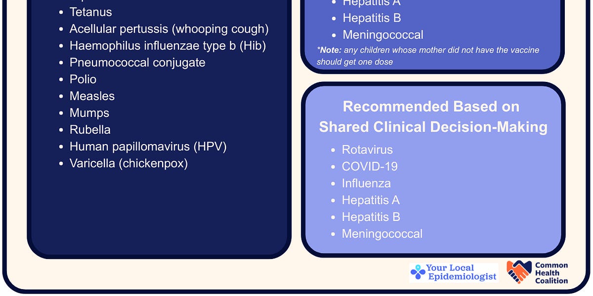
RFK Jr. unilaterally made sweeping changes to the routine vaccination schedule for children in the United States. This change isn’t based on new data or new evidence, but rather on political and ideological…

The game is slated for a 7…

Dell has brought back its popular XPS laptop lineup a year after retiring the premium brand, as it looks to drum up demand in a sagging personal computer market.
The company unveiled its thinnest laptops yet, XPS14 and…