Bob Rich’s frozen food business was so successful that he bought the first naming rights to an NFL Stadium in 1973. With the Buffalo Bills’ home set to be demolished after this season, his son, Bob Jr. looks back on the cold realities of running a $5.8 billion family business.
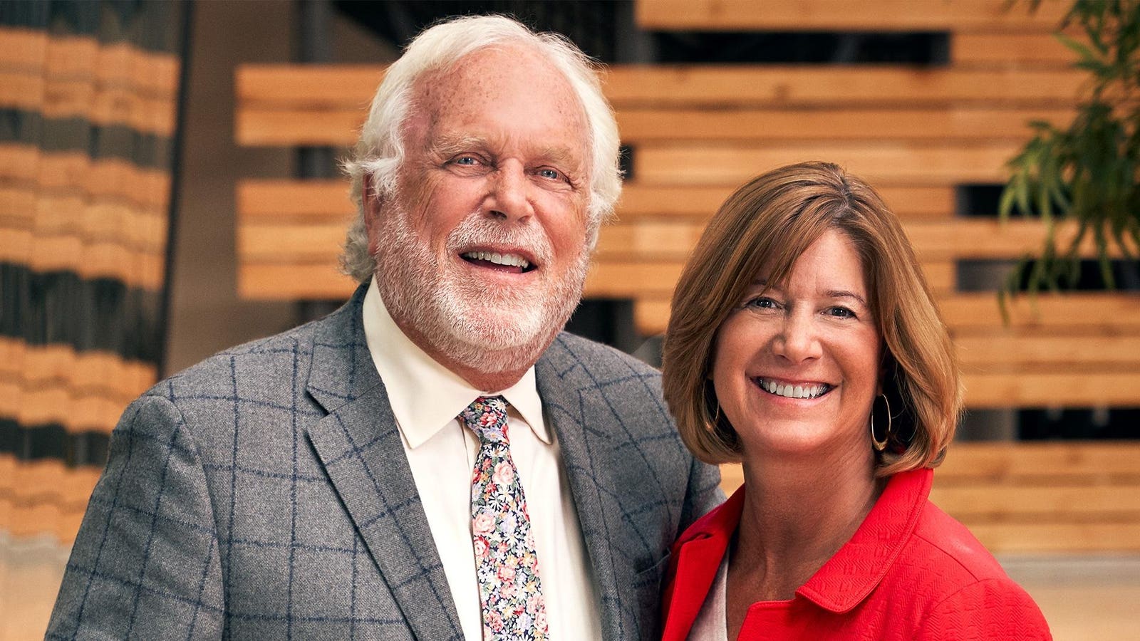
Whenever someone offers to acquire Rich Products—Buffalo, New York’s $5.8 billion (annual sales) food giant that you’ve probably never heard of—its senior chairman and son of the founder, Bob Rich Jr., has a form letter ready for his assistant to send back.
The response gets sent often, according to Rich: “We say, Dear blank, thank you for your interest in our company. Rich Products is not for sale. Yours Truly.”
His assistant often asks if he wants to know who she’s sending the letter back to, but he actually doesn’t. “I don’t really care,” the 84-year-old billionaire says, chuckling. “How bad is that?”
“Our biggest priority is that we want to remain a privately held company for eternity,” adds his wife, Mindy, 68, who is chairman of Rich’s and its board.
Rich’s north star is keeping the business under 100% family control, as he says, “to have the freedom to make decisions quickly and move ahead with more speed.” His father, Bob Sr., invented the first non-dairy whipped topping in 1945—three years before the better-known (and dairy-based) Reddi-Wip came to market—and Rich’s signature whipped topping is now sold in more than 100 countries. It remains one of the top products for an expansive food conglomerate—which Forbes values at north of $7 billion—whose range of products include cookies sold at supermarket bakeries, cold foam offered at coffee shops, pizza dough for independent and chain pizzerias, as well as SeaPak frozen seafood and Carvel ice cream cakes. Its longtime customers include Walmart, Kroger, and Dunkin’, Publix, Sodexo and more.
“Growth for us is not exponential. It’s not a straight line. It goes step by step,” says Rich, whose fortune Forbes estimates at $6.5 billion, based on his stake in the business and other investments.
The company expects to grow annual revenue to $10 billion by 2030, and the plan to get there includes more “breakthrough” products designed for restaurants and wholesalers that alleviate labor woes as well as reformulating some bestsellers for the MAHA era.
Dreams of Fields: In 1973, Bob Rich Sr. paid $1.5 million for the naming rights to the Buffalo Bills’ stadium, the first such deal in NFL history.
Jeff Goode/Toronto Star/Getty Images
And there’s another massive change ahead for Rich’s: At the end of this NFL season, the Buffalo Bills’ stadium—which became the first to sell naming rights to a business in 1973 when Bob Sr. spent $1.5 million for a 25-year contract, the only one to make a bid—will be demolished. It bore the Rich family name until 1997, when it was renamed for team founder Ralph Wilson until 2015.
“Now there are about 500 stadiums around the world that have sold their naming rights. It was a crazy decision,” Rich says, “that was okay.”
Bob and Mindy, avid Bills fans, say they are excited for the new stadium, just as they are excited about what’s on the horizon for Rich’s as it moves into its next 80 years. “I saw someone walking around last week wearing a shirt that said, ‘We still call it Rich Stadium,’ which I laughed about,” says Bob. “It’ll be a point of pride for everybody, including us.”
The son of a Buffalo dairyman, Robert E. Rich delivered milk for his father during summers in high school and when he graduated in 1935 he started his own dairy business. It soon became one of the largest in the region. Then, during World War II, he served as a milk administrator while dairy was rationed and got inspired when a chance call from a hospital purchasing agent mentioned how they were using soybean-based milks and creams from Henry Ford’s George Washington Carver Laboratory. After a tour of the facility, Rich was granted rights to their manufacturing system for a symbolic $1 fee. And he set out to develop a dessert topping—less fattening and harder to spoil, and, above all, cheaper to make than whipped cream—to the masses.
His frozen blue cans of Rich’s non-dairy whipped topping were a hit—he had $29,900 in sales that first year in 1945 (or about $540,000 today)—and as World War II rations came to an end and post-war grocery spending boomed, so did sales. By 1952, sales topped $1 million (or $12 million today) for the first time. And the business prevailed even after attracting 40 different lawsuits from the dairy industry claiming he was counterfeiting cream. Rich didn’t let any of that stop him, and he quickly brought the treat around the world.
“My father used to joke that his office was the tray of an airplane,” says Bob Jr., who joined the business full-time in 1963 after summers and afterschool hours spent on the family loading docks. Bob Jr. had to be wooed by his father. His interests had been elsewhere, after playing hockey as the backup goalie for Buffalo’s American Hockey League franchise, a failed 1964 Olympics hockey tryout and interviews with the Air Force and the CIA. But Bob Sr. offered his son the chance to build a plant in Canada and oversee a $1 million budget (about $10 million today) as president of the company’s first international division.
Just Desserts: Invented in 1945, Rich’s Whip Topping was less fattening and cheaper to make than whipped cream
Rich Products
The father and son had a competitive relationship at first. But they soon realized they were teammates after the first 5,000 pounds of topping from Bob Jr.’s new Canadian factory wouldn’t whip, and Bob Jr. had to swallow his pride and ask his dad for help.
Rich’s first major acquisition came in 1976, the year that company annual sales topped $100 million for the first time. The business purchased SeaPak frozen seafood for $11.5 million—and it cemented the business’s strategy of useing acquisitions to grow. Bob Jr. became president of Rich’s two years later, and has added 60 brands through acquisitions since. He also bought Buffalo’s struggling Triple A baseball team to make sure the franchise stayed in the city and has owned the Buffalo Bisons, the minor league franchise of the Toronto Blue Jays, since 1983.
Later that year, Bob Jr. met Mindy, 16 years younger, at a Buffalo Bisons baseball game. It turned out that she also grew up in a family-owned food business—one based in Cincinnati— that sold donuts and other products like extruded crunchy onion rings.
The two wed—it was Bob Jr.’s third marriage—and Mindy started working at Rich’s “the day we got back from our honeymoon” in 1985—in the company’s internal entertainment department. (And it wasn’t until years later that she realized that, after her family sold its business amid problems, some of the brands changed hands a few times, and the onion ring brand even ended up being owned by Rich’s.) “Having grown up in the food industry, it didn’t make sense when we got married for me to work anywhere else,” says Mindy. By 1996, annual sales topped $1 billion.
Bob Jr. took over as chairman in 2006, after his father passed away at 92. Bob Sr. had spent 61 years at the helm of the company, and until his death he always had a dog-eared piece of paper in his pocket with the company’s annual sales. Rich’s had made a profit every year it was in business at the time (and that’s still true today). Forbes estimated annual sales in the final year of his life at $2.4 billion.
Bob Jr. inherited a fortune worth at least $1.5 billion. His younger brother, David, who became a priest and had moved to Jackson, Mississippi to work for an Anglican Church, inherited the rest of the family stake, worth hundreds of millions. Their sister, Joanna, whose husband sued his father-in-law twice and lost both times, was cut out of the will.
As Rich’s became a $3 billion (annual sales) company in 2013, the business went on a new acquisition spree, adding patented smoothie machine brand F’Real Foods as well as three wholesale bakery businesses.
With that kind of growth, the Riches had to make a concerted effort to stop talking about work at home, and even vowed that they would never speak of work while spending time on their fishing boat. “I’d say we were successful 80% of the time,” Mindy recalls. Rich has written several novels about fishing, and 2015’s Looking Through Water about an estranged father and son at a fly fishing tournament was turned into a movie with Michael Douglas that was released in September.
Buffalo Billing: Until his death at 92, Bob Rich Sr. always had a paper in his pocket with the company’s annual sales. In 2006, the final year of his life, it was $2.4 billion.
Rich Products
In 2021, when annual sales were $4 billion, Rich decided to replace himself as chairman of the board, which he had run for the past 15 years, and determined Mindy was the perfect person to replace him. “It’s given me the opportunity to step into a new role as a senior chairman and brought new joy watching Mindy bring her personality to the forefront,” Rich says.
“Our approach to being transparent and authentic during the challenging times has helped us build trust,” she adds. “You can’t always paint a rosy picture when the picture is maybe not as rosy as you’d like it to be.”
One thing the Riches agree on is Bob Sr.’s guiding principle—remaining private: “We realized that publicly held companies couldn’t have the stability that we could in a well-run privately held company that has continuity of leadership and direction.”
That unwavering commitment to being family-owned doesn’t mean that the family needs to have Rich’s be family-run in the future. For years, they had a rule in place that any of his four children who want to work at Rich’s must first get a job and a promotion at another company.
The heir apparent would be Ted Rich, 56, Bob’s second-eldest son who started at Rich’s in sales in 1995 at 26 years old, and is now the chief growth officer. But Ted, who is also on Rich’s board and leads the family council, demurs when asked if he’s next in line: “Every day I wake up and just think about the importance of stewardship,” he says. “I’m just happy to be a part of it and offer my leadership where I can, candidly. I will continue to support and offer my leadership in any way possible.”
“I you’re not moving forward, it’s not going to work,” adds Ted. “You can’t stand still in business.”
Rich’s CEO Richard Ferranti, 65, describes Bob and Mindy’s leadership style as “simple but powerful.” Referencing one of their core beliefs that “you can’t do good business with bad people,” he shares a moment that drove this ideal home to him a few years ago. Ferranti had been pursuing a large acquisition that would have reshaped Rich’s portfolio and significantly expanded its customer base. “On paper, it was a game-changer,” he says.
But late in the due-diligence process, they uncovered two serious issues. As Ferranti recalls, “While this company’s explanations and mitigation plans met legal and regulatory requirements, what stood out was their lack of genuine care and concern for the impact on customers and reputation. That gave us a window into the management team’s values, and since we planned to retain most of them, it was a deal-breaker. Walking away from something that big was difficult and easy at the same time.”
Another important aspect of what Rich’s doesn’t compromise on is its location. Rich says the business is often asked to move its headquarters to “wonderful warm climate cities” often with tax incentives or other funding offered. But he doesn’t think twice.
“We are a Buffalo company,” he says. “We’re going to fight for our community. And, as everybody says—last one to leave, turn out the lights. If that happens, it’ll probably be us.”
More from Forbes