- Saudi Arabia’s New Power Play Is Exporting A.I. to the World The New York Times
- Blackstone, Saudi AI Firm Humain Ink $3 Billion Data Center Deal Bloomberg.com
- Saudi Arabia to Become Hotspot for AI Data Centres Data Centre Magazine
- Enterprise AI platform HUMAIN ONE launches with strategic partners EY, Groq and Replit Consultancy-me.com
- Saudi AI firm Humain targets dual listing on Tadawul, NYSE in 4 years, says CEO Arab News PK
Category: 3. Business
-
Saudi Arabia’s New Power Play Is Exporting A.I. to the World – The New York Times
-

Royal Caribbean sees cruise demand accelerate – but here’s why the stock is dropping
By James Rogers
Royal Caribbean beat profit expectations and raised its full-year outlook, but revenue missed the mark, as customers are still waiting until the last minute to book cruises
Cruise operator Royal Caribbean Group reported third-quarter results before market open.
Royal Caribbean Group shares fell Tuesday as the cruise operator’s revenue again came up short, despite better-than-expected quarterly profit and an improved full-year outlook.
Consumers are still spending on affordable luxuries such as cruises, Royal Caribbean (RCL) said, noting that demand is accelerating. The company also saw higher-than-expected close-in demand during the third quarter, indicating that consumers waited till the last minute to make bookings.
Last month, rival Carnival Corp.’s (CCL) third-quarter results broke several records amid strong cruise demand.
In a statement, Royal Caribbean Chief Executive Jason Liberty highlighted the company’s “strong booked position,” which he said gives it confidence about 2026 and beyond.
“Consumers continue to prioritize experiences and make room in their budgets for meaningful vacations,” Liberty said during a conference call to discuss the results. Citing Royal Caribbean’s research, the CEO added that roughly three-quarters of consumers intend to spend the same or more on vacations over the next 12 months, a level that has remained consistent for several quarters.
Third-quarter revenue rose to $5.14 billion from $4.89 billion in the prior year’s quarter, but that was just below the average analyst revenue estimate compiled by FactSet of $5.17 billion. That marked the fifth straight quarterly revenue miss.
Investors didn’t seem happy, as Royal Caribbean’s stock fell nearly 9% on Tuesday. Ahead of the results, the stock had already fallen 12.5% since it closed at a record $365.84 on Aug. 28.
Within the company’s total revenue, passenger-ticket revenue rose 4.8% to $3.64 billion. Analysts surveyed by FactSet were looking for $3.66 billion. Onboard and other revenue rose 6.1% to $1.502 billion, just above the FactSet consensus estimate of $1.498 billion.
Net yields, a measure of revenue per available cruise day, rose 2.8%, but that was below expectations for a 3.2% increase.
Net income for the quarter rose to $1.58 billion from $1.11 billion in the prior year’s comparable quarter. Adjusted earnings per share, which excludes special items, rose to $5.75 from $5.20, beating the FactSet consensus estimate of $5.69.
For 2025, Royal Caribbean raised its adjusted EPS outlook to a range of $15.58 to $15.63 from its prior guidance of a range of $15.41 to $15.55. The company maintained its full-year outlook for net yields, which are expected to increase 3.5% to 4%.
For the fourth quarter, Royal Caribbean expects adjusted earnings between $2.74 and $2.79, below the FactSet consensus estimate of $2.90. Net yields are expected to increase 2.6% to 3.1%, driven by both ticket and onboard spending. Analysts surveyed by FactSet are looking for a 3.9% increase.
The company’s results weighed on shares of rivals Carnival, which slid more than 5%, and Norwegian Cruise Line Holdings Ltd. (NCLH), which also fell about 5%.
Royal Caribbean shares have risen 26.3% in 2025, outpacing the S&P 500 index’s SPX gain of 17%.
-James Rogers
This content was created by MarketWatch, which is operated by Dow Jones & Co. MarketWatch is published independently from Dow Jones Newswires and The Wall Street Journal.
(END) Dow Jones Newswires
10-28-25 1849ET
Copyright (c) 2025 Dow Jones & Company, Inc.
Continue Reading
-

Teck’s 2025 QB Operations Site Visit November 3, 2025
Vancouver, B.C. – Teck Resources Limited (TSX: TECK.A and TECK.B, NYSE: TECK) (“Teck”) President and Chief Executive Officer, Jonathan Price and members of Teck’s executive management team will be presenting on Monday, November 3, 2025 from 10:55 a.m. to 1:30 p.m. Eastern / 7:55 a.m. to 10:30 a.m. Pacific time as part of Teck’s QB Operations Site Visit.
A webcast to view the event will be held as follows:
Date: Monday, November 3, 2025
Time: 10:55 a.m. ET / 7:55 a.m. PT
Listen-Only Webcast: hereAn archive of the webcast will be available at teck.com within 24 hours.
About Teck
Teck is a leading Canadian resource company focused on responsibly providing metals essential to economic development and the energy transition. Teck has a portfolio of world-class copper and zinc operations across North and South America and an industry-leading copper growth pipeline. We are focused on creating value by advancing responsible growth and ensuring resilience built on a foundation of stakeholder trust. Headquartered in Vancouver, Canada, Teck’s shares are listed on the Toronto Stock Exchange under the symbols TECK.A and TECK.B and the New York Stock Exchange under the symbol TECK. Learn more about Teck at www.teck.com or follow @TeckResources.Investor Contact:
Ellen Lai
Coordinator, Investor Relations
604.699.4257
ellen.lai@teck.comMedia Contact:
Dale Steeves
Director, External Communications
236.987.7405
dale.steeves@teck.com25-28-TR
Continue Reading
-
AI-Based ECG Analysis Significantly Improves STEMI Detection, Reduces False Activations
Artificial intelligence (AI)-based echocardiogram (ECG) analysis significantly improved STEMI detection, reduced false activations and enhanced recognition of nonconventional presentations, according to research presented at TCT 2025 and simultaneously published in JACC: Cardiovascular Interventions. These findings support the integration of AI-based ECG analysis into acute chest pain pathways.
In one of the first large, real-world evaluations of an AI-based ECG model for STEMI triage in the emergency setting, investigators retrospectively analyzed the Queen of Hearts algorithm (PMcardio) in 1,032 patients with suspected STEMI who triggered cardiac catheterization laboratory activation at three geographically diverse PCI centers from January 2020 through May 2024. Patients were identified using activation logs maintained for ACC’s Chest Pain – MI Registry and ACC’s CathPCI Registry.
Index ECGs underwent both standard triage and blinded retrospective AI ECG analysis, which was trained to detect benign mimics and acute coronary occlusion. Of note, the reference standard was an angiographically confirmed culprit lesion with positive enzymes, and diagnostic accuracy, subgroup analyses and false-positive activation (FPA) classification were compared.
Of included patients (all ≥18 years old), 601 (58%) had confirmed STEMI. The AI ECG model outperformed standard triage by reducing FPA rates (8% vs. 42%), demonstrating higher index sensitivity (92% vs. 71%) and improving specificity (81% vs. 29%) (all p<0.001). Additionally, the AI ECG model area under the curve was 0.94, and it maintained consistent performance across clinically challenging subgroups, and reclassified 91% of biomarkers correctly.
In presenting the findings, Timothy D. Henry, MD, FACC, said, “These results indicate that AI-enhanced STEMI diagnosis at the first medical contact has the potential to shorten time to treatment and reduce false activations. This technology may be especially valuable in optimizing the transfer of STEMI patients from non-PCI centers to ensure timely and appropriate care.” However, he adds that moving forward, prospective implementation studies are needed to confirm real-world effectiveness.
In accompanying editorial comment, Mohamad Adnan Alkhouli, MD, FACC, and Abdullah Al-Abcha, MD, note the “remarkable progress” of the integration of AI into cardiovascular medicine. They add that the authors “should be commended for developing an operational AI model aimed at addressing one of the most complex and error-prone aspects of interventional cardiology practice – STEMI activation.”
Clinical Topics:
Acute Coronary Syndromes, Invasive Cardiovascular Angiography and Intervention, Interventions and ACS, Interventions and Imaging, Angiography, Nuclear Imaging
Keywords:
Transcatheter Cardiovascular Therapeutics, TCT25, Acute Coronary Syndrome, AngiographyContinue Reading
-
Here’s why Oreo maker Mondelez gave Wall Street a cloudier outlook for the year
By Claudia Assis
Mondelez says it reached ‘peak cost’
Oreo maker Mondelez said it expects revenue growth of about 4%, versus a 5% growth projection it gave markets in July
Mondelez International Inc., the maker of Oreo cookies, Ritz crackers and Sour Patch Kids candy, said late Tuesday it reached “peak costs” and dialed down its expectations for the year.
Chicago-based Mondelez (MDLZ) said that it expected “challenging conditions” to continue in some markets, although it was encouraged by a recent moderation in cocoa prices and signs of a strong cocoa crop this year. Mondelez’s brands also include chocolate and candy maker Cadbury, Toblerone and Brazil’s Lacta chocolates.
“Our teams are focused on executing clear plans for volume improvement, significantly increasing growth investments, and driving meaningful cost efficiencies,” Chief Executive Dirk Van de Put said in a statement.
The company said it expects revenue growth of about 4%, versus a 5% growth projection it gave markets in July. It said its adjusted per-share earnings would drop about 15%, whereas it called for a decline of around 10% in EPS in July.
Mondelez repeated its assertion that the outlook was given “in the context of greater-than-usual volatility, including due to geopolitical, trade and regulatory uncertainty and commodity prices.” It added that it also does not reflect any potential tariff changes to the U.S.-Mexico-Canada trade agreement.
Revenues rose in Latin America, Europe, Asia, the Middle East and Africa but declined in North America, Mondelez said. The company reported adjusted earnings of 73 cents a share on revenue of $9.7 billion, right around Wall Street expectations.
The stock dived nearly 5% in the after-hours session Tuesday, after ending the regular trading day down 2.4%.
Related: Some people are spending $200 on Halloween candy – and others are skipping the holiday – as ‘greedy’ trick-or-treaters spoil the fun
-Claudia Assis
This content was created by MarketWatch, which is operated by Dow Jones & Co. MarketWatch is published independently from Dow Jones Newswires and The Wall Street Journal.
(END) Dow Jones Newswires
10-28-25 1756ET
Copyright (c) 2025 Dow Jones & Company, Inc.
Continue Reading
-
Press Briefing Transcript: Asia and Pacific Department Regional Economic Outlook – International Monetary Fund (IMF)
- Press Briefing Transcript: Asia and Pacific Department Regional Economic Outlook International Monetary Fund (IMF)
- IMF urges Asia to cut trade barriers to beat US tariffs Reuters
- Reducing Non-Tariff Barriers Can Boost ASEAN GDP By 4.3 Pct Over Long Run – IMF Bernama
- APEC in Numbers: Asia-Pacific remains vital to global economic growth news.cgtn.com
- Reversal of weak dollar may test Asia’s resilience to tariffs, IMF says Yahoo Finance
Continue Reading
-
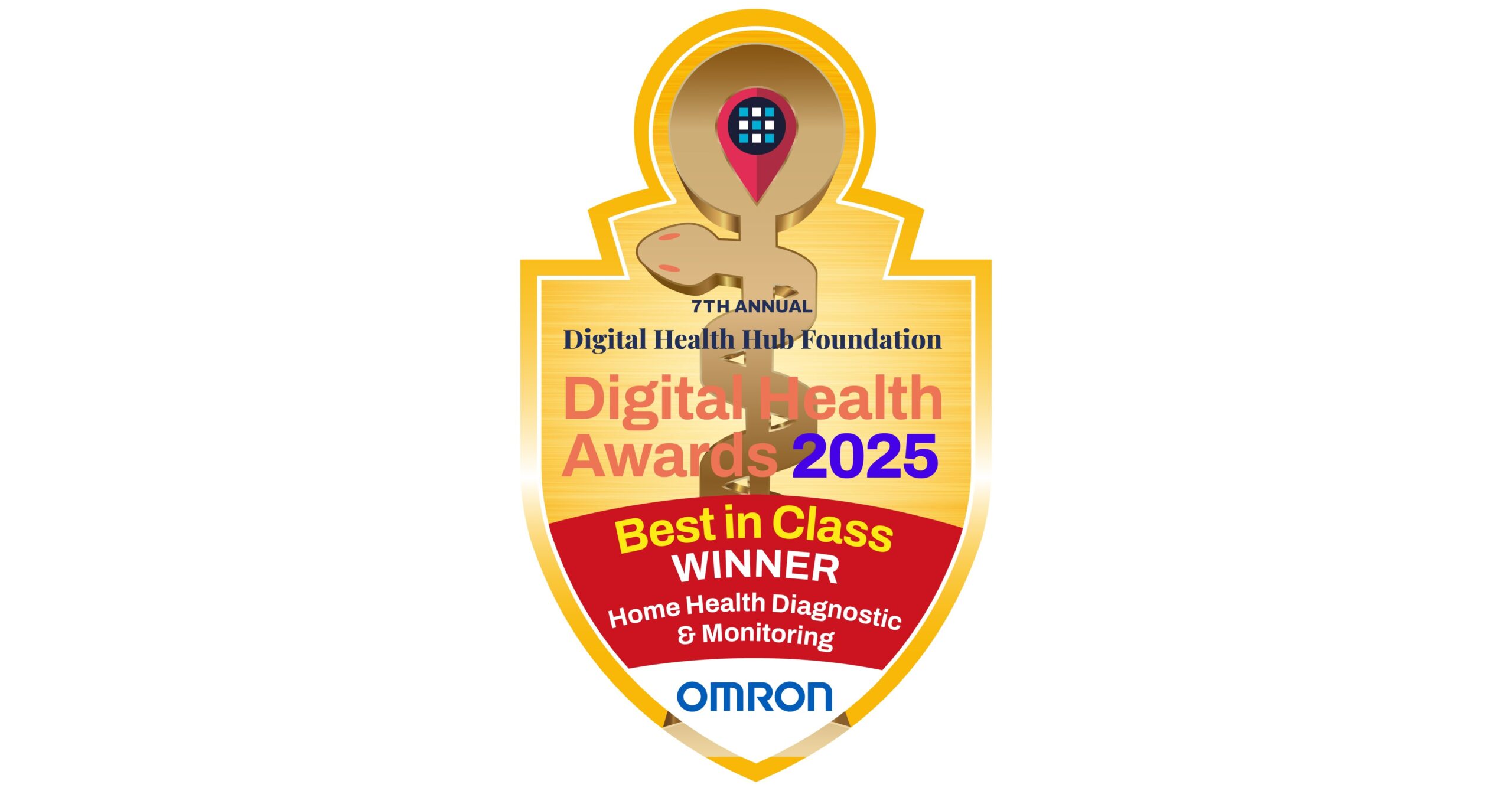
OMRON Healthcare Wins Best in Class in the 2025 Digital Health Awards at HLTH
Expert judges determining the best of the best in healthcare innovation recognized OMRON Healthcare for its blood pressure monitors with AI-powered AFib detection
HOFFMAN ESTATES, Ill., Oct. 28, 2025 /PRNewswire/ — Judges in the prestigious Digital Health Hub Foundation 2025 Digital Health Awards recognized OMRON Healthcare as the Best in Class Winner in the Home Health Diagnostics & Monitoring category for its breakthrough blood pressure monitors with AI-powered AFib detection.
OMRON won Best in Class in the Digital Health Hub Foundation Digital Health Awards for its breakthrough blood pressure monitors with AI-powered AFib detection.
The Digital Health Awards heralded OMRON’s evolution of the blood pressure monitoring experience. OMRON blood pressure monitors featuring IntelliSense™ AFib detection, such as the 10 Series, automatically screen for atrial fibrillation during every blood pressure measurement.
The Digital Health Awards, which honor the best of the best in healthcare innovation, heralded OMRON’s evolution of the blood pressure monitoring experience. In a medical device first, OMRON blood pressure monitors featuring IntelliSense™ AFib detection automatically screen for atrial fibrillation during every blood pressure measurement and can provide early detection of elevated risk for a high stroke1, heart failure2, and dementia risk3.
“Our OMRON Healthcare team is thrilled to accept this Best in Class Digital Health Award,” said Alice Koehler, Managing Director of OMRON Healthcare North America. “The digital health category is rapidly maturing and there was a record-setting number of entrants in the competition this year. Thank you to the judges for recognizing our achievement in innovation that empowers people to take charge of their health.”
“OMRON applied more than 50 years of collaboration with doctors and service to consumers to develop this breakthrough technology,” said Koehler. “The latest AHA and ACC blood pressure guidelines urge regular blood pressure monitoring at home and earlier intervention on hypertension, especially for those over age 40 and for expectant moms throughout pregnancy.”
“Atrial fibrillation (AFib) is widely undiagnosed. The health risks of AFib increase for those with high blood pressure and women in menopause. With OMRON IntelliSense™ AFib detection, we are helping people act on this condition earlier than ever before,” added Koehler.
OMRON blood pressure monitors with IntelliSense™ AFib detection include the OMRON Platinum BP5465, OMRON 10 Series® BP7465, , OMRON Gold BP5360, and OMRON 7 Series® BP7360. These clinically validated blood pressure monitors are listed on the U.S. Blood Pressure Validated Device Listing (VDL), and sync with the OMRON connect app which tracks user readings, analyzes trends and habits, presents insights to explain data, and provides daily reminders and progress reports.
OMRON’s IntelliSense™ AFib detection incorporates over 300 mathematical indices into a machine-learning algorithm that analyzes the pressure pulse wave generated as the blood pressure monitor cuff inflates. A study published in the October 2024 Heart Rhythm Journal4 found that OMRON blood pressure monitors with the Intellisense™ AFib algorithm can detect disturbances specific to AFib with remarkable accuracy.
The FDA granted OMRON medical device authorization for Intellisense™ AFib via the agency’s De Novo classification5, a regulatory pathway for first-of-their-kind innovations.6
The Digital Health Hub Foundation Awards spotlight the most innovative healthtech companies transforming healthcare through technology and recognizing the companies making the biggest impact. Each year, the competition attracts groundbreaking companies from around the world, with the most impactful advancing to the final stage.
The 7th Annual Digital Health Awards announced the Best in Class and Rising Star winners across all categories during the Grand Finale at HLTH USA in Las Vegas on October 20, 2025. The awards were presented across multiple live stages and celebrated before an audience of global healthcare leaders, investors and innovators.
“This year’s winners represent the best of what digital health has to offer, innovation that is not only transformative, but practical, scalable, and ready to make an impact today. We are proud to celebrate these companies for their vision and commitment to improving healthcare,” said Mark H. Goldstein, Chairman of the Digital Health Hub Foundation.
About the Digital Health Hub Foundation
The Digital Health Hub Foundation’s mission is to support the world’s most innovative healthcare companies in scaling and growing. Since 2017, the organization’s 30,000-member community has been dedicated to fostering innovation, including early-to-late-stage healthcare companies, industry providers, payors, mentors, and investors. Through their annual awards, the Digital Health Hub Foundation brings together the healthcare industry to celebrate and validate the best of the best in health technology.About OMRON Healthcare, Inc.
OMRON Healthcare, Inc., is the world’s leading manufacturer and distributor of personal heart health products and an innovator in technologies supporting respiratory and pain management care. With over 50 years of medical device category leadership, OMRON is passionate about empowering people to take charge of their health at home through precise technology. Its market-leading products include a full range of home blood pressure monitors, nebulizers and TENS devices. The company’s mission is Going for Zero, the elimination of heart attacks and strokes. With more than 400 million devices sold globally, OMRON provides the world’s most recommended blood pressure monitors by healthcare professionals. OMRON Healthcare strives to improve lives and contribute to a better society by developing innovations that help people prevent, treat, and manage their medical conditions. The company provides products and services in over 130 countries. For more information, visit OmronHealthcare.com.1 CDC AFib and Stroke https://www.cdc.gov/heart-disease/about/atrial-fibrillation.html?CDC_AAref_Val=https://www.cdc.gov/heartdisease/atrial_fibrillation.htm
2 AFib and Heart Failure https://www.hopkinsmedicine.org/health/conditions-and-diseases/atrial-fibrillation/afib-complications
3 PubMed: AFib and dementia https://pmc.ncbi.nlm.nih.gov/articles/PMC10568548/
4 Heart Rhythm Journal, October 2024 https://www.heartrhythmjournal.com/article/S1547-5271(24)02520-7/fulltext
5 FDA De Novo https://www.fda.gov/medical-devices/premarket-submissions-selecting-and-preparing-correct-submission/de-novo-classification-request
6 FDA De Novo AI First https://www.fda.gov/news-events/press-announcements/fda-authorizes-marketing-first-device-uses-artificial-intelligence-help-detect-potential-signs-colonSOURCE OMRON Healthcare
Continue Reading
-

Elon Musk launched Grokipedia. Here’s how it compares to Wikipedia
Elon Musk has launched Grokipedia, a crowdsourced online encyclopedia that the billionaire seeks to position as a rival to Wikipedia.
Writing on social media, Musk said that Grokipedia.com is “now live” and its goal is the “truth, the whole truth and nothing but the truth.”
Musk has previously criticized Wikipedia for being filled with “propaganda” and called for people to stop donating to the site, which is run by a nonprofit. In September he announced that his artificial intelligence company xAI was working on Grokipedia.
READ MORE: Why does the AI-powered chatbot Grok post false, offensive things on X?
The Grokipedia site has a minimalist appearance with little beyond a search bar where users can type in queries. It states that it has 885,279 articles. Wikipedia, meanwhile, says it has more than 7 million articles in English.
Like Wikipedia, users can search for articles on various topics such as Taylor Swift, the baseball World Series, or Buckingham Palace.
While Wikipedia is written and edited by volunteers, it’s unclear how exactly Grokipedia articles are put together. Reports suggest the site is powered by the same xAI model that underpins Musk’s Grok chatbot, but some articles are seemingly adapted from Wikipedia.
As a huge trove of well-constructed sentences with little restriction on how it’s used, Wikipedia has been a key source used to train AI chatbots, including Grok’s rivals ChatGPT and Google’s Gemini. That’s one reason Republican lawmakers in the U.S. Congress said they launched an investigation in August of alleged “manipulation efforts” in Wikipedia’s editing process that could inject bias and undermine neutral points of view on its platform and the AI systems that rely on it.
READ MORE: Citing ‘disruptive’ edits, Wikipedia bars anonymous entries from Congress-linked computers
Wikipedia encourages its volunteer editors to cite nearly every sentence or paragraph with a primary source, and sentences not verified can be challenged and removed. Some of Grokipedia’s entries are thinly sourced, such as an entry on the Chola Dynasty of southern India that has three linked sources, compared to Wikipedia’s that has 113 linked sources plus dozens of referenced books.
Grokipedia’s entry on Wikipedia accuses the site of having “systemic ideological biases — particularly a left-leaning slant in coverage of political figures and topics.”
The San Francisco-based Wikimedia Foundation didn’t immediately respond to a request for comment Tuesday.
A free press is a cornerstone of a healthy democracy.
Support trusted journalism and civil dialogue.
Continue Reading
-
Kulicke and Soffa Industries, Inc. Announces CEO Transition
Kulicke and Soffa Industries, Inc. Announces CEO Transition
Lester Wong to Serve as Interim CEO; Board of Directors Initiates Process to Identify Permanent Successor; Dr. Fusen Chen to Retire for Health Reasons
SINGAPORE, Oct. 28, 2025 /PRNewswire/ — Kulicke and Soffa Industries, Inc. (NASDAQ: KLIC) (“Kulicke & Soffa”, “K&S”, “we” or the “Company”) today announced that Dr. Fusen Chen has agreed to retire from his position as President and CEO, and as a member of the Board, effective December 1, 2025, due to health reasons. Following the effective date, Dr. Chen will serve as an advisor to the Board for a 12-month period. The Board has initiated a process to identify the Company’s next permanent CEO. The search will include internal and external candidates.
Effective immediately, the Board of Directors has appointed Lester Wong, the Company’s current Executive Vice President, Finance and IT and Chief Financial Officer, as the Company’s Interim CEO. As Dr. Chen transitions from his role and in leading up to his retirement, he will support Lester Wong in ensuring a smooth transition and handover. Mr. Wong will also continue in his existing roles during this transitional period.
“We are profoundly grateful to Fusen for his visionary leadership, unwavering commitment, and remarkable achievements during his nine-year tenure as CEO and President,” said Peter Kong, Chairman of the K&S Board. “Fusen’s dedication to innovation and operational excellence has enabled K&S to grow and create value for shareholders. His leadership has solidified the Company’s core served market positions while accelerating its position at the forefront of advanced packaging and dispense technologies. His legacy will continue to inspire our organization for years to come. On behalf of the Board and the entire K&S family, I extend our heartfelt thanks and wish Fusen good health and success in his next chapter.”
“It has been a great honor to serve as Kulicke & Soffa’s CEO,” said Dr. Fusen Chen, President and CEO of K&S. “I thank our incredible team for their support. Together, we have navigated unprecedented global challenges with resilience and agility while building on the 75-year track record of innovation to unlock new opportunities across our markets, drive significant growth and create value for our shareholders. I am proud to have been a part of this progress and have the utmost confidence in K&S’ future.”
Mr. Kong added, “The Board has commenced a comprehensive search to identify the right leader to continue driving K&S’ strong momentum, expand our market share, further enhance our diverse portfolio and help our customers address critical challenges in the industry. As Executive Vice President and CFO, Lester has played an instrumental role in the Company’s success and we are confident that he will continue to drive K&S’ growth and innovation during this interim period.”
Mr. Kong concluded, “Together with the Board, I will assist Lester and the Executive Leadership Team in ensuring the continuity of leadership, stability and strategic focus of the Company. We believe this collaborative approach is critical as the Company is ushering through a period of exciting growth, driven by continued core-market improvement and capacity expansion, coupled with strong demand in sectors like AI, electric vehicles, and power semiconductors. We are investing in next-generation solutions such as ATPremier MEM PlusTM and expanding our capabilities in fluxless thermocompression bonding and chiplet integration. With a solid financial foundation and a clear strategic roadmap, we are well-positioned to support our customers’ evolving needs and deliver long-term value.”
“I look forward to assuming the interim CEO position and working closely with Peter, the Board and the entire Executive Leadership Team as the Board conducts its search for a permanent CEO to lead K&S into its next phase,” said Lester Wong, Executive Vice President, Finance and IT and CFO of K&S. “I am confident in the clear strategy we’ve established, which positions us to continue driving our leadership across semiconductor assembly technology and unlock meaningful value for our shareholders.”
K&S Reaffirms its Fourth Quarter Fiscal 2025 Outlook
In connection with today’s announcement, the Company reaffirmed its fourth quarter fiscal 2025 outlook. The Company intends to announce its fourth quarter fiscal 2025 financial results on November 19, 2025.
About Lester Wong
Mr. Wong, 59, has been the Company’s Chief Financial Officer since December 2018, and was promoted to Executive Vice President, from Senior Vice President, on January 1, 2022. Prior to that, Mr. Wong served as General Counsel & Senior Vice President, Legal Affairs beginning in September 2011. Mr. Wong has over 20 years of legal leadership at U.S. listed and Asian-based companies, with a wealth of experience in global corporate law. Previously, Mr. Wong served as General Counsel and held other senior legal positions at publicly traded companies including Gigamedia Limited and CDC Corporation.
About Kulicke & Soffa
Kulicke & Soffa is a global leader in semiconductor assembly technology, advancing device performance across automotive, compute, industrial, memory and communications markets. Founded on innovation in 1951, K&S is uniquely positioned to overcome increasingly dynamic process challenges – creating and delivering long-term value by aligning technology with opportunity.
Contacts
Kulicke & Soffa
Marilyn Sim
Public Relations
+65-6880 9309
msim@kns.comKulicke & Soffa
Joseph Elgindy
Finance
+1-215-784-7500
investor@kns.comSOURCE Kulicke & Soffa Industries, Inc.
Continue Reading
-

Booking Holdings beats results estimates on steady travel demand, shares up
(Reuters) -Online travel agent Booking Holdings beat Wall Street estimates for third-quarter revenue and profit on Tuesday, helped by more customers bundling their reservations on its platform and steady travel demand.
Shares of the company rose nearly 5% in after-hours trading.
“While there remains some uncertainty in the macroeconomic and geopolitical backdrop, we are pleased to see continued momentum with steady travel demand trends in our business so far in the fourth quarter,” the company said in a statement.
Gross bookings for the third quarter came in at $49.7 billion, up 14% from last year.
The Norwalk, Connecticut-based company reported a quarterly adjusted profit of $99.50 per share, compared with analysts’ estimates of $95.66 per share, according to data compiled by LSEG.
Total revenue for the quarter ended September 30 was $9.01 billion, also above expectations of $8.72 billion.
(Reporting by Aishwarya Jain and Utkarsh Shetti in Bengaluru; Editing by Alan Barona)
Continue Reading