The Japanese carmaker Nissan is to team up with its Chinese electric vehicle rival BYD in an attempt to offset their carbon emissions and avoid EU penalties for 2025, it has confirmed.
It is part of a wider offsetting scheme the EU has sanctioned for the car industry that could help manufacturers of combustion engine cars head off an estimated £13bn in fines.
Nissan said in a statement: “Nissan has formed a pool with BYD for its CO2 fleet emissions in Europe for the 2025 calendar year. The scope of the agreement covers passenger vehicles within EU markets and will contribute to Nissan’s commitment towards zero emissions in a sustainable way, while continuing to support the EU’s 2050 decarbonisation target.”
It added that it had entered into the agreement to “ensure the business is better able to comply with EU regulations and continue the transition towards our own goal of zero emissions”.
Chinese exports of EVs to the EU are already posing an existential crisis to the European car industry but are now, like Tesla, helping traditional car firms meet their decarbonisation targets courtesy of an EU regulation that in effect allows car firms to “pool” emissions.
The EU has already extended the period for compliance with emissions rules from one year to three years, fuelling fears this will further delay the already slow take-up of EVs in the EU, particularly in southern Europe, but also in key states such as France and Germany.
Fredrik Eklund, responsible for carbon credits trading at the Chinese-owned Swedish brand Polestar, which only makes electric vehicles, said: “It risks delaying the transition from legacy cars to EVs. We are already seeing car manufacturers pushing at the 2027 expiry date, but from our point of view and from the point of view of society, we really don’t want to delay this.”
Under the rules, car manufacturers have to meet emissions targets of 93.6g of CO2 per kilometre.
But under the car pooling arrangement, car manufacturers can pay electric car companies to use their zero emissions record to average out the pollution from sales of their combustion engine cars to avoid fines.
The industry in the past has said the 2025 emissions targets could have led to as much as €15bn (£13.03bn) in fines.
The latest car pooling agreement, confirmed by Nissan, mirrors that of other companies who have teamed up with other big name electric car brands including Tesla and Polestar.
skip past newsletter promotion
after newsletter promotion
Polestar has a pooling arrangement with Mercedes-Benz, Volvo and Smart cars, while Tesla’s zero emissions record is being mopped up by Toyota, Ford, Mazda, Alfa Romeo and Suzuki.
The price car companies are paying EV firms to offset their emissions remains confidential. But in January it was reported that carbon credit sales accounted for almost 3% of Tesla’s $72bn (£54bn) total revenue in the first nine months of last year – just over £1.6bn.
The car industry is now fighting for a softening of the EU’s 2035 target for banning the sale of new combustion engine cars, arguing that the public is still not prepared to make the switch in sufficient numbers, citing lack of infrastructure in southern and central Europe as part of the problem.
Newton K, Strasser A, Kayagaki N, Dixit VM. Cell death. Cell. 2024;187(2):235–56. https://doi.org/10.1016/j.cell.2023.11.044.
Article
PubMed
Google Scholar
Green DR, Victor B. The pantheon of the fallen: why are there so many forms of cell death? Trends Cell Biol. 2012;22(11):555–6. https://doi.org/10.1016/j.tcb.2012.08.008.
Article
PubMed
PubMed Central
Google Scholar
Dixon SJ, Lemberg KM, Lamprecht MR, et al. Ferroptosis: an iron-dependent form of nonapoptotic cell death. Cell. 2012;149(5):1060–72. https://doi.org/10.1016/j.cell.2012.03.042.
Article
PubMed
PubMed Central
Google Scholar
Bannai S, Kitamura E. Transport interaction of L-cystine and L-glutamate in human diploid fibroblasts in culture. J Biol Chem. 1980;255(6):2372–6.
Article
PubMed
Google Scholar
Fotiadis D, Kanai Y, Palacín M. The SLC3 and SLC7 families of amino acid transporters. Mol Aspects Med. 2013;34(2–3):139–58. https://doi.org/10.1016/j.mam.2012.10.007.
Article
PubMed
Google Scholar
Koppula P, Zhang Y, Zhuang L, Gan B. Amino acid transporter SLC7A11/xCT at the crossroads of regulating redox homeostasis and nutrient dependency of cancer. Cancer Commun (Lond). 2018;38(1):12. https://doi.org/10.1186/s40880-018-0288-x.
Article
PubMed
Google Scholar
Mou Y, Wang J, Wu J, He D, Zhang C, Duan C, et al. Ferroptosis, a new form of cell death: opportunities and challenges in cancer. J Hematol Oncol. 2019;12(1):34. https://doi.org/10.1186/s13045-019-0720-y.
Article
PubMed
PubMed Central
Google Scholar
Liu J, Xia X, Huang P, xCT:. A critical molecule that links cancer metabolism to redox signaling. Mol Ther. 2020;28(11):2358–66. https://doi.org/10.1016/j.ymthe.2020.08.021.
Article
PubMed
PubMed Central
Google Scholar
Koppula P, Zhuang L, Gan B. Cystine transporter SLC7A11/xCT in cancer: ferroptosis, nutrient dependency, and cancer therapy. Protein Cell. 2021;12(8):599–620. https://doi.org/10.1007/s13238-020-00789-5.
Article
PubMed
Google Scholar
Liu X, Nie L, Zhang Y, et al. Actin cytoskeleton vulnerability to disulfide stress mediates Disulfidptosis. Nat Cell Biol. 2023;25(3):404–14. https://doi.org/10.1038/s41556-023-01091-2.
Article
PubMed
PubMed Central
Google Scholar
Lee N, Park SJ, Lange M, et al. Selenium reduction of ubiquinone via SQOR suppresses ferroptosis. Nat Metab. 2024;6(2):343–58. https://doi.org/10.1038/s42255-024-00974-4.
Article
PubMed
PubMed Central
Google Scholar
Jiang X, Stockwell BR, Conrad M. Ferroptosis: mechanisms, biology and role in disease. Nat Rev Mol Cell Biol. 2021;22(4):266–82. https://doi.org/10.1038/s41580-020-00324-8.
Article
PubMed
PubMed Central
Google Scholar
Lin Q, Zhou H, Zeng J, et al. Bioactive polysaccharides mediate ferroptosis to modulate tumor immunotherapy. Int J Biol Macromol. 2024. https://doi.org/10.1016/j.ijbiomac.2024.135147.
Article
PubMed
Google Scholar
Mao C, Wang M, Zhuang L, Gan B. Metabolic cell death in cancer: ferroptosis, cuproptosis, disulfidptosis, and beyond. Protein Cell. 2024;15(9):642–60. https://doi.org/10.1093/procel/pwae003.
Article
PubMed
PubMed Central
Google Scholar
Chen X, Kang R, Kroemer G, Tang D. Broadening horizons: the role of ferroptosis in cancer. Nat Rev Clin Oncol. 2021;18(5):280–96. https://doi.org/10.1038/s41571-020-00462-0.
Article
PubMed
Google Scholar
Tang D, Chen X, Kang R, Kroemer G. Ferroptosis: molecular mechanisms and health implications. Cell Res. 2021;31(2):107–25. https://doi.org/10.1038/s41422-020-00441-1.
Article
PubMed
Google Scholar
Tong X, Tang R, Xiao M, et al. Targeting cell death pathways for cancer therapy: recent developments in necroptosis, pyroptosis, ferroptosis, and Cuproptosis research. J Hematol Oncol. 2022;15(1):174. https://doi.org/10.1186/s13045-022-01392-3.
Article
PubMed
PubMed Central
Google Scholar
Lang X, Green MD, Wang W, et al. Radiotherapy and immunotherapy promote tumoral lipid oxidation and ferroptosis via synergistic repression of SLC7A11. Cancer Discov. 2019;9(12):1673–85. https://doi.org/10.1158/2159-8290.CD-19-0338.
Article
PubMed
PubMed Central
Google Scholar
Li Y, Yan J, Zhao Q, Zhang Y, Zhang Y. ATF3 promotes ferroptosis in sorafenib-induced cardiotoxicity by suppressing Slc7a11 expression. Front Pharmacol. 2022;13:904314. https://doi.org/10.3389/fphar.2022.904314.
Article
PubMed
PubMed Central
Google Scholar
Bassi MT, Gasol E, Manzoni M, et al. Identification and characterisation of human xCT that co-expresses, with 4F2 heavy chain, the amino acid transport activity system Xc-. Pflugers Arch. 2001;442(2):286–96. https://doi.org/10.1007/s004240100537.
Article
PubMed
Google Scholar
Bridges CC, Kekuda R, Wang H, et al. Structure, function, and regulation of human cystine/glutamate transporter in retinal pigment epithelial cells. Invest Ophthalmol Vis Sci. 2001;42(1):47–54.
PubMed
Google Scholar
Kim JY, Kanai Y, Chairoungdua A, et al. Human cystine/glutamate transporter: cDNA cloning and upregulation by oxidative stress in glioma cells. Biochim Biophys Acta. 2001;1512(2):335–44.
Article
PubMed
Google Scholar
Sato H, Tamba M, Kuriyama-Matsumura K, Okuno S, Bannai S. Molecular cloning and expression of human xCT, the light chain of amino acid transport system Xc-. Antioxid Redox Signal. 2000;2(4):665–71. https://doi.org/10.1089/ars.2000.2.4-665.
Article
PubMed
Google Scholar
Li S, Lu Z, Sun R, et al. The role of SLC7A11 in cancer: friend or foe? Cancers (Basel). 2022;14(13):3059. https://doi.org/10.3390/cancers14133059.
Article
PubMed
Google Scholar
Yang J, Zhou Y, Xie S, et al. Metformin induces ferroptosis by inhibiting ufmylation of SLC7A11 in breast cancer. J Exp Clin Cancer Res. 2021;40(1):206. https://doi.org/10.1186/s13046-021-02012-7.
Article
PubMed
PubMed Central
Google Scholar
Badgley MA, Kremer DM, Maurer HC, et al. Cysteine depletion induces pancreatic tumor ferroptosis in mice. Science. 2020;368(6486):85–9. https://doi.org/10.1126/science.aaw9872.
Article
PubMed
PubMed Central
Google Scholar
Hong T, Lei G, Chen X, et al. PARP inhibition promotes ferroptosis via repressing SLC7A11 and synergizes with ferroptosis inducers in BRCA-proficient ovarian cancer. Redox Biol. 2021;42:101928. https://doi.org/10.1016/j.redox.2021.101928.
Article
PubMed
PubMed Central
Google Scholar
Park JW, Kilic O, Deo M, et al. CIC reduces xCT/SLC7A11 expression and glutamate release in glioma. Acta Neuropathol Commun. 2023;11(1):13. https://doi.org/10.1186/s40478-023-01507-y.
Article
PubMed
PubMed Central
Google Scholar
Long Y, Tao H, Karachi A, et al. Dysregulation of glutamate transport enhances Treg function that promotes VEGF blockade resistance in glioblastoma. Cancer Res. 2020;80(3):499–509. https://doi.org/10.1158/0008-5472.CAN-19-1577.
Article
PubMed
Google Scholar
Yuan L, Li S, Chen Q, et al. EBV infection-induced GPX4 promotes chemoresistance and tumor progression in nasopharyngeal carcinoma. Cell Death Differ. 2022;29(8):1513–27. https://doi.org/10.1038/s41418-022-00939-8.
Article
PubMed
PubMed Central
Google Scholar
Combs JA, DeNicola GM. The non-essential amino acid cysteine becomes essential for tumor proliferation and survival. Cancers (Basel). 2019;11(5):678. https://doi.org/10.3390/cancers11050678.
Article
PubMed
Google Scholar
Carlisle AE, Lee N, Matthew-Onabanjo AN, et al. Selenium detoxification is required for cancer-cell survival. Nat Metab. 2020;2(7):603–11. https://doi.org/10.1038/s42255-020-0224-7.
Article
PubMed
PubMed Central
Google Scholar
Lee N, Carlisle AE, Kim D. Examining xCT-mediated selenium uptake and Selenoprotein production capacity in cells. Methods Enzymol. 2022;662:1–24. https://doi.org/10.1016/bs.mie.2021.10.002.
Article
PubMed
Google Scholar
Goji T, Takahara K, Negishi M, Katoh H. Cystine uptake through the cystine/glutamate antiporter xCT triggers glioblastoma cell death under glucose deprivation. J Biol Chem. 2017;292(48):19721–32. https://doi.org/10.1074/jbc.M117.814392.
Article
PubMed
PubMed Central
Google Scholar
Timmerman LA, Holton T, Yuneva M, et al. Glutamine sensitivity analysis identifies the xCT antiporter as a common triple-negative breast tumor therapeutic target. Cancer Cell. 2013;24(4):450–65. https://doi.org/10.1016/j.ccr.2013.08.020.
Article
PubMed
PubMed Central
Google Scholar
Shin CS, Mishra P, Watrous JD, et al. The glutamate/cystine xCT antiporter antagonizes glutamine metabolism and reduces nutrient flexibility. Nat Commun. 2017;8:15074. https://doi.org/10.1038/ncomms15074.
Article
PubMed
PubMed Central
Google Scholar
Romero R, Sayin VI, Davidson SM, et al. Keap1 loss promotes Kras-driven lung cancer and results in dependence on glutaminolysis. Nat Med. 2017;23(11):1362–8. https://doi.org/10.1038/nm.4407.
Article
PubMed
PubMed Central
Google Scholar
Bannai S. Induction of cystine and glutamate transport activity in human fibroblasts by diethyl maleate and other electrophilic agents. J Biol Chem. 1984;259(4):2435–40.
Article
PubMed
Google Scholar
Bannai S, Kitamura E. Adaptive enhancement of cystine and glutamate uptake in human diploid fibroblasts in culture. Biochim Biophys Acta. 1982;721(1):1–10. https://doi.org/10.1016/0167-4889(82)90017-9.
Article
PubMed
Google Scholar
Bannai S, Sato H, Ishii T, Taketani S. Enhancement of glutathione levels in mouse peritoneal macrophages by sodium arsenite, cadmium chloride and glucose/glucose oxidase. Biochim Biophys Acta. 1991;1092(2):175–9. https://doi.org/10.1016/0167-4889(91)90153-o.
Article
PubMed
Google Scholar
Pakos-Zebrucka K, Koryga I, Mnich K, Ljujic M, Samali A, Gorman AM. The integrated stress response. EMBO Rep. 2016;17(10):1374–95. https://doi.org/10.15252/embr.201642195.
Article
PubMed
PubMed Central
Google Scholar
Ye P, Mimura J, Okada T, et al. Nrf2- and ATF4-dependent upregulation of xCT modulates the sensitivity of T24 bladder carcinoma cells to proteasome inhibition. Mol Cell Biol. 2014;34(18):3421–34. https://doi.org/10.1128/MCB.00221-14.
Article
PubMed
PubMed Central
Google Scholar
Ye Y, Chen A, Li L, et al. Repression of the antiporter SLC7A11/glutathione/glutathione peroxidase 4 axis drives ferroptosis of vascular smooth muscle cells to facilitate vascular calcification. Kidney Int. 2022;102(6):1259–75. https://doi.org/10.1016/j.kint.2022.07.034.
Article
PubMed
Google Scholar
Wang L, Liu Y, Du T, et al. ATF3 promotes erastin-induced ferroptosis by suppressing system Xc. Cell Death Differ. 2020;27(2):662–75. https://doi.org/10.1038/s41418-019-0380-z.
Article
PubMed
Google Scholar
Zhao X, Chen C, Qiu H, et al. The landscape of ATF3 in tumors: metabolism, expression regulation, therapy approach, and open concerns. Pharmacol Res. 2025;214:107666. https://doi.org/10.1016/j.phrs.2025.107666.
Article
PubMed
Google Scholar
Wang Y, Yang L, Zhang X, et al. Epigenetic regulation of ferroptosis by H2B monoubiquitination and p53. EMBO Rep. 2019;20(7):e47563. https://doi.org/10.15252/embr.201847563.
Article
PubMed
PubMed Central
Google Scholar
Zhang Y, Koppula P, Gan B. Regulation of H2A ubiquitination and SLC7A11 expression by BAP1 and PRC1. Cell Cycle. 2019;18(8):773–83. https://doi.org/10.1080/15384101.2019.1597506.
Article
PubMed
PubMed Central
Google Scholar
Tsuchihashi K, Okazaki S, Ohmura M, et al. The EGF receptor promotes the malignant potential of glioma by regulating amino acid transport system xc(-). Cancer Res. 2016;76(10):2954–63. https://doi.org/10.1158/0008-5472.CAN-15-2121.
Article
PubMed
PubMed Central
Google Scholar
Liu L, He J, Sun G, et al. The N6-methyladenosine modification enhances ferroptosis resistance through inhibiting SLC7A11 mRNA deadenylation in hepatoblastoma. Clin Transl Med. 2022;12(5):e778. https://doi.org/10.1002/ctm2.778.
Article
PubMed
PubMed Central
Google Scholar
Zhu Y, Zhang C, Huang M, Lin J, Fan X, Ni T. TRIM26 induces ferroptosis to inhibit hepatic stellate cell activation and mitigate liver fibrosis through mediating SLC7A11 ubiquitination. Front Cell Dev Biol. 2021;9:644901. https://doi.org/10.3389/fcell.2021.644901.
Article
PubMed
PubMed Central
Google Scholar
Li S, Lu Z, Sun R, et al. The role of SLC7A11 in cancer: friend or foe? Cancers (Basel). 2022;14(13):3059. https://doi.org/10.3390/cancers14133059.
Article
PubMed
Google Scholar
Ishimoto T, Nagano O, Yae T, et al. CD44 variant regulates redox status in cancer cells by stabilizing the xCT subunit of system xc(-) and thereby promotes tumor growth. Cancer Cell. 2011;19(3):387–400. https://doi.org/10.1016/j.ccr.2011.01.038.
Article
PubMed
Google Scholar
Gan W, Dai X, Dai X, et al. LATS suppresses mTORC1 activity to directly coordinate Hippo and mTORC1 pathways in growth control. Nat Cell Biol. 2020;22(2):246–56. https://doi.org/10.1038/s41556-020-0463-6.
Article
PubMed
PubMed Central
Google Scholar
Zhang W, Feng J, Ni Y, et al. The role of SLC7A11 in diabetic wound healing: novel insights and new therapeutic strategies. Front Immunol. 2024;15:1467531. https://doi.org/10.3389/fimmu.2024.1467531.
Article
PubMed
PubMed Central
Google Scholar
Gu Y, Albuquerque CP, Braas D, et al. mTORC2 regulates amino acid metabolism in cancer by phosphorylation of the Cystine-Glutamate antiporter xCT. Mol Cell. 2017;67(1):128–e1387. https://doi.org/10.1016/j.molcel.2017.05.030.
Article
PubMed
PubMed Central
Google Scholar
Stockwell BR, Friedmann Angeli JP, Bayir H, et al. Ferroptosis: a regulated cell death nexus linking metabolism, redox biology, and disease. Cell. 2017;171(2):273–85. https://doi.org/10.1016/j.cell.2017.09.021.
Kagan VE, Mao G, Qu F, et al. Oxidized arachidonic and adrenic PEs navigate cells to ferroptosis. Nat Chem Biol. 2017;13(1):81–90. https://doi.org/10.1038/nchembio.2238.
Article
PubMed
Google Scholar
Chu B, Kon N, Chen D, et al. ALOX12 is required for p53-mediated tumour suppression through a distinct ferroptosis pathway. Nat Cell Biol. 2019;21(5):579–91. https://doi.org/10.1038/s41556-019-0305-6.
Article
PubMed
PubMed Central
Google Scholar
Jiang L, Kon N, Li T, et al. Ferroptosis as a p53-mediated activity during tumour suppression. Nature. 2015;520(7545):57–62. https://doi.org/10.1038/nature14344.
Article
PubMed
PubMed Central
Google Scholar
Jennis M, Kung CP, Basu S, et al. An African-specific polymorphism in the TP53 gene impairs p53 tumor suppressor function in a mouse model. Genes Dev. 2016;30(8):918–30. https://doi.org/10.1101/gad.275891.115.
Article
PubMed
PubMed Central
Google Scholar
Zhang Y, Shi J, Liu X, et al. BAP1 links metabolic regulation of ferroptosis to tumour suppression. Nat Cell Biol. 2018;20(10):1181–92. https://doi.org/10.1038/s41556-018-0178-0.
Article
PubMed
PubMed Central
Google Scholar
Liu T, Jiang L, Tavana O, Gu W. The deubiquitylase OTUB1 mediates ferroptosis via stabilization of SLC7A11. Cancer Res. 2019;79(8):1913–24. https://doi.org/10.1158/0008-5472.CAN-18-3037.
Article
PubMed
PubMed Central
Google Scholar
Reck M, Carbone DP, Garassino M, Barlesi F. Targeting KRAS in non-small-cell lung cancer: recent progress and new approaches. Ann Oncol. 2021;32(9):1101–10. https://doi.org/10.1016/j.annonc.2021.06.001.
Article
PubMed
Google Scholar
Xiong HJ, Yu HQ, Zhang J, et al. Elevated FBXL6 activates both wild-type KRAS and mutant KRASG12D and drives HCC tumorigenesis via the ERK/mTOR/PRELID2/ROS axis in mice. Mil Med Res. 2023;10(1):68. https://doi.org/10.1186/s40779-023-00501-8.
Article
PubMed
PubMed Central
Google Scholar
Mueller S, Engleitner T, Maresch R, et al. Evolutionary routes and KRAS dosage define pancreatic cancer phenotypes. Nature. 2018;554(7690):62–8. https://doi.org/10.1038/nature25459.
Article
PubMed
PubMed Central
Google Scholar
Hu K, Li K, Lv J, et al. Suppression of the SLC7A11/glutathione axis causes synthetic lethality in KRAS-mutant lung adenocarcinoma. J Clin Invest. 2020;130(4):1752–66. https://doi.org/10.1172/JCI124049.
Article
PubMed
PubMed Central
Google Scholar
Lim JKM, Delaidelli A, Minaker SW, et al. Cystine/glutamate antiporter xCT (SLC7A11) facilitates oncogenic RAS transformation by preserving intracellular redox balance. Proc Natl Acad Sci U S A. 2019;116(19):9433–42. https://doi.org/10.1073/pnas.1821323116.
Article
PubMed
PubMed Central
Google Scholar
Chen X, Li J, Kang R, Klionsky DJ, Tang D. Ferroptosis: machinery and regulation. Autophagy. 2021;17(9):2054–81. https://doi.org/10.1080/15548627.2020.1810918.
Article
PubMed
Google Scholar
Wang W, Green M, Choi JE, et al. CD8 + T cells regulate tumour ferroptosis during cancer immunotherapy. Nature. 2019;569(7755):270–4. https://doi.org/10.1038/s41586-019-1170-y.
Article
PubMed
PubMed Central
Google Scholar
Kim DH, Kim WD, Kim SK, Moon DH, Lee SJ. TGF-β1-mediated repression of SLC7A11 drives vulnerability to GPX4 Inhibition in hepatocellular carcinoma cells. Cell Death Dis. 2020;11(5):406. https://doi.org/10.1038/s41419-020-2618-6.
Article
PubMed
PubMed Central
Google Scholar
Dai E, Han L, Liu J, et al. Autophagy-dependent ferroptosis drives tumor-associated macrophage polarization via release and uptake of oncogenic KRAS protein. Autophagy. 2020;16(11):2069–83. https://doi.org/10.1080/15548627.2020.1714209.
Article
PubMed
PubMed Central
Google Scholar
Bi G, Liang J, Zhao M, et al. MiR-6077 promotes cisplatin/pemetrexed resistance in lung adenocarcinoma via CDKN1A/cell cycle arrest and KEAP1/ferroptosis pathways. Mol Ther. 2022;28:366–86. https://doi.org/10.1016/j.omtn.2022.03.020.
Article
Google Scholar
Qin K, Zhang F, Wang H, et al. CircRNA circSnx12 confers cisplatin chemoresistance to ovarian cancer by inhibiting ferroptosis through a miR-194-5p/SLC7A11 axis. BMB Rep. 2023;56(2):184–9. https://doi.org/10.5483/BMBRep.2022-0175.
Article
PubMed
PubMed Central
Google Scholar
Sun C, Liu P, Pei L, Zhao M, Huang Y. Propofol inhibits proliferation and augments the anti-tumor effect of doxorubicin and paclitaxel partly through promoting ferroptosis in triple-negative breast cancer cells. Front Oncol. 2022;12:837974. https://doi.org/10.3389/fonc.2022.837974.
Article
PubMed
PubMed Central
Google Scholar
Yadav P, Sharma P, Sundaram S, Venkatraman G, Bera AK, Karunagaran D. SLC7A11/ xCT is a target of miR-5096 and its restoration partially rescues miR-5096-mediated ferroptosis and anti-tumor effects in human breast cancer cells. Cancer Lett. 2021;522:211–24. https://doi.org/10.1016/j.canlet.2021.09.033.
Article
PubMed
Google Scholar
Chen X, Kang R, Kroemer G, Tang D. Ferroptosis in infection, inflammation, and immunity. J Exp Med. 2021;218(6):e20210518. https://doi.org/10.1084/jem.20210518.
Article
PubMed
PubMed Central
Google Scholar
Mi T, Kong X, Chen M, Guo P, He D. Inducing disulfidptosis in tumors: potential pathways and significance. MedComm (2020). 2024;5(11):e791. https://doi.org/10.1002/mco2.791
Yan Y, Teng H, Hang Q, et al. Slc7a11 expression level dictates differential responses to oxidative stress in cancer cells. Nat Commun. 2023;14(1):3673. https://doi.org/10.1038/s41467-023-39401-9.
Article
PubMed
PubMed Central
Google Scholar
Joly JH, Delfarah A, Phung PS, Parrish S, Graham NA. A synthetic lethal drug combination mimics glucose deprivation-induced cancer cell death in the presence of glucose. J Biol Chem. 2020;295(5):1350–65. https://doi.org/10.1074/jbc.RA119.011471.
Article
PubMed
Google Scholar
Liu T, Ren Y, Wang Q, et al. Exploring the role of the disulfidptosis-related gene SLC7A11 in adrenocortical carcinoma: implications for prognosis, immune infiltration, and therapeutic strategies. Cancer Cell Int. 2023;23(1):259. https://doi.org/10.1186/s12935-023-03091-6.
Article
PubMed
PubMed Central
Google Scholar
Zhao D, Meng Y, Dian Y, et al. Molecular landmarks of tumor disulfidptosis across cancer types to promote disulfidptosis-target therapy. Redox Biol. 2023;68:102966. https://doi.org/10.1016/j.redox.2023.102966.
Article
PubMed
PubMed Central
Google Scholar
Li J, Yu T, Sun J, et al. Integrated analysis of disulfidptosis-related immune genes signature to boost the efficacy of prognostic prediction in gastric cancer. Cancer Cell Int. 2024;24(1):112. https://doi.org/10.1186/s12935-024-03294-5.
Article
PubMed
PubMed Central
Google Scholar
Xia Q, Yan Q, Wang Z, et al. Disulfidptosis-associated lncRNAs predict breast cancer subtypes. Sci Rep. 2023;13(1):16268. https://doi.org/10.1038/s41598-023-43414-1.
Article
PubMed
PubMed Central
Google Scholar
Liu L, Liu J, Lyu Q, et al. Disulfidptosis-associated lncRNAs index predicts prognosis and chemotherapy drugs sensitivity in cervical cancer. Sci Rep. 2023;13(1):12470. https://doi.org/10.1038/s41598-023-39669-3.
Article
PubMed
PubMed Central
Google Scholar
Korangath P, Teo WW, Sadik H, et al. Targeting glutamine metabolism in breast cancer with aminooxyacetate. Clin Cancer Res. 2015;21(14):3263–73. https://doi.org/10.1158/1078-0432.CCR-14-1200.
Article
PubMed
PubMed Central
Google Scholar
Stine ZE, Schug ZT, Salvino JM, Dang CV. Targeting cancer metabolism in the era of precision oncology. Nat Rev Drug Discov. 2022;21(2):141-162. doi:10.1038/s41573-021-00339-6
Shao N, Qiu H, Liu J, et al. Targeting lipid metabolism of macrophages: a new strategy for tumor therapy. J Adv Res. 2025;68:99–114. https://doi.org/10.1016/j.jare.2024.02.009.
Article
PubMed
Google Scholar
Zhao X, Zhao J, Li D, et al. Akkermansia muciniphila: a potential target and pending issues for oncotherapy. Pharmacol Res. 2023;196:106916. https://doi.org/10.1016/j.phrs.2023.106916.
Article
PubMed
Google Scholar
Liu X, Zhuang L, Gan B. Disulfidptosis: disulfide stress-induced cell death. Trends Cell Biol. 2024;34(4):327–37. https://doi.org/10.1016/j.tcb.2023.07.009.
Article
PubMed
Google Scholar
Qiu H, Shao N, Liu J, et al. Amino acid metabolism in tumor: new shine in the fog? Clin Nutr. 2023;42(8):1521–30. https://doi.org/10.1016/j.clnu.2023.06.011.
Article
PubMed
Google Scholar
Liu J, Shao N, Qiu H, et al. Intestinal microbiota: A Bridge between intermittent fasting and tumors. Biomed Pharmacother. 2023;167:115484. https://doi.org/10.1016/j.biopha.2023.115484.
As part of the deal, Eli Lilly will acquire Adverum’s lead candidate ixo-vec, an intravitreal gene therapy for wAMD treatment. Credit: Tada Images via Shutterstock.com.
Eli Lilly has agreed to acquire eye disease specialist Adverum Biotechnologies, bucking a recent trend of big pharma companies deciding to steer clear of the cell and gene therapy sector.
Eli Lilly has offered Adverum $3.56 per share in cash, including an additional $8.91 in milestone payments. The latter depends on US approval of the biotech’s lead gene therapy candidate, ixo-vec, within seven years and achieving more than $1bn in annual global sales within ten years. This brings the total consideration to $12.47 a share, valuing the deal at a possible $261.7m.
Discover B2B Marketing That Performs
Combine business intelligence and editorial excellence to reach engaged professionals across 36 leading media platforms.
Find out more
The share offer agreed on 24 October reflects a nearly 15% discount from the $4.18 closing price on 23 October.
For Adverum, the potential buyout from Eli Lilly provides financial respite. The biotech has been struggling for cash in recent times – holding $44.4m to its name in July 2025. The lack of capital had increased jeopardy for ixo-vec, an intravitreal gene therapy that advanced into a Phase III trial (NCT06856577) for the treatment of wet age-related macular degeneration (wAMD) in March 2025.
Indeed, Eli Lilly stated that without a $65m loan given to Adverum to continue ongoing clinical trials, the biotech would only be able to finance itself through October before having to wind down operations.
Despite having to help fund ixo-vec’s development, which has been granted fast track and regenerative medicine advanced therapy (RMAT) designations by the US Food and Drug Administration (FDA), Eli Lilly could use the candidate to enter the lucrative wAMD market. The AMD sector, which also includes the dry form, is expected to reach $27.5bn across 7MM by 2031 (7MM: US, France, Germany, Italy, Spain, UK, and Japan), according to GlobalData analysis.
There is no gene therapy approved with a wAMD indication, with current treatments working via the anti–vascular endothelial growth factor (VEGF) mechanism, such as Regeneron’s blockbuster Eylea (aflibercept). The therapy is administered every four weeks for the first five months, followed by a single injection every two months. For Eli Lilly’s soon-to-be acquired ixo-vec, this could offer patients a one-and-done treatment.
Lilly molecule discovery group vice-president Andrew Adams said: “Ixo-vec has the potential to transform wAMD treatment from a paradigm of chronic care with repeated intravitreal injections to a convenient one-time therapy.”
Adverum CEO Laurent Fischer: “[Lilly’s] scientific depth and global reach offer the opportunity to accelerate our vision to deliver a transformative one-and-done therapy that can potentially restore and preserve vision for millions of patients living with wAMD.”
Lilly bucks big pharma trend
This is not the first time in 2025 that Eli Lilly has swooped in to rescue a cash-strapped biotech specialising in gene therapies. In April, the big pharma signed a licensing deal worth up to $1.4bn for Sangamo Therapeutics’ neurology-targeting gene therapy.
However, Lilly’s recent deals, which includes a $1.3bn acquisition of RNA-based gene therapy developer Rznomics in May 2025, goes against the grain of big pharma generally opting to retreat from the cell and gene therapy sector.
Earlier this month, Galapagos wound down its cell and gene therapy division after failing to sell the unit. Japanese pharma Takeda also abandoned its cell therapy research, pivoting instead towards small molecules, biologics and antibody-drug conjugates (ADCs).
In addition, Gilead Sciences’ Kite Pharma terminated its cell therapy collaboration with Shoreline in September 2025, ending a research partnership valued at $2.3bn.
Cell & Gene Therapy coverage on Pharmaceutical Technology is supported by Cytiva.
Editorial content is independently produced and follows the highest standards of journalistic integrity. Topic sponsors are not involved in the creation of editorial content.
Sign up for our daily news round-up!
Give your business an edge with our leading industry insights.
Pharmaceutical Technology Excellence Awards – The Benefits of Entering
Gain the recognition you deserve! The Pharmaceutical Technology Excellence Awards celebrate innovation, leadership, and impact. By entering, you showcase your achievements, elevate your industry profile, and position yourself among top leaders driving pharmaceutical advancements. Don’t miss your chance to stand out—submit your entry today!
I recently had the pleasure of visiting the lovely mountain town of Lugano, Switzerland, whose appeal lies in that it is basically Italy but administered by the Swiss. That’s according to Tether CEO Paolo Ardoino, one of the prime backers of Plan B, a Bitcoin conference where I hosted a discussion on the growing trend of nation states embracing the original cryptocurrency.
The event had an upbeat vibe—not surprising since everyone there worshipped Bitcoin—but it was also clear there was trouble in paradise. It turns out there is a growing schism over Bitcoin’s codebase, and whether it should be modified to permit the blockchain to include more non-financial data.
The notion of including data unrelated to Bitcoin transactions is hardly new and, indeed, the very first block on the blockchain includes a reference to a newspaper headline about bank bailouts. Now, though, Bitcoin’s biggest and most influential group of coders, known as Core, are planning to tweak their software in order to significantly lift the restrictions on how much non-payment information can be included in a block.
For the Core crowd, this is a simple and pragmatic way to promote new uses for Bitcoin and, in the process, drum up extra fees for miners at a time when the blockchain’s lottery payment is 3.125 Bitcoins, and set to halve again in 2028. A fast-growing rival faction, though, wants nothing to do with the scheme and is promoting a Bitcoin client software of its own called Knots.
That faction’s software is led by an influential Bitcoin developer, who is a devout Catholic and reportedly named it Knots after the “whip of knots” Jesus used to drive money changers from a temple. According to a lawyer I spoke with on the Knots side, the software is necessary to protect the blockchain from what he decried as spammers and “scam adjacency” projects that promote things like Bitcoin NFTs.
If you’ve encountered Bitcoiners in person or online, you’re aware they’re not known for their tact. That is true of prominent figures from Bitcoin’s early days who have been denouncing each other on stage in Lugano and on X. These high profile partisans include Peter Todd and Jameson Lopp for the Core faction, and Nick Szabo and Luke Dashjr for the rival Knots sect.
This latest schism (you can read a helpful breakdown here) hearkens back to the Bitcoin block size wars that raged from 2015 to 2017, and ultimately saw the “small blockers”—who favored keeping Bitcoin blocks at 1MB—prevail over rivals who claimed boosting the blocks to 2MB or more would be more commercially viable. That fight produced bad blood that has lasted to this day.
In the current fight, Knots is still the smaller faction, but has already become the client of choice for over 20% of Bitcoin node operators. Its growing popularity lies not only in Knots’ position on expanding the blockchain, but from a perception that the Core crowd has grown arrogant and out-of-touch with Bitcoin’s core values. The Core folks, meanwhile, dismiss the Knots faction as lying trouble-makers.
I lack the authority to weigh in on much of this, other than to observe that this latest battle for the soul of Bitcoin reinforces what I’ve said for years: Bitcoin is a marvelous technology, but also a religion. And with any religion, there will be divisions between old-line believers and more modern adherents. Happily for the crowd in Lugano, there was a moment of unity that came with the unveiling of a restored Satoshi Nakamoto statue on the city’s beautiful lakefront. Bitcoin’s factions may be at war but there’s no doubt they still worship a common god.
Jeff John Roberts jeff.roberts@fortune.com @jeffjohnroberts
DECENTRALIZED NEWS
If you can’t beat ‘em, join ‘em: JPMorgan Chase’s CEO continues to soften his longtime anti-crypto stance as his bank announced that it will let borrowers use Bitcoin and Ethereum for loan collateral by the end of year. (Bloomberg)
COIN upgrade: Coinbase’s forthcoming crypto token could be worth $12 billion to $34 billion, said a JPM analyst, who cited the token and the slowing growth of DEXes as reasons to upgrade the stock ahead of third-quarter earnings this week. (DL News)
Here we ICO again? In assessing Coinbase’s $375 million acquisition of Echo, which was founded by crypto influencer Cobie and helps token projects raise funds, one journalist speculated it could inaugurate the return of 2016-style initial coin offerings. (Bloomberg)
DAT doesn’t add up: Following a Fortune exposé pointing to potential insider trading ahead of public company pivots to digital asset treasuries, a new report provides evidence that insiders tied to some popular DATs are using share sales to circumvent token lockups. (Unchained)
Trump picks a CFTC chair: The White House selected longtime lawyer and crypto guy Mike Selig to lead the agency. The choice of Selig, which came after the Winklevii helped torpedo the original frontrunner, was hailed by industry vets who are eager to finalize a key bill that will divide responsibilities between the SEC and CFTC. (Politico)
MAIN CHARACTER OF THE WEEK
Changpeng Zhao, cofounder of Binance.
Samsul Said—Bloomberg/Getty Images
CZ was the easy choice for main character of the week after finally securing a Presidential pardon. Critics, pointing to a $2 billion deal involving the Trump family’s stablecoin and Binance, blasted the pardon as massively corrupt while many on Crypto Twitter claimed it was fair since CZ—who pleaded guilty—had allegedly been the target of a political prosecution.
MEME O’ THE MOMENT
In Lugano, Switzerland, Bitcoiners unveiled a refurbished statue of Satoshi Nakamoto.
@Globalstats11
Bitcoin devotees seeking to make a pilgrimage have a growing number of options. In addition to the refurbished Satoshi statue unveiled in Lugano, there is one in Budapest as well. Can a formal shrine—or perhaps a Bitcoin theme park—be far behind?
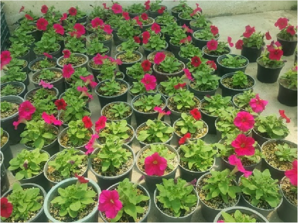
Chatterjee S. Petunia. Commercial flowers, vol. 4. New Delhi: Daya Publishing House, A Division of Astral International Pvt. Ltd.; 2022. p. 55.
Google Scholar
Guo G, Xiao J, Jeong BR. Iron source and medium pH affect nutrient uptake and pigment content in Petunia hybrida ‘madness red’ cultured in vitro. Int J Mol Sci. 2022;23:8943. https://doi.org/10.3390/ijms23168943.
Article
PubMed
PubMed Central
Google Scholar
Velez Bermudez IC, Schmidt W. Iron sensing in plant. Front Plant Sci. 2023;14:1145510. https://doi.org/10.3389/fpls.2023.1145510.
Article
PubMed
PubMed Central
Google Scholar
Ansari A, Amiri J, Norouzi P, Fattahi M, Easouli-Sadaghiani MH, Alipour H. Assessing the efficacy of different nano-iron sources for alleviating alkaline soil challenges in Goji berry trees (Lycium barbarum L). BMC Plant Biol. 2024;24:1153. https://doi.org/10.1186/s12870-024-05870-3.
Article
PubMed
PubMed Central
Google Scholar
Yang S, Xu Y, Tang Z, Jin S, Yang S. The impact of alkaline stress on plant growth and its alkaline resistance mechanisms. Int J Mol Sci. 2024;25(24):13719. https://doi.org/10.3390/ijms252413719.
Article
PubMed
PubMed Central
Google Scholar
Savchenko T, Tikhonov K. Oxidative stress-induced alteration of plant central metabolism. Life. 2021;11:304. https://doi.org/10.3390/life11040304.
Article
PubMed
PubMed Central
Google Scholar
Bontpart T, Weiss A, Vile D, Gérard F, Lacombe B, Reichheld JP, et al. Growing on calcareous soils and facing climate change. Trends Plant Sci. 2024;29(12):1319–30. https://doi.org/10.1016/j.tplants.2024.03.013.
Article
PubMed
Google Scholar
Tamir G, Zilkah S, Dai N, Shawahna R, Cohen S, Bar-Tal A. Combined effects of CaCO3 and the proportion of N-NH4+ among the total applied inorganic N on the growth and mineral uptake of rabbiteye blueberry. J Soil Sci Plant Nutr. 2021;21:35–48. https://doi.org/10.1007/s42729-020-00339-2.
Article
Google Scholar
Kumar K, Jaiswal A, Koppolu UMK, Kumar KRR. Alkaline stress disrupts growth, biochemistry, and ion homeostasis of Chickpea (Cicer arietinum L.) roots. Front Agron. 2024;6:1497054. https://doi.org/10.3389/fagro.2024.1497054.
Article
Google Scholar
Zhao Y, Chen Y, Liu S, Li F, Sun M, Liang Z, et al. Bicarbonate rather than high pH in growth medium induced Fe-deficiency chlorosis in dwarfing rootstock quince A (Cydonia oblonga Mill.) but did not impair Fe nutrition of vigorous rootstock Pyrus betulifolia. Front Plant Sci. 2023;14:1237327. https://doi.org/10.3389/fpls.2023.1237327.
Article
PubMed
PubMed Central
Google Scholar
Saleem A, Zulfiqar A, Saleem MZ, Ali B, Saleem MH, Ali S, et al. Alkaline and acidic soil constraints on iron accumulation by rice cultivars in relation to several physio-biochemical parameters. BMC Plant Biol. 2023;23(1):397. https://doi.org/10.1186/s12870-023-04400-x.
Article
PubMed
PubMed Central
Google Scholar
Liang G. Iron uptake, signaling, and sensing in plants. Plant Commun. 2022;3(5):100349. https://doi.org/10.1016/j.xplc.2022.100349.
Article
PubMed
PubMed Central
Google Scholar
Ning X, Lin M, Huang G, Mao J, Gao Z, Wang X. Research progress on iron absorption, transport, and molecular regulation strategy in plants. Front Plant Sci. 2023;14:1190768. https://doi.org/10.3389/fpls.2023.1190768.
Article
PubMed
PubMed Central
Google Scholar
Li J, Cao X, Jia X, Liu L, Cao H, Qin W, et al. Iron deficiency leads to chlorosis through impacting chlorophyll synthesis and nitrogen metabolism in Areca catechu L. Front Plant Sci. 2021a;12:710093. https://doi.org/10.3389/fpls.2021.710093.
Article
PubMed
PubMed Central
Google Scholar
Trofimov K, Mankotia S, Ngigi M, Baby D, Satbhai SB, Bauer P. Shedding light on iron nutrition: exploring intersections of transcription factor cascades in light and iron deficiency signaling. J Exp Bot. 2025;76:787–802. https://doi.org/10.1093/jxb/erae324.
Article
PubMed
Google Scholar
Khalil S, Strah R, Lodovici A, Vojta P, Ziegler J, Novak MP, Zanin L, Tomasi N, Forneck A, Griesser M. Lime-induced iron deficiency stimulates a stronger response in tolerant grapevine rootstocks compared to low iron availability. Plant Stress. 2025;16:100841. https://doi.org/10.1016/j.stress.2025.100841.
Article
Google Scholar
Martín-Barranco A, Thomine S, Vert G, Zelazny E. A quick journey into the diversity of iron uptake strategies in photosynthetic organisms. Plant Signal Behav. 2021;16(11):1975088. https://doi.org/10.1080/15592324.2021.1975088.
Article
PubMed
PubMed Central
Google Scholar
Amooaghaie R, Roohollahi S. Effect of sodium Nitroprusside on responses of Melissa officinalis to bicarbonate exposure and direct Fe deficiency stress. Photosynthetica. 2017;55(1):153–63. https://doi.org/10.1007/s11099-016-0240-8.
Article
Google Scholar
Wang N, Dong X, Chen Y, Ma B, Yao C, Ma F, et al. Direct and bicarbonate-induced iron deficiency differently affect iron translocation in Kiwifruit roots. Plants. 2020;9:1578. https://doi.org/10.3390/plants9111578.
Article
PubMed
PubMed Central
Google Scholar
Marschner H, Römheld V. Strategies of plants for acquisition of iron. Plant Soil. 1994;165:375–88. https://doi.org/10.1007/BF00008069.
Article
Google Scholar
Kobayashi T, Nakanishi H, Nishizawa NK. Recent insights into iron homeostasis and their application in graminaceous crops. Proc Jpn Acad Ser B. 2010;86:900–13. https://doi.org/10.2183/pjab.86.900.
Article
Google Scholar
Nozoye T, Nagasaka S, Kobayashi T, Takahashi M, Sato Y, Sato Y, et al. Phytosiderophore efflux transporters are crucial for iron acquisition in graminaceous plants. J Biol Chem. 2011;286:5446–54. https://doi.org/10.1074/jbc.M110.180026.
Article
PubMed
Google Scholar
Wagner ALS, Araniti F, Ishii-Iwamoto EL, Abenavoli MR. Resveratrol exerts beneficial effects on the growth and metabolism of Lactuca sativa L. Plant Physiol Biochem. 2022;171:26–37. https://doi.org/10.1016/j.plaphy.2021.12.023.
Article
Google Scholar
Rao MJ, Zheng B. The role of polyphenols in abiotic stress tolerance and their antioxidant properties to scavenge reactive oxygen species and free radicals. Antioxidants. 2025;14(1):74. https://doi.org/10.3390/antiox14010074.
Article
PubMed
PubMed Central
Google Scholar
Zheng X, Chen H, Su Q, Wang C, Sha G, Ma C, et al. Resveratrol improves the irondeficiency adaptation of Malus baccata seedlings by regulating iron absorption. BMC Plant Biol. 2021;21(1):433. https://doi.org/10.1186/s12870-021-03215-y.
Article
PubMed
PubMed Central
Google Scholar
Šamec D, Karalija E, Šola I, Vujčić Bok V, Salopek-Sondi B. The role of polyphenols in abiotic stress response: the influence of molecular structure. Plants. 2021;10(1):118. https://doi.org/10.3390/plants10010118.
Article
PubMed
PubMed Central
Google Scholar
Jian J, Su W, Liu Y, Wang M, Chen X, Wang E, et al. Effects of saline–alkali composite stress on the growth and soil fixation capacity of four herbaceous plants. Agronomy. 2024;14(7):1556. https://doi.org/10.3390/agronomy14071556.
Article
Google Scholar
López-Pérez M, Acosta J, Pérez-Labrada F. Iron nutrition management in calcisol soils as a tool to mitigate chlorosis and promote crop quality – An overview. J Appl Biol Biotechnol. 2023;12(1):17–29. https://doi.org/10.7324/JABB.2024.157536.
Article
Google Scholar
Mehrotra R, Rajesh KV, Anirban P. Iron deficiency chlorosis in aromatic grasses—A review. Environ Chall. 2022;9:100646. https://doi.org/10.1016/j.envc.2022.100646.
Article
Google Scholar
Liu X, Niu H, Li J, Jiang D, Chen R, Zhang R, et al. Higher endogenous abscisic acid confers greater tolerance to saline-alkaline stress in Petunia hybrida. Environ Exp Bot. 2024;228:106035. https://doi.org/10.1016/j.envexpbot.2024.106035.
Article
Google Scholar
Murata Y, Itoh Y, Iwashita T, Namba K. Transgenic petunia with the iron(III)phytosiderophore transporter gene acquires tolerance to iron deficiency in alkaline environments. PLoS ONE. 2015;10:e0120227. https://doi.org/10.1371/journal.pone.0120227.
Article
PubMed
PubMed Central
Google Scholar
Jelali N, Wasli H, Youssef RB, Hessini K, Cardoso SM. Iron deficiency modulates secondary metabolite biosynthesis and antioxidant potential in Sulla carnosa L. primed with Salicylic acid. Appl Sci. 2022;12(20):10351. https://doi.org/10.3390/app122010351.
Article
Google Scholar
Sun Z, Wang T, Li J, Zheng X, Ge H, Sha G, et al. Resveratrol enhances the tolerance of Malus hupehensis to potassium deficiency stress. Front Plant Sci. 2024;15:1503463. https://doi.org/10.3389/fpls.2024.1503463.
Article
PubMed
PubMed Central
Google Scholar
Li T, Li Y, Sun Z, Xi X, Sha G, Ma C, et al. Resveratrol alleviates the KCl salinity stress of Malus hupehensis Rhed. Front Plant Sci. 2021b;12:650485. https://doi.org/10.3389/fpls.2021.650485.
Article
PubMed
PubMed Central
Google Scholar
Hoagland DR, Arnon DI. The waterculture method for growing plants without soil. Berkeley (CA): California Agricultural Experiment Station; 1950. Circular No. 347. 32.
Sonneveld C, Straver N. Nutrient solutions for vegetables and flowers grown in water or substrates. Naaldwijk (Netherlands): Glasshouse Crops Research Station; 1999. p. 43.
Google Scholar
Poorter H, Niinemets Ü, Poorter L, Wright IJ, Villar R. Causes and consequences of variation in leaf mass per area (LMA): a metaanalysis. New Phytol. 2009;182(3):565–88. https://doi.org/10.1111/j.1469-8137.2009.02830.x.
Article
PubMed
Google Scholar
Pang W, Crow WT, Luc JE, McSorley R, GiblinDavis RM, Kenworthy KE, et al. Comparison of water displacement and WinRHIZO software for plant root parameter assessment. Plant Dis. 2011;95(10):1308–10. https://doi.org/10.1094/PDIS-01-11-0026.
Article
PubMed
Google Scholar
Markwell J, Osterman JC, Mitchell JL. Calibration of the Minolta SPAD-502 leaf chlorophyll meter. Photosynth Res. 1995;46:467–72. https://doi.org/10.1007/BF00032301.
Article
PubMed
Google Scholar
Lichtenthaler HK. Chlorophylls and carotenoids: pigments of photosynthetic biomembranes. Methods Enzymol. 1987;148:350–82. https://doi.org/10.1016/0076-6879(87)48036-1.
Article
Google Scholar
Lutts S, Kinet JM, Bouharmont J. Changes in plant response to NaCl during development of rice (Oryza sativa L.) varieties differing in salinity resistance. J Exp Bot. 1995;46(12):1843–52. https://doi.org/10.1093/jxb/46.12.1843.
Article
Google Scholar
Horst JH, Cakmak I. Effects of aluminum on lipid peroxidation, superoxide dismutase, catalase, and peroxidase activities in root tips of soybean (Glycine max). Physiol Plant. 1991;83:463–8. https://doi.org/10.1111/j.1399-3054.1991.tb00121.x.
Article
Google Scholar
Velikova V, Yordanov I, Edreva A. Oxidative stress and some antioxidant systems in acid rain-treated bean plants: protective role of exogenous polyamines. Plant Sci. 2000;151(1):59–66. https://doi.org/10.1016/S0168-9452(99)00197-1.
Article
Google Scholar
Ojeda M, Schaffer B, Davies FS. Root and leaf ferric chelate reductase activity in pond Apple and soursop. J Plant Nutr. 2004;27:1381–93. https://doi.org/10.1081/PLN-200025836.
Article
Google Scholar
Grieve CM, Grattan SR. Rapid assay for determination of water-soluble quaternary ammonium compounds. Plant Soil. 1983;70(3):303–7. https://doi.org/10.1007/BF02374789.
Article
Google Scholar
Ohayama T, Ito M, Kobayashi K, Araki S, Yasuyoshi S, Sasaki O, et al. Analytical procedures of N, P and K content in plant and manure materials using H₂SO₄–H₂O₂ Kjeldahl digestion method. Bull Fac Agric Niigata Univ. 1991;43:111–20.
Google Scholar
Ryan J, Estefan G, Rashid A. Soil and plant analysis: laboratory manual. Aleppo (Syria): ICARDA; 2001.
Google Scholar
Mizukoshi K, Nishiwaki T, Ohtake N, Minagawa R, Kobayashi K, Ikarashi T, et al. Determination of tungstate concentration in plant materials by HNO₃–HClO₄ digestion and colorimetric method using thiocyanate. Plant Anal Methods. 1994;46:51–6.
Google Scholar
Ghazanshahi J. Soil and plant analysis. Tehran (Iran): Motarjem; 2006. p. 311.
Google Scholar
Ahmed N, Zhang B, Chachar Z, Li J, Xiao G, Wang Q, et al. Micronutrients and their effects on horticultural crop quality, productivity and sustainability. Sci Hortic. 2024;323:112512. https://doi.org/10.1016/j.scienta.2023.112512.
Article
Google Scholar
Khan F, Siddique AB, Shabala S, Zhou M, Zhao C. Phosphorus plays key roles in regulating plants’ physiological responses to abiotic stresses. Plants. 2023;12(15):2861. https://doi.org/10.3390/plants12152861.
Article
PubMed
PubMed Central
Google Scholar
Therby-Vale R, Lacombe B, Rhee SY, Nussaume L, Rouached H. Mineral nutrient signaling controls photosynthesis: focus on iron deficiency-induced chlorosis. Trends Plant Sci. 2022;27(5):502–9. https://doi.org/10.1016/j.tplants.2021.11.005.
Article
PubMed
Google Scholar
Hasanuzzaman M, Bhuyan MHMB, Parvin K, Bhuiyan TF, Anee TI, Nahar K, et al. Regulation of ROS metabolism in plants under environmental stress: a review of recent experimental evidence. Int J Mol Sci. 2020a;21(22):8695. https://doi.org/10.3390/ijms21228695.
Article
PubMed
PubMed Central
Google Scholar
Hong Y, Boiti A, Vallone D, Foulkes NS. Reactive oxygen species signaling and oxidative stress: transcriptional regulation and evolution. Antioxidants. 2024;13(3):312. https://doi.org/10.3390/antiox13030312.
Article
PubMed
PubMed Central
Google Scholar
Saito A, Shinjo S, Ito D, Doi Y, Sato A, Wakabayashi Y, et al. Enhancement of photosynthetic iron-use efficiency is an important trait of Hordeum vulgare for adaptation of photosystems to iron deficiency. Plants. 2021;10(2):234. https://doi.org/10.3390/plants10020234.
Article
PubMed
PubMed Central
Google Scholar
Marschner P. Marschner’s mineral nutrition of higher plants. 3rd ed. San Diego: Academic; 2012. https://doi.org/10.1016/C2009-0-63043-9.
Book
Google Scholar
Zheng L, Yamaji N, Ma JF. Iron transport and distribution in plants: research progress and future perspectives. Plant Cell Physiol. 2022;63(2):185–93. https://doi.org/10.1093/pcp/pcab164.
Article
Google Scholar
Giehl RF, Lima JE, von Wirén N. Localized iron supply triggers lateral root elongation in Arabidopsis by altering the AUX1-mediated auxin distribution. Plant Cell. 2012;24(1):33–49. https://doi.org/10.1105/tpc.111.092973.
Article
PubMed
PubMed Central
Google Scholar
Yang C, Shi D, Wang D. Comparative effects of salt and alkali stresses on growth, osmotic adjustment and ionic balance of an alkali-resistant halophyte Suaeda glauca (Bge). Plant Growth Regul. 2008;56:179–90. https://doi.org/10.1007/s10725-008-9299-y.
Article
Google Scholar
Sun X, Zhu C, Li B, Ning W, Yin J. Combining physiology and transcriptome to reveal mechanisms of Hosta ‘golden cadet’ in response to alkali stress. Plants. 2025;14(4):593. https://doi.org/10.3390/plants14040593.
Article
PubMed
PubMed Central
Google Scholar
Yang Y, Ian J, Qiu X, Wang G, Zong J. Effects of combined saline-alkali stress on physiological and biochemical characteristics of OT hybrid Lily. J Nanjing Univ. 2022;46(4):117. https://doi.org/10.12302/j.issn.1000-2006.202105041.
Article
Google Scholar
Gao Q, Zheng R, Lu J, Li X, Wang D, Cai X, et al. Trends in the potential of stilbenes to improve plant stress tolerance: insights of plant defense mechanisms in response to biotic and abiotic stressors. J Agric Food Chem. 2024;72(14):7655–71. https://doi.org/10.1021/acs.jafc.4c00326.
Article
PubMed
Google Scholar
Vélez-Bermúdez IC, Schmidt W. Plant strategies to mine iron from alkaline substrates. Plant Soil. 2023;483:1–25. https://doi.org/10.1007/s11104-022-05746-1.
Article
Google Scholar
Rottet S, Förster B, Hee WY, Rourke LM, Price GD, Long BM. Engineered accumulation of bicarbonate in plant chloroplasts: known knowns and known unknowns. Front Plant Sci. 2021;12:727118. https://doi.org/10.3389/fpls.2021.727118.
Article
PubMed
PubMed Central
Google Scholar
Bhat MA, Mishra AK, Shah SN, Bhat MA, Jan S, Rahman S, et al. Soil and mineral nutrients in plant health: a prospective study of iron and phosphorus in the growth and development of plants. Curr Issues Mol Biol. 2024;46(6):5194–222. https://doi.org/10.3390/cimb46060312.
Article
PubMed
PubMed Central
Google Scholar
Rengasamy P, Lacerda C, Gheyi H. Salinity, sodicity and alkalinity. Subsoil constraints for crop production. Cham: Springer; 2022. pp. 75–94. https://doi.org/10.1007/978-3-031-00317-2_4.
Chapter
Google Scholar
Zagoskina NV, Zubova MY, Nechaeva TL, Kazantseva VV, Goncharuk EA, Katanskaya VM, et al. Polyphenols in plants: structure, biosynthesis, abiotic stress regulation, and practical applications. Int J Mol Sci. 2023;24(18):13874. https://doi.org/10.3390/ijms241813874.
Article
PubMed
PubMed Central
Google Scholar
Chauhan J, Prathibha MD, Singh P, Choyal P, Mishra UN, Saha D, et al. Plant photosynthesis under abiotic stresses: damages, adaptive, and signaling mechanisms. Plant Stress. 2023;10:100296. https://doi.org/10.1016/j.stress.2023.100296.
Article
Google Scholar
Graziano M, Lamattina L. Nitric oxide and iron in plants: an emerging and converging story. Trends Plant Sci. 2005;10:4–8. https://doi.org/10.1016/j.tplants.2004.12.004.
Article
PubMed
Google Scholar
Tripathy BC, Oelmüller R. Reactive oxygen species generation and signaling in plants. Plant Signal Behav. 2012;7(12):1621–33. https://doi.org/10.4161/psb.22455.
Article
PubMed
PubMed Central
Google Scholar
Apel K, Hirt H. Reactive oxygen species: metabolism, oxidative stress, and signal transduction. Annu Rev Plant Biol. 2004;55:373–99. https://doi.org/10.1146/annurev.arplant.55.031903.141701.
Article
PubMed
Google Scholar
Ahuja I, Kissen R, Bones AM. Phytoalexins in defense against pathogens. Trends Plant Sci. 2012;17(2):73–90. https://doi.org/10.1016/j.tplants.2011.11.002.
Article
PubMed
Google Scholar
Jeandet P, Douillet-Breuil AC, Bessis R, Debord S, Sbaghi M, Adrian M. Phytoalexins from the vitaceae: biosynthesis, phytoalexin gene expression in Transgenic plants, antifungal activity, and metabolism. J Agric Food Chem. 2013;51(20):6109–15. https://doi.org/10.1021/jf011429s.
Article
Google Scholar
Kong Q, Zheng S, Li W, Liang H, Zhou L, Yang H, et al. Performance of Camellia oleifera seedlings under alkali stress improved by spraying with types of exogenous biostimulants. Agriculture. 2025;15(3):274. https://doi.org/10.3390/agriculture15030274.
Article
Google Scholar
Arcas A, López-Rayo S, Gárate A, Lucena JJ. A critical review of methodologies for evaluating iron fertilizers based on iron reduction and uptake by strategy i plants. Plants. 2024;13(6):819. https://doi.org/10.3390/plants13060819.
Article
PubMed
PubMed Central
Google Scholar
Kobayashi T, Nishizawa NK. Iron uptake, translocation, and regulation in higher plants. Annu Rev Plant Biol. 2012;63:131–52. https://doi.org/10.1146/annurev-arplant-042811-105522.
Article
PubMed
Google Scholar
Santi S, Schmidt W. Dissecting iron deficiency-induced proton extrusion in Arabidopsis roots. New Phytol. 2009;183(4):1072–84. https://doi.org/10.1111/j.1469-8137.2009.02901.x.
Article
PubMed
Google Scholar
Hsieh EJ, Waters BM. Alkaline stress and iron deficiency regulate iron uptake and riboflavin synthesis gene expression differently in root and leaf tissue: implications for iron deficiency chlorosis. J Exp Bot. 2016;67(19):5671–85. https://doi.org/10.1093/jxb/erw328.
Article
PubMed
PubMed Central
Google Scholar
Ashraf M, Foolad MR. Roles of glycine betaine and proline in improving plant abiotic stress resistance. Environ Exp Bot. 2007;59(2):206–16. https://doi.org/10.1016/j.envexpbot.2005.12.006.
Article
Google Scholar
Zhu XG, Long SP, Ort DR. Improving photosynthetic efficiency for greater yield. Annu Rev Plant Biol. 2016;61:235–61. https://doi.org/10.1146/annurev-arplant-042809-112206.
Article
Google Scholar
Truong VL, Jun M, Jeong WS. Role of resveratrol in regulation of cellular defense systems against oxidative stress. Biofactors. 2018;44(1):36–49. https://doi.org/10.1002/biof.1399.
Article
PubMed
Google Scholar
D’Introno A, Paradiso A, Scoditti E, D’Amico L, De Paolis A, Carluccio MA, et al. Antioxidant and anti-inflammatory properties of tomato fruits synthesizing different amounts of Stilbenes. Plant Biotechnol J. 2009;7(5):422–9. https://doi.org/10.1111/j.1467-7652.2009.00409.x.
Article
PubMed
Google Scholar
Shi Y, Guo S, Zhao X, Xu M, Xu J, Xing G, Ahammed GJ. Comparative physiological and transcriptomics analysis revealed crucial mechanisms of silicon-mediated tolerance to iron deficiency in tomato. Front Plant Sci. 2022;13:1094451. https://doi.org/10.3389/fpls.2022.1094451.
Article
PubMed
PubMed Central
Google Scholar
Johan PD, Ahmed OH, Omar L, Hasbullah NA. Phosphorus transformation in soils following co-application of charcoal and wood ash. Agronomy. 2021;11(10):2010. https://doi.org/10.3390/agronomy11102010.
Article
Google Scholar
Santoro V, Schiavon M, Celi L. Role of soil abiotic processes on phosphorus availability and plant responses with a focus on Strigolactones in tomato plants. Plant Soil. 2024;494:1–49. https://doi.org/10.1007/s11104-023-06266-2.
Article
Google Scholar
Zhao H, Zhang W, Zhang L. Interactive effects of iron deficiency and other mineral nutrients on plants. Plant Soil. 2014;382(1–2):1–19. https://doi.org/10.1007/s11104-014-2152-1.
Article
Google Scholar
Wdowiak A, Podgórska A, Szal B. Calcium in plants: an important element of cell physiology and structure, signaling, and stress responses. Acta Physiol Plant. 2024;46:108. https://doi.org/10.1007/s11738-024-03733-w.
Article
Google Scholar
Zhang X, Zhang D, Sun W, Wang T. The adaptive mechanism of plants to iron deficiency via iron uptake, transport, and homeostasis. Int J Mol Sci. 2019;20(10):2424. https://doi.org/10.3390/ijms20102424.
Article
PubMed
PubMed Central
Google Scholar
Ahmed N, Zhang B, Bozdar B, Chachar S, Rai M, Li J, et al. The power of magnesium: unlocking the potential for increased yield, quality, and stress tolerance of horticultural crops. Front Plant Sci. 2023;14:1285512. https://doi.org/10.3389/fpls.2023.1285512.
Article
PubMed
PubMed Central
Google Scholar
Cakmak I. Enrichment of cereal grains with zinc: agronomic or genetic biofortification? Plant Soil. 2008;302(1–2):1–17. https://doi.org/10.1007/s11104-007-9466-3.
Article
Google Scholar
Rai S, Singh PK, Mankotia S, Swain J, Satbhai SB. Iron homeostasis in plants and its crosstalk with copper, zinc, and manganese. Plant Stress. 2021;1:100008. https://doi.org/10.1016/j.stress.2021.100008.
Article
Google Scholar
Shaver TM, Westfall D, Ronaghi M. Zinc fertilizer solubility and its effects on zinc bioavailability over time. J Plant Nutr. 2007;30:123–33. https://doi.org/10.1080/01904160601055145.
Article
Google Scholar
Garcia-Caparros P, Ciriello M, Rouphael Y, Giordano M. The role of organic extracts and inorganic compounds as alleviators of drought stress in plants. Horticulturae. 2025;11(1):91. https://doi.org/10.3390/horticulturae11010091.
Article
Google Scholar
Jeandet P. Phytoalexins. Current progress and future prospects. Mol. 2015;20(2):2770–4. https://doi.org/10.3390/molecules20022770.
Article
Google Scholar
Chang X, Heene E, Qiao F, Nick P. The phytoalexin Resveratrol regulates the initiation of hypersensitive cell death in Vitis cell. PLoS ONE. 2011;6(10):e26405. https://doi.org/10.1371/journal.pone.0026405.
Article
PubMed
PubMed Central
Google Scholar
Stanton C, Sanders D, Kraemer U, Podar D. Zinc in plants: integrating homeostasis and biofortification. Mol Plant. 2022;15(1):65–85. https://doi.org/10.1016/j.molp.2021.12.008.
Article
PubMed
Google Scholar
Xu L, Wang X. A comprehensive review of phenolic compounds in horticultural plants. Int J Mol Sci. 2025;26:5767. https://doi.org/10.3390/ijms26125767.
General characteristics of malignancy-PJP patients
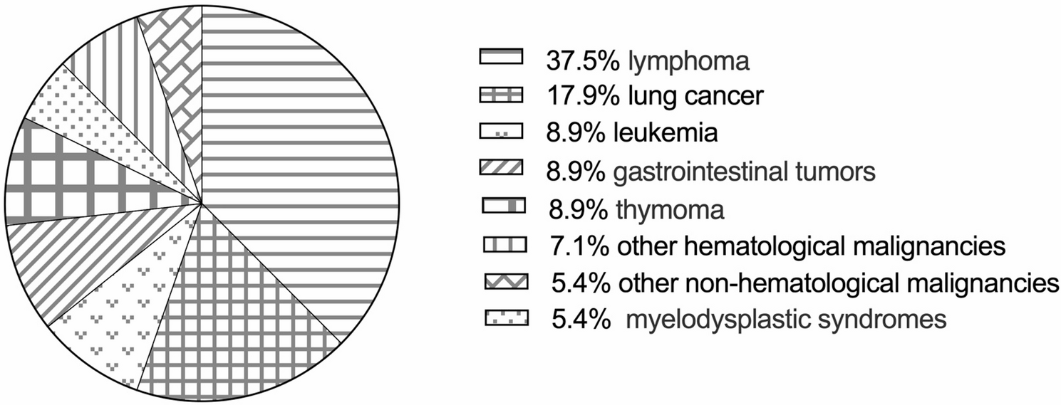
Fifty-six patients with an identified diagnosis of malignancy-PJP were enrolled in our study after a detailed medical record review. Thirty-four patients were male (60.7%), 22 patients were female (39.3%), and the mean age was 63 (52, 68) years. The underlying malignancies are shown in Fig. 1. Most patients had solid malignancies (45, 80.4%), and 11 (19.6%) had non-solid malignancies. According to the involved system, 23 (41.1%) patients had non-hematological malignancies, and 33 (58.9%) had hematological malignancies.
Fig. 1
The underlying malignancies of enrolled 56 malignancy-PJP patients. Other hematological malignancies: multiple myeloma and aplastic anemia; other non-hematological malignancies: prostate cancer, nasopharyngeal cancer, and breast cancer
The main clinical manifestations of PJP were fever (52, 92.9%), cough (47, 83.9%), expectoration (41, 73.2%), and dyspnea (47, 83.9%). Bilateral (56, 100%), ground-glass opacities (GGOs) (48, 85.7%), and patches (45, 80.4%) were the most common chest CT manifestations. Consolidations (24, 42.9%), nodular (24, 42.9%), and pleural thickening (32, 57.1%) were observed on some chest CTs of patients with malignancy-PJP. Low peripheral CD4+ T-cell [125.0 (66.0, 207.0)/µL] counts were common in patients with malignancy-PJP.
Some patients were complicated with other infections, such as CMV (25, 44.6%), bacterial HAP (23, 41.1%), oral candida infection (6, 10.7%), aspergillus infection (6, 10.7%), and Nocardia infection (2, 3.6%). Most patients experienced respiratory failure (47, 83.9%), approximately half of the patients needed intensive care unit (ICU) support, and 29 patients (51.8%) died.
After PJP diagnosis, most patients (50, 89.3%) were prescribed 15 mg/kg/d trimethoprim-sulfamethoxazole (TMP-SMX). More than one-third of our patients (21, 37.5%) were also prescribed a combination of second-line anti-PJP medications, such as caspofungin, clindamycin and primaquine.
Differences in the clinical characteristics and prognosis between PJP patients with non-hematological and hematological malignancies
According to the involved system, the 56 patients were divided into a non-hematological malignancy group and a hematological malignancy group. The differences in clinical characteristics, laboratory test results (Table 1) and imaging findings (Table 2) between the two groups were analyzed.
Table 1 The clinical characteristics between non-hematological malignancy-PJP group and hematological malignancy-PJP group
Table 2 The chest CT features in non-hematological malignancy-PJP group and hematological malignancy-PJP group
There were no significant differences in age, sex or comorbidities between the two groups. Compared with patients in the non-hematological malignancy group, more patients in the hematological malignancy group needed invasive mechanical ventilation support (60.6% vs. 43.5%, p = 0.03). Patients in the hematological malignancy group were more prone to respiratory failure and higher mortality, but the difference was not statistically significant. The time from diagnosis of oncological disease to PJP infection [72 (38.0, 112.5) days vs. 153 (92.5, 223.5) days, p < 0.01] and the time from chemotherapy to PJP infection [79.0 (46.5, 415.5) days vs. 229.0 (116.0, 677.5) days, p = 0.04] were shorter in the hematological malignancy group than in the non-hematological malignancy group. In terms of chest CT features, pleural thickening was more common in the non-hematological malignancy group than in the hematological malignancy group (73.9% vs. 45.5%, p = 0.03). However, there were no significant differences in the minimal albumin level, peripheral lymphocyte count or inflammatory marker levels between the two groups.
Differences between the survival and non-survival groups of patients with malignancy-PJP
The 56 patients were divided into a survival group (27 patients) and a non-survival group (29 patients) according to their clinical outcome. Compared with those in the survival group, more patients in the non-survival group were complicated with CMV (62.1% vs. 25.9%, p < 0.01) and bacterial HAP (58.6% vs. 22.2%, p < 0.01). However, there were no significant differences in clinical symptoms, chest CT features, chemotherapy before PJP infection or anti-PJP treatment between the two groups.
In terms of laboratory test results, in the non-survival group, the peripheral lymphocyte count [0.4 (0.3, 0.7) × 109/L vs. 0.8 (0.5, 1.4) × 109/L, p < 0.01], platelet count [138.0 (74.0, 197.5) × 109/L vs. 212.0 (160.8, 265.3) × 109/L, p < 0.01], minimal albumin level [21.7 ± 5.3 g/L vs. 26.6 ± 4.6 g/L, p < 0.001], T-cell count [307.0 (151.0, 377.0)/µL vs. 447.0 (245.5, 920.5)/µL, p = 0.01) and CD4+ T-cell count [123.0 (37.0, 163.0)/µL vs. 146.0 (97.0, 417.0)/µL, p = 0.03] were significantly lower than those in the survival group. However, D-dimer [8.3 (2.0, 15.6) mg/L vs. 1.9 (0.9, 6.3) mg/L, p = 0.01], high-sensitivity C-reactive protein [107.0 (36.3, 191.3) mg/L vs. 42.2 (6.9, 87.0) mg/L, p < 0.01] and lactate dehydrogenase [588.0 (441.0, 789.5) U/L vs. 319.0 (255.0, 481.0) U/L, p < 0.01] levels were greater in the non-survival group than in the survival group.
Prognostic analysis for patients with malignancy-PJP
As shown in Table 3, univariate Cox regression analysis revealed that non-solid malignancies, decreased lymphocyte count, CMV viremia, bacterial HAP, and pneumomediastinum were associated with non-survival. Subsequent multivariate Cox regression analysis revealed that non-solid malignancies (HR = 2.77, χ2 = 4.83, p = 0.03, 95% CI: 1.12–6.89), CMV viremia (HR = 3.33, χ2 = 8.93, p < 0.01, 95% CI: 1.51–7.33), bacterial HAP (HR = 2.21, χ2 = 4.10, p = 0.04, 95% CI: 1.03–4.77) and pneumomediastinum (HR = 2.50, χ2 = 3.96, p < 0.05, 95% CI: 1.01–6.14) were independent risk factors associated with poor survival in patients with malignancy-PJP.
Table 3 Univariable and multivariable Cox regression analysis of survival associated risk factors for patients with malignancy-PJP
Kaplan‒Meier analysis (Fig. 2) was performed to explore the impact of the different types of underlying malignancies on the cumulative survival of malignancy-PJP patients. The results revealed that there was no significant difference in survival between patients with non-hematological malignancies and those with hematological malignancies. Compared with that of patients with solid malignancies, the survival rate of patients with non-solid malignancies (p < 0.05) was significantly lower.
Fig. 2
Kaplan-Meier analysis of malignancy-PJP patients on 60-day. A with hematological malignancies and with non-hematological malignancies; B with solid malignancies and with non-solid malignancies
Our study provides novel insights into the relationship between the IBI and the risk of 30-day readmission following elective PCI. By leveraging a comprehensive retrospective cohort, we have demonstrated that higher IBI values are significantly correlated with an increased risk of readmission, independent of traditional risk factors. This correlation was particularly pronounced in older, male patients and those with diabetes, highlighting the potential utility of IBI in risk stratification for these vulnerable populations. Our multivariate analysis revealed that a one-unit increase in IBI is associated with a 41% increase in the risk of 30-day readmission (OR 1.41, 95% CI 1.19–1.67, p < 0.001). This means that for every unit increase in IBI, the likelihood of a patient being readmitted within 30 days increases significantly. For example, a patient with an IBI of 2 compared to a patient with an IBI of 1 would have a 41% higher risk of readmission. This increased risk is likely due to the role of inflammation in promoting plaque instability, thrombus formation, and other adverse cardiovascular events that can lead to hospital readmission.
When compared to other studies, our findings are consistent with those of Li et al. [9], who demonstrated the association between inflammatory markers and the risk of hospitalization for heart failure post-myocardial infarction. However, our study extends these insights by showing that an integrated inflammatory index, rather than a single biomarker, is associated with readmission, emphasizing the complexity of inflammatory processes in cardiovascular disease [10]. The association between inflammation and cardiovascular outcomes, including post-PCI readmission, is well-established in the literature [11, 12]. Our findings are consistent with those of recent studies that have implicated inflammation in the pathogenesis of adverse cardiovascular events [13]. For instance, a study by Xie et al. [14] confirmed the predictive value of C-reactive protein, a key component of IBI, for cardiovascular events. Our study extends these insights by showing that an integrated inflammatory index, rather than a single biomarker, is associated with readmission, emphasizing the complexity of inflammatory processes in cardiovascular disease.
The potential mechanisms underlying the association between IBI and readmission are multifaceted. Inflammation is known to play a role in plaque rupture and thrombus formation, which can lead to acute coronary syndromes and potentially readmission [15]. Also, local or systemic inflammation has been proven to be a possible mechanism underlying the development of coronary slow flow phenomenon [16, 17]. Many patients experience recurrent episodes of angina due to the coronary slow flow phenomenon, leading to frequent hospitalizations [18]. Furthermore, inflammation may also contribute to the development of heart failure, a common cause of readmission following PCI [19]. By integrating multiple inflammatory biomarkers, IBI may provide a more comprehensive assessment of the inflammatory state and its impact on post-PCI outcomes.
The stronger correlation observed in older patients and those with diabetes may reflect the heightened inflammatory state often observed in these patient groups [20, 21]. Diabetes is known to induce a chronic low-grade inflammatory state, which could potentiate the association between IBI and readmission [22]. Similarly, aging is associated with an increased inflammatory burden, which may contribute to the observed association [23]. These findings underscore the importance of considering IBI in the context of patient-specific risk factors when assessing the risk of readmission. The stronger correlation observed in males may reflect sex-specific differences in inflammatory responses to PCI [24]. Emerging evidence suggests that sex hormones modulate inflammation, with males exhibiting higher levels of certain inflammatory markers compared to females [25]. This could potentially explain the enhanced association between IBI and readmission in male patients. Additionally, the higher IBI in males may also be indicative of a more aggressive inflammatory process post-PCI, which could lead to a higher likelihood of complications and subsequent readmission [26].
IL−6 is a well-established inflammatory marker that has been extensively studied in the context of cardiovascular disease. Recent studies have shown that elevated IL−6 levels are associated with increased risk of adverse outcomes following PCI. For instance, high levels of IL−6 have been linked to larger infarct sizes and higher mortality rates in patients with ST-segment elevation myocardial infarction [27]. Additionally, IL−6 has been identified as an independent predictor of non-target lesion progression in patients after coronary stenting [28]. In our study, we collected data on IL−6 levels to provide additional supporting evidence for the effectiveness of IBI. The significant difference in IL−6 levels between the readmitted and non-readmitted groups aligns with the observed trends in IBI, further validating its role as a comprehensive measure of inflammation. The inclusion of IL−6 in our data collection was intended to demonstrate that it shares a similar trend with IBI, thereby reinforcing the validity of IBI as a predictor of readmission risk.
The implications of our findings for clinical practice are significant. By identifying patients with higher IBI values as being at increased risk of readmission, clinicians may be able to target these individuals for more intensive post-discharge monitoring and intervention. This could potentially lead to a reduction in readmission rates and associated healthcare costs, as well as improved patient outcomes.
It is important to note that our study is not without limitations. As a retrospective cohort study, it is subject to the inherent biases and limitations of such designs. First, Our study is limited by the lack of standardized adjudication of readmission urgency or etiology, which precluded stratification into urgent vs. non-urgent or cardiac vs. non-cardiac categories. Future prospective studies with dedicated adjudication committees are needed to validate these findings in such contexts. Secondly, Second, geographical factors and variations in healthcare practices, as well as disparities in the availability and utilization of primary care, can significantly influence readmission rates. Our study population is drawn from a specific region, which may not be representative of other areas with different healthcare systems, patient demographics, or clinical practices. For instance, regions with limited access to primary care or specialized cardiovascular services may experience higher readmission rates due to inadequate post-discharge follow-up and management. Notably, we excluded patients who experienced major procedural complications, which were defined as complications necessitating additional interventions or treatments beyond standard PCI, such as vascular perforation, acute stent thrombosis, or significant bleeding requiring transfusion. While this exclusion was intended to focus on the elective PCI population and minimize confounding from procedures that became emergent, it may introduce selection bias. Future prospective studies are needed to validate our findings and to explore the potential of IBI as a predictive tool in a broader range of patient populations and clinical settings.
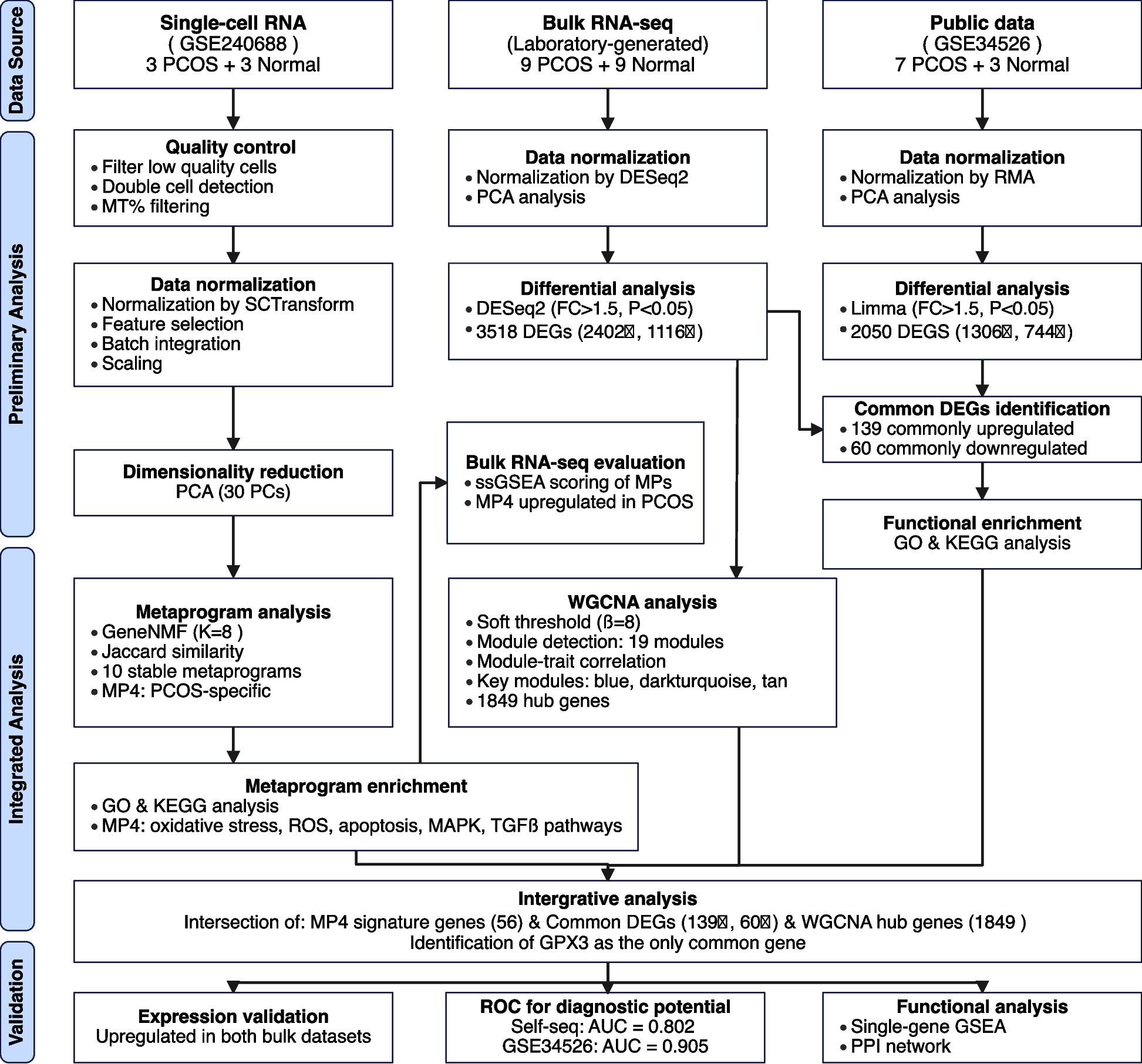
Figure 1 depicts our analytical workflow integrating three datasets: single-cell RNA-seq (GSE240688), bulk RNA-seq from our laboratory cohort, and a validation dataset (GSE34526). This integrative transcriptomic approach combines non-negative matrix factorization, differential expression analysis, and co-expression network analysis to bridge single-cell and bulk transcriptomic findings, ultimately identifying key regulatory genes in PCOS pathophysiology.
Fig. 1
Flowchart of the Study Design and Analytical Workflow
Metaprogram analysis reveals molecular signatures and cellular heterogeneity in PCOS granulosa cells
NMF was applied to deconvolve transcriptional programs in single-cell datasets, yielding 10 stable metaprograms (MPs, Fig. 2). To establish the optimal factorization dimensionality, we systematically scanned k values from 5 to 20, guided by quantitative evaluation of intra-sample cluster separation using silhouette coefficient analysis to ensure robust program delineation. The selected k = 8 demonstrated balanced performance, achieving both high-resolution separation of transcriptional programs within individual samples and preservation of biologically interpretable modules. This parameterization generated 48 sample-specific expression programs (8 per sample across six specimens), which were aggregated into 10 consensus metaprograms via cosine similarity-based hierarchical clustering. The resultant block-diagonal similarity matrix structure revealed conserved transcriptional modules exhibiting cross-sample reproducibility. Metaprogram refinement employed stringent dual criteria: genes recurrently detected in sample-level programs (confidence ≥ 0.5) and accounting for ≥ 80% of cumulative loading variance (weight threshold = 0.8), ensuring both technical robustness and biological coherence.
Fig. 2
Metaprogram Analysis Reveals Molecular Signatures and Cellular Heterogeneity in PCOS Granulosa Cells. A PCA of single-cell RNA-seq samples. B Heatmap showing the expression of 10 identified MPs across samples. C Differential expression of MPs between PCOS and normal granulosa cells. D t-SNE visualization of six cell clusters and corresponding MP expression patterns in PCOS and normal samples. E Secondary clustering of granulosa cells based on MP expression, identifying four distinct GC subtypes and their corresponding MP expression profiles. F Expression patterns of key markers across the four GC subtypes. G GO, HALLMARK, and KEGG pathway enrichment analysis for MP4, highlighting pathways related to oxidative stress and stress-response signaling
Analysis of metaprogram distribution revealed distinct patterns associated with disease status. Metaprograms MP1-3 and MP8-10 were stably expressed across all samples, indicating their involvement in fundamental cellular processes independent of disease state. In contrast, MP4-7 demonstrated PCOS-specific expression patterns.
Gene set enrichment analysis revealed distinct biological functions for each metaprogram. Notably, MP4 showed significant enrichment in pathways related to cellular response to endogenous stimuli, oxygen-containing compounds, programmed cell death, reactive oxygen species, hypoxia, mitogen-activated protein kinase (MAPK) signaling, transforming growth factor-beta (TGF-β) signaling, and Wingless/Integrated (WNT) signaling pathways. This functional profile strongly implicates MP4 in oxidative stress responses and stress-induced signaling cascades known to be dysregulated in PCOS.
To characterize cellular Heterogeneity, we performed unsupervised clustering using the first 30 principal components with a resolution parameter of 0.2, resulting in distinct cell clusters visualized by t-SNE. UCell score calculation for all 10 metaprograms revealed significant associations between specific metaprograms and cell clusters: MP2 scores were significantly higher in cluster 0 (0.17 ± 0.09 vs 0.05 ± 0.05, p < 0.001); MP4 scores were elevated in cluster 2 (0.25 ± 0.09 vs 0.07 ± 0.05, p < 0.001); and MP8 was predominantly expressed in cluster 3 (0.22 ± 0.06 vs 0.11 ± 0.05, p < 0.001). Based on these patterns, we designated cluster 0 as MP2 granulosa cells (GCs), cluster 2 as MP4 GCs, cluster 3 as MP8 GCs, and remaining cells as other GCs. Notably, the proportion of MP4 GCs was significantly higher in PCOS samples compared to normal controls as determined by Wilcoxon rank-sum test with p = 0.0046, suggesting that expansion of MP4-expressing granulosa cells may be a characteristic feature of PCOS pathophysiology.
Differential expression analysis identifies common dysregulated genes in PCOS
Table 1 presents the clinical and endocrine characteristics of study participants. PCOS and control groups were successfully matched with no significant differences in age (29.00 ± 3.84 vs. 29.78 ± 3.31 years, p = 0.651), BMI (22.03 ± 3.12 vs. 23.12 ± 3.11 kg/m2, p = 0.47), and TSH levels (1.86 ± 1.03 vs. 2.05 ± 1.31 mIU/L, p = 0.732). As expected for PCOS pathophysiology, patients exhibited significantly elevated basal luteinizing hormone (LH, 7.36 ± 2.07 vs. 3.02 ± 0.75 IU/L, p < 0.001), testosterone (1.08 ± 0.43 vs. 0.65 ± 0.19 nmol/L, p = 0.014), anti-Müllerian hormone (AMH) levels (6.96 ± 2.21 vs. 3.80 ± 1.42 ng/mL, p = 0.002), and antral follicle counts (24 vs. 14, p < 0.001) compared to controls. During ovarian stimulation, PCOS patients yielded significantly more oocytes (20 vs. 13, p = 0.024) and mature oocytes (15 vs. 11, p = 0.022), consistent with their enhanced follicular development potential. These findings confirm the distinct hormonal and reproductive characteristics of our PCOS population while validating the effectiveness of our matching strategy for potential confounding variables.
Table 1 Clinical Characteristics and Hormonal Profiles of PCOS Patients and Control Subjects
Differential expression analysis was performed on two independent datasets utilizing different transcriptomic platforms: our laboratory-generated bulk RNA-seq dataset (9 PCOS and 9 normal samples) and the publicly available GSE34526 microarray dataset (7 PCOS and 3 normal samples) (Fig. 3). Principal component analysis confirmed clear separation between PCOS and control samples after normalization in both datasets. To account for the fundamental differences between these technologies, we employed platform-specific analytical approaches. For our RNA-seq dataset, DESeq2 analysis with criteria of |FoldChange|> 1.5 and P < 0.05 identified 3,518 differentially expressed genes (DEGs), including 2,402 upregulated and 1,116 downregulated genes in PCOS samples. For the GSE34526 microarray dataset, Limma analysis with the same fold-change and significance thresholds identified 2,050 DEGs (1,306 upregulated and 744 downregulated).
Fig. 3
Differential Expression Analysis Identifies Common Dysregulated Genes in PCOS. A Analysis of the laboratory-generated bulk RNA-seq dataset: PCA plot of samples (left), heatmap of DEGs (middle), and volcano plot highlighting upregulated and downregulated genes (right). B Analysis of the GSE34526 dataset: PCA plot of samples (left), heatmap of DEGs (middle), and volcano plot (right). C Venn diagram showing the overlap of DEGs between the two datasets. D GO and KEGG pathway enrichment analysis of commonly upregulated genes. E GO and KEGG pathway enrichment analysis of commonly downregulated genes
To identify consistently dysregulated genes across different patient cohorts, we determined the intersection of DEGs from both datasets, considering the direction of expression changes. This stringent approach yielded 139 commonly upregulated and 60 commonly downregulated genes across both datasets. Functional enrichment analysis of common upregulated genes identified 13 KEGG pathways and 236 GO terms, while common downregulated genes were enriched in 5 KEGG pathways and 21 GO terms. Upregulated genes were significantly associated with pathways related to cellular response to stress, inflammatory processes, and signaling cascades. Downregulated genes were enriched in metabolic pathways and cellular homeostasis processes. These patterns suggest that PCOS is characterized by enhanced stress response mechanisms coupled with impaired metabolic functions.
WGCNA identifies co-expression modules associated with PCOS
To identify co-expressed gene networks associated with PCOS, we performed WGCNA on the laboratory-generated dataset (Fig. 4). Hierarchical clustering of samples confirmed appropriate grouping without outliers. A soft threshold power of 8 was selected based on scale-free topology criteria (R2 = 0.9) and mean connectivity analysis, ensuring optimal network construction while preserving biological relevance. Using dynamic tree cutting with a minimum module size of 100 genes and a cut Height of 0.4, we identified 19 distinct co-expression modules.
Fig. 4
WGCNA Identifies Co-expression Modules Associated with PCOS. A Sample dendrogram and trait heatmap illustrating clustering of samples and their association with PCOS. B Analysis of scale independence and mean connectivity to determine the optimal soft threshold for network construction. C Cluster dendrogram of genes, showing module assignment based on hierarchical clustering. D Module-trait relationships, indicating correlations between module eigengenes and PCOS status. E Scatter plots showing module membership correlation with PCOS status for the blue, darkturquoise, and tan modules
Correlation analysis between module eigengenes and PCOS status revealed significant associations for several modules. Among these, the blue, darkturquoise, and tan modules exhibited the strongest correlations with disease status (correlation coefficient > 0.3, P < 0.05). To identify key regulatory genes within the PCOS-associated modules, we calculated the correlation between individual genes and both module membership (MM) and gene significance for PCOS (GS). By applying thresholds of MM > 0.3 and GS > 0.3, we identified 1,849 hub genes with strong connections to both their respective modules and PCOS status. These hub genes represent potential master regulators of the transcriptional networks dysregulated in PCOS.
Metaprogram validation in bulk RNA-seq data confirms single-cell findings
To validate the relevance of single-cell-derived metaprograms at the tissue level, we first analyzed their distribution across granulosa cell subsets in single-cell RNA-seq data (Fig. 5A). Metaprogram composition varied across different granulosa cell clusters, with distinct enrichment patterns in PCOS and normal samples.
Fig. 5
Metaprogram Validation in Bulk RNA-Seq Data Confirms Single-Cell Findings. A Stacked bar plot showing the distribution of MPs across different granulosa cell populations in single-cell RNA-seq data. B Differential expression of MPs in bulk RNA-seq data, comparing PCOS and normal samples. ns = not significant, * p < 0.05, ** p < 0.01. C Deconvolution analysis of 193 GTEx ovary samples showing the relative proportions of MP2 GCs, MP4 GCs, and MP8 GCs
To further bridge the gap between single-cell and bulk transcriptomic analyses, we employed single-sample Gene Set Enrichment Analysis (ssGSEA) to score each metaprogram in bulk RNA-seq samples (Fig. 5B). This approach quantified the activity of each transcriptional program in both PCOS and normal cohorts. Comparative analysis of metaprogram ssGSEA scores revealed significant differences in MP2, MP4, MP5, MP6, and MP7 activity. Consistent with single-cell findings, MP2 exhibited higher activity in normal samples, while MP4, MP5, MP6, and MP7 were upregulated in PCOS samples. The differential activity of these metaprograms in bulk tissue samples corroborates our single-cell findings and further supports the pathological relevance of these transcriptional programs in PCOS. In particular, the consistent upregulation of MP4 across both single-cell and bulk analyses reinforces its potential role as a key driver of PCOS pathophysiology.
To validate that the identified granulosa cell subtypes represent genuine biological entities rather than clustering artifacts, we performed deconvolution analysis on 193 GTEx v10 ovary bulk RNA-seq samples. The analysis successfully detected all three major granulosa cell subtypes (MP2, MP4, and MP8 GCs) across the tissue samples (Fig. 5C).
The deconvolution results revealed consistent patterns of cell type proportions across samples. MP4 GCs constituted the predominant subtype in most samples, typically representing 60–80% of the granulosa cell population. MP8 GCs showed intermediate abundance (approximately 10–30%), while MP2 GCs were consistently detected at lower proportions (5–15%). This abundance hierarchy (MP4 > MP8 > MP2) was remarkably stable across the majority of samples, with only minor variations observed in individual cases.
The successful detection of these cellular subtypes in independent bulk tissue samples, with reproducible relative abundance patterns, provides strong evidence that our single-cell-defined metaprograms correspond to biologically meaningful cell states rather than technical artifacts.
Integrative transcriptomic approach identifies key regulator in PCOS pathophysiology
To identify high-confidence key regulators involved in PCOS pathophysiology, we performed an integrative analysis combining three complementary approaches: MP4 signature genes from single-cell analysis, common differentially expressed genes across bulk datasets, and hub genes from WGCNA modules (Fig. 6). This stringent multi-dimensional filtering strategy identified GPX3 as the only gene that consistently emerged across all three analytical approaches. The convergence of these independent methods strongly suggests its central role in PCOS-associated transcriptional dysregulation, particularly in relation to oxidative stress responses.
Fig. 6
Integrative Transcriptomic Analysis Identifies GPX3 as a Key Regulator in PCOS. A Venn diagram showing the intersection of DEGs, WGCNA hub genes, and MP4 signature genes (upregulated). B Venn diagram showing the intersection of DEGs, WGCNA hub genes, and MP4 signature genes (downregulated). C Left: Box plot displaying GPX3 expression differences between PCOS and normal samples in the laboratory-generated dataset. Right: ROC curve assessing the diagnostic value of GPX3 in the same dataset. D Left: Box plot showing GPX3 expression differences in the GSE34526 dataset. Right: ROC curve from the GSE34526 dataset, validating the diagnostic potential of GPX3. E Single-gene GSEA of GPX3, revealing its association with metabolic and mitochondrial pathways, including the citrate cycle, insulin signaling, glucose metabolism, and mitochondrial protein degradation (NES < 0, adjusted P < 0.001 for all pathways)
Expression analysis confirmed significant upregulation of GPX3 in PCOS samples compared to normal controls across both the laboratory-generated dataset and the GSE34526 validation dataset. ROC curve analysis demonstrated strong discriminatory power of GPX3 between PCOS and normal samples in both the laboratory-generated dataset (AUC = 0.802) and the GSE34526 validation dataset (AUC = 0.905), highlighting its potential as a clinically relevant biomarker for PCOS diagnosis.
Examination of GPX3 expression at the single-cell level revealed specific distribution patterns across granulosa cell subpopulations. Single-gene Gene Set Enrichment Analysis identified 818 significantly enriched pathways (|Normalized Enrichment Score, NES|> 1, p.adjust < 0.05, q.value < 0.2), with those related to glucose metabolism, mitochondrial protein degradation, insulin signaling, citrate cycle, and TCA cycle prominently represented. These enrichment patterns suggest that GPX3 dysregulation may impact fundamental metabolic processes and energy homeostasis, which are known to be perturbed in PCOS.
Multi-level GPX3 regulatory network analysis reveals potential mechanisms in PCOS
To establish a comprehensive understanding of the functional relevance of GPX3 in PCOS pathophysiology, we performed integrative multi-levels analysis constructing a complex regulatory network (Fig. 7). The protein–protein interaction network based on MP4 signature genes revealed GPX3 in a network comprising 49 proteins with multiple functional connections. Within this network, GPX3 demonstrated direct interactions with several proteins involved in redox homeostasis and related cellular processes.
Fig. 7
Multi-level Regulatory Network Analysis of GPX3 in PCOS. A PPI network of GPX3 and its associated proteins. B Integrated regulatory elements of GPX3 including: ceRNA network prediction showing GPX3-miRNA-lncRNA interactions; Transcription factor binding site prediction; Drug-gene interaction prediction for potential therapeutic targets
Most notably, GPX3 showed significant connections with selenoprotein P (SELENOP), a major selenium transport protein that works synergistically with GPX3 in the selenium-dependent antioxidant system, providing essential selenium cofactors for glutathione peroxidase activity. Similarly, glutathione S-transferase alpha 1 (GSTA1) exhibited direct interaction with GPX3, suggesting coordinated roles in glutathione metabolism and detoxification of reactive oxygen species. These interactions highlight GPX3’s central position in cellular antioxidant defense mechanisms.
Additionally, GPX3 directly interacts with SLC40A1 (ferroportin), an iron exporter critical for preventing iron-catalyzed oxidative damage, connecting iron homeostasis with antioxidant defense in granulosa cells. Interactions between GPX3 and both THBS1 and F3 suggest linkages between oxidative stress and PCOS-related coagulation and inflammatory pathways. Additionally, the associations with extracellular matrix proteins COL1A1 and CCN2 indicate involvement in oxidative stress-induced matrix remodeling. The connection with GDNF suggests novel neuroendocrine regulatory mechanisms influenced by oxidative status in PCOS pathophysiology.
Our miRNA-mRNA interaction analysis identified several microRNAs potentially regulating GPX3 expression, including has-miR-4644, hsa-miR-4306 and hsa-miR-185-5p, both predicted with high confidence scores. Further exploration through miRNA-lncRNA association analysis uncovered a complex layer of epigenetic regulation, with multiple long non-coding RNAs (lncRNAs) including XIST, UCA1, SNHG14, AC073896.4, MALAT1, NEAT1, and AC005082.1 potentially modulating these miRNA-mediated effects on GPX3 expression.
Transcription factor binding site analysis revealed that GPX3 expression may be regulated by several key transcription factors implicated in ovarian function, including SREBF1, HINFP, E2F1, STAT3, PPARG, MEF2A, FOXL1, and JUND. This suggests multiple potential mechanisms for transcriptional dysregulation of GPX3 in PCOS pathogenesis.
Furthermore, drug-gene interaction queries identified several compounds potentially targeting the GPX3-associated pathway, including DOXORUBICIN HYDROCHLORIDE, DAUNORUBICIN LIPOSOMAL, CL_AMIDINE, and COMPOUND 14B, providing potential therapeutic avenues for further investigation. Collectively, this integrative analysis positions GPX3 within a complex regulatory network essential for redox homeostasis in ovarian function, with multiple layers of regulation that may be disrupted in PCOS pathophysiology.
Ahmad F, Das D, Kommaddi RP, Diwakar L, Gowaikar R, Rupanagudi KV, Bennett DA, Ravindranath V (2018) Isoform-specific hyperactivation of calpain-2 occurs presymptomatically at the synapse in Alzheimer’s disease mice and correlates with memory deficits in human subjects. Sci Rep 8:13119. https://doi.org/10.1038/s41598-018-31073-6
Article
CAS
PubMed
PubMed Central
Google Scholar
Aldridge JE, Horibe T, Hoogenraad NJ (2007) Discovery of genes activated by the mitochondrial unfolded protein response (mtUPR) and cognate promoter elements. PLoS ONE 2:e874. https://doi.org/10.1371/journal.pone.0000874
Article
CAS
PubMed
PubMed Central
Google Scholar
Alldred MJ, Che S, Ginsberg SD (2008) Terminal continuation (TC) RNA amplification enables expression profiling using minute RNA input obtained from mouse brain. Int J Mol Sci 9:2091–2104
CAS
PubMed
PubMed Central
Google Scholar
Alldred MJ, Ibrahim KW, Pidikiti H, Chiosis G, Mufson EJ, Stutzmann GE, Ginsberg SD (2024) Down syndrome frontal cortex layer III and layer V pyramidal neurons exhibit lamina specific degeneration in aged individuals. Acta Neuropathol Commun 12:182. https://doi.org/10.1186/s40478-024-01891-z
Article
CAS
PubMed
PubMed Central
Google Scholar
Alldred MJ, Penikalapati SC, Lee SH, Heguy A, Roussos P, Ginsberg SD (2021) Profiling basal forebrain cholinergic neurons reveals a molecular basis for vulnerability within the Ts65Dn model of Down syndrome and Alzheimer’s disease. Mol Neurobiol. https://doi.org/10.1007/s12035-021-02453-3
Article
PubMed
PubMed Central
Google Scholar
Allen TG, Abogadie FC, Brown DA (2006) Simultaneous release of glutamate and acetylcholine from single magnocellular “cholinergic” basal forebrain neurons. J Neurosci 26:1588–1595. https://doi.org/10.1523/JNEUROSCI.3979-05.2006
Article
CAS
PubMed
PubMed Central
Google Scholar
Anderson MJ (2006) Distance-based tests for homogeneity of multivariate dispersions. Biometrics 62:245–253. https://doi.org/10.1111/j.1541-0420.2005.00440.x
Article
PubMed
Google Scholar
Anderson MJ (2001) A new method for non-parametric multivariate analysis of variance. Austral Ecol 26:32–46
Google Scholar
Arriagada PV, Growdon JH, Hedley-Whyte ET, Hyman BT (1992) Neurofibrillary tangles but not senile plaques parallel duration and severity of Alzheimer’s disease. Neurology 42:631–639
CAS
PubMed
Google Scholar
Baumann K, Mandelkow EM, Biernat J, Piwnica-Worms H, Mandelkow E (1993) Abnormal Alzheimer-like phosphorylation of tau-protein by cyclin-dependent kinases cdk2 and cdk5. FEBS Lett 336:417–424
CAS
PubMed
Google Scholar
Beck JS, Madaj Z, Cheema CT, Kara B, Bennett DA, Schneider JA, Gordon MN, Ginsberg SD, Mufson EJ, Counts SE (2022) Co-expression network analysis of frontal cortex during the progression of Alzheimer’s disease. Cereb Cortex. https://doi.org/10.1093/cercor/bhac001
Article
PubMed
PubMed Central
Google Scholar
Beck JS, Mufson EJ, Counts SE (2015) Evidence for mitochondrial UPR gene activation in familial and sporadic Alzheimer’s disease. Curr Alzheimer Res 13:610
Google Scholar
Bennett DA, Buchman AS, Boyle PA, Barnes LL, Wilson RS, Schneider JA (2018) Religious orders study and rush memory and aging project. J Alzheimers Dis 64:S161–S189. https://doi.org/10.3233/JAD-179939
Article
PubMed
PubMed Central
Google Scholar
Bennett DA, Wilson RS, Schneider JA, Evans DA, Beckett LA, Aggarwal NT, Barnes LL, Fox JH, Bach J (2002) Natural history of mild cognitive impairment in older persons. Neurology 59:198–205
CAS
PubMed
Google Scholar
Berchtold NC, Coleman PD, Cribbs DH, Rogers J, Gillen DL, Cotman CW (2013) Synaptic genes are extensively downregulated across multiple brain regions in normal human aging and Alzheimer’s disease. Neurobiol Aging 34:1653–1661. https://doi.org/10.1016/j.neurobiolaging.2012.11.024
Article
CAS
PubMed
Google Scholar
Bonda DJ, Castellani RJ, Zhu X, Nunomura A, Lee HG, Perry G, Smith MA (2011) A novel perspective on tau in Alzheimer’s disease. Curr Alzheimer Res 8:639–642. https://doi.org/10.2174/156720511796717131
Article
CAS
PubMed
PubMed Central
Google Scholar
Botella-Lopez A, Burgaya F, Gavin R, Garcia-Ayllon MS, Gomez-Tortosa E, Pena-Casanova J, Urena JM, Del Rio JA, Blesa R, Soriano E et al (2006) Reelin expression and glycosylation patterns are altered in Alzheimer’s disease. Proc Natl Acad Sci U S A 103:5573–5578. https://doi.org/10.1073/pnas.0601279103
Article
CAS
PubMed
PubMed Central
Google Scholar
Bowser R, Kordower JH, Mufson EJ (1997) A confocal microscopic analysis of galaninergic hyperinnervation of cholinergic basal forebrain neurons in Alzheimer’s disease. Brain Pathol 7:723–730
CAS
PubMed
Google Scholar
Braak H, Alafuzoff I, Arzberger T, Kretzschmar H, Del Tredici K (2006) Staging of Alzheimer disease-associated neurofibrillary pathology using paraffin sections and immunocytochemistry. Acta Neuropathol 112:389–404. https://doi.org/10.1007/s00401-006-0127-z
Article
PubMed
PubMed Central
Google Scholar
Braak H, Braak E (1991) Neuropathological stageing of Alzheimer-related changes. Acta Neuropathol 82:239–259. https://doi.org/10.1007/bf00308809
Article
CAS
PubMed
Google Scholar
Braak H, Del Tredici K (2004) Alzheimer’s disease: intraneuronal alterations precede insoluble amyloid-beta formation. Neurobiol Aging 25:713–718. https://doi.org/10.1016/j.neurobiolaging.2003.12.015. (discussion 743-716)
Article
PubMed
Google Scholar
Carvalho C, Santos MS, Oliveira CR, Moreira PI (2015) Alzheimer’s disease and type 2 diabetes-related alterations in brain mitochondria, autophagy and synaptic markers. Biochim Biophys Acta 1852:1665–1675. https://doi.org/10.1016/j.bbadis.2015.05.001
Article
CAS
PubMed
Google Scholar
Castillo-Carranza DL, Gerson JE, Sengupta U, Guerrero-Munoz MJ, Lasagna-Reeves CA, Kayed R (2014) Specific targeting of tau oligomers in Htau mice prevents cognitive impairment and tau toxicity following injection with brain-derived tau oligomeric seeds. J Alzheimers Dis 40(Suppl 1):S97–S111. https://doi.org/10.3233/JAD-132477
Article
CAS
PubMed
Google Scholar
Chu Y, Cochran EJ, Bennett DA, Mufson EJ, Kordower JH (2001) Down-regulation of trkA mRNA within nucleus basalis neurons in individuals with mild cognitive impairment and Alzheimer’s disease. J Comp Neurol 437:296–307
CAS
PubMed
Google Scholar
Clarke MTM, Brinkmalm A, Foiani MS, Woollacott IOC, Heller C, Heslegrave A, Keshavan A, Fox NC, Schott JM, Warren JD et al (2019) CSF synaptic protein concentrations are raised in those with atypical Alzheimer’s disease but not frontotemporal dementia. Alzheimers Res Ther 11:105. https://doi.org/10.1186/s13195-019-0564-2
Article
CAS
PubMed
PubMed Central
Google Scholar
Combs B, Mueller RL, Morfini G, Brady ST, Kanaan NM (2019) Tau and axonal transport misregulation in tauopathies. Adv Exp Med Biol 1184:81–95. https://doi.org/10.1007/978-981-32-9358-8_7
Article
CAS
PubMed
PubMed Central
Google Scholar
Counts SE, Alldred MJ, Che S, Ginsberg SD, Mufson EJ (2013) Synaptic gene dysregulation within hippocampal CA1 pyramidal neurons in mild cognitive impairment. Neuropharmacology 79C:172–179. https://doi.org/10.1016/j.neuropharm.2013.10.018
Article
CAS
Google Scholar
Counts SE, Alldred MJ, Che S, Ginsberg SD, Mufson EJ (2014) Synaptic gene dysregulation within hippocampal CA1 pyramidal neurons in mild cognitive impairment. Neuropharmacology 79:172–179. https://doi.org/10.1016/j.neuropharm.2013.10.018
Article
CAS
PubMed
Google Scholar
Counts SE, Che S, Ginsberg SD, Mufson EJ (2011) Gender differences in neurotrophin and glutamate receptor expression in cholinergic nucleus basalis neurons during the progression of Alzheimer’s disease. J Chem Neuroanat 42:111–117. https://doi.org/10.1016/j.jchemneu.2011.02.004
Article
CAS
PubMed
PubMed Central
Google Scholar
Counts SE, He B, Che S, Ginsberg SD, Mufson EJ (2009) Galanin fiber hyperinnervation preserves neuroprotective gene expression in cholinergic basal forebrain neurons in Alzheimer’s disease. J Alzheimers Dis 18:885–896
CAS
PubMed
PubMed Central
Google Scholar
Counts SE, He B, Che S, Ginsberg SD, Mufson EJ (2008) Galanin hyperinnervation upregulates choline acetyltransferase expression in cholinergic basal forebrain neurons in Alzheimer’s disease. Neurodegener Dis 5:228
CAS
PubMed
PubMed Central
Google Scholar
Counts SE, He B, Che S, Ikonomovic MD, DeKosky ST, Ginsberg SD, Mufson EJ (2007) Alpha7 nicotinic receptor up-regulation in cholinergic basal forebrain neurons in Alzheimer disease. Arch Neurol 64:1771–1776
PubMed
Google Scholar
Counts SE, He B, Nadeem M, Wuu J, Scheff SW, Mufson EJ (2012) Hippocampal drebrin loss in mild cognitive impairment. Neurodegener Dis 10:216–219. https://doi.org/10.1159/000333122
Article
CAS
PubMed
PubMed Central
Google Scholar
Counts SE, Mufson EJ (2005) The role of nerve growth factor receptors in cholinergic basal forebrain degeneration in prodromal Alzheimer disease. J Neuropathol Exp Neurol 64:263–272
CAS
PubMed
Google Scholar
Counts SE, Nadeem M, Lad SP, Wuu J, Mufson EJ (2006) Differential expression of synaptic proteins in the frontal and temporal cortex of elderly subjects with mild cognitive impairment. J Neuropathol Exp Neurol 65:592–601
CAS
PubMed
Google Scholar
Counts SE, Nadeem M, Wuu J, Ginsberg SD, Saragovi HU, Mufson EJ (2004) Reduction of cortical TrkA but not p75(NTR) protein in early-stage Alzheimer’s disease. Ann Neurol 56:520–531
CAS
PubMed
Google Scholar
Cowan CM, Mudher A (2013) Are tau aggregates toxic or protective in tauopathies? Front Neurol 4:114. https://doi.org/10.3389/fneur.2013.00114
Article
PubMed
PubMed Central
Google Scholar
da Rocha TJ, Silva Alves M, Guisso CC, de Andrade FM, Camozzato A, de Oliveira AA, Fiegenbaum M (2018) Association of GPX1 and GPX4 polymorphisms with episodic memory and Alzheimer’s disease. Neurosci Lett 666:32–37. https://doi.org/10.1016/j.neulet.2017.12.026
Article
CAS
PubMed
Google Scholar
Davis KL, Mohs RC, Marin D, Purohit DP, Perl DP, Lantz M, Austin G, Haroutunian V (1999) Cholinergic markers in elderly patients with early signs of Alzheimer disease. JAMA 281:1401–1406
CAS
PubMed
Google Scholar
de Vries LE, Jongejan A, Monteiro Fortes J, Balesar R, Rozemuller AJM, Moerland PD, Huitinga I, Swaab DF, Verhaagen J (2024) Gene-expression profiling of individuals resilient to Alzheimer’s disease reveals higher expression of genes related to metallothionein and mitochondrial processes and no changes in the unfolded protein response. Acta Neuropathol Commun 12:68. https://doi.org/10.1186/s40478-024-01760-9
Article
CAS
PubMed
PubMed Central
Google Scholar
DeTure MA, Dickson DW (2019) The neuropathological diagnosis of Alzheimer’s disease. Mol Neurodegener 14:32. https://doi.org/10.1186/s13024-019-0333-5
Article
PubMed
PubMed Central
Google Scholar
Dhar SS, Liang HL, Wong-Riley MT (2009) Nuclear respiratory factor 1 co-regulates AMPA glutamate receptor subunit 2 and cytochrome c oxidase: tight coupling of glutamatergic transmission and energy metabolism in neurons. J Neurochem 108:1595–1606
CAS
PubMed
PubMed Central
Google Scholar
Dhar SS, Wong-Riley MT (2009) Coupling of energy metabolism and synaptic transmission at the transcriptional level: role of nuclear respiratory factor 1 in regulating both cytochrome c oxidase and NMDA glutamate receptor subunit genes. J Neurosci 29:483–492
CAS
PubMed
PubMed Central
Google Scholar
Du F, Yu Q, Kanaan NM, Yan SS (2022) Mitochondrial oxidative stress contributes to the pathological aggregation and accumulation of tau oligomers in Alzheimer’s disease. Hum Mol Genet 31:2498–2507. https://doi.org/10.1093/hmg/ddab363
Article
CAS
PubMed
PubMed Central
Google Scholar
Dujardin S, Commins C, Lathuiliere A, Beerepoot P, Fernandes AR, Kamath TV, De Los Santos MB, Klickstein N, Corjuc DL, Corjuc BT et al (2020) Tau molecular diversity contributes to clinical heterogeneity in Alzheimer’s disease. Nat Med 26:1256–1263. https://doi.org/10.1038/s41591-020-0938-9
Article
CAS
PubMed
PubMed Central
Google Scholar
Falke E, Nissanov J, Mitchell TW, Bennett DA, Trojanowski JQ, Arnold SE (2003) Subicular dendritic arborization in Alzheimer’s disease correlates with neurofibrillary tangle density. Am J Pathol 163:1615–1621. https://doi.org/10.1016/S0002-9440(10)63518-3
Article
PubMed
PubMed Central
Google Scholar
Figueiro-Silva J, Gruart A, Clayton KB, Podlesniy P, Abad MA, Gasull X, Delgado-Garcia JM, Trullas R (2015) Neuronal pentraxin 1 negatively regulates excitatory synapse density and synaptic plasticity. J Neurosci 35:5504–5521. https://doi.org/10.1523/JNEUROSCI.2548-14.2015
Article
CAS
PubMed
PubMed Central
Google Scholar
Floyd RA (1999) Antioxidants, oxidative stress, and degenerative neurological disorders. Proc Soc Exp Biol Med 222:236–245
CAS
PubMed
Google Scholar
Fukutani Y, Cairns NJ, Shiozawa M, Sasaki K, Sudo S, Isaki K, Lantos PL (2000) Neuronal loss and neurofibrillary degeneration in the hippocampal cortex in late-onset sporadic Alzheimer’s disease. Psychiatry Clin Neurosci 54:523–529. https://doi.org/10.1046/j.1440-1819.2000.00747.x
Article
CAS
PubMed
Google Scholar
Furuya TK, Silva PN, Payao SL, Bertolucci PH, Rasmussen LT, De Labio RW, Braga IL, Chen ES, Turecki G, Mechawar N et al (2012) Analysis of SNAP25 mRNA expression and promoter DNA methylation in brain areas of Alzheimer’s Disease patients. Neuroscience 220:41–46. https://doi.org/10.1016/j.neuroscience.2012.06.035
Article
CAS
PubMed
Google Scholar
Gene Ontology C (2021) The gene ontology resource: enriching a GOld mine. Nucleic Acids Res 49:D325–D334. https://doi.org/10.1093/nar/gkaa1113
Article
CAS
Google Scholar
Geula C, Nagykery N, Nicholas A, Wu CK (2008) Cholinergic neuronal and axonal abnormalities are present early in aging and in Alzheimer disease. J Neuropathol Exp Neurol 67:309–318. https://doi.org/10.1097/NEN.0b013e31816a1df3
Article
PubMed
Google Scholar
Giannakopoulos P, von Gunten A, Kovari E, Gold G, Herrmann FR, Hof PR, Bouras C (2007) Stereological analysis of neuropil threads in the hippocampal formation: relationships with Alzheimer’s disease neuronal pathology and cognition. Neuropathol Appl Neurobiol 33:334–343. https://doi.org/10.1111/j.1365-2990.2007.00827.x
Article
CAS
PubMed
Google Scholar
Ginsberg SD (2008) Transcriptional profiling of small samples in the central nervous system. Methods Mol Biol 439:147–158
CAS
PubMed
PubMed Central
Google Scholar
Ginsberg SD, Alldred MJ, Counts SE, Cataldo AM, Neve RL, Jiang Y, Wuu J, Chao MV, Mufson EJ, Nixon RA et al (2010) Microarray analysis of hippocampal CA1 neurons implicates early endosomal dysfunction during Alzheimer’s disease progression. Biol Psychiatry 68:885–893
CAS
PubMed
PubMed Central
Google Scholar
Ginsberg SD, Che S (2004) Combined histochemical staining, RNA amplification, regional, and single cell cDNA analysis within the hippocampus. Lab Invest 84:952–962
CAS
PubMed
Google Scholar
Ginsberg SD, Che S, Counts SE, Mufson EJ (2006) Shift in the ratio of three-repeat tau and four-repeat tau mRNAs in individual cholinergic basal forebrain neurons in mild cognitive impairment and Alzheimer’s disease. J Neurochem 96:1401–1408
CAS
PubMed
Google Scholar
Ginsberg SD, Che S, Counts SE, Mufson EJ (2006) Single cell gene expression profiling in Alzheimer’s disease. NeuroRx 3:302–318
CAS
PubMed
PubMed Central
Google Scholar
Ginsberg SD, Che S, Wuu J, Counts SE, Mufson EJ (2006) Down regulation of trk but not p75NTR gene expression in single cholinergic basal forebrain neurons mark the progression of Alzheimer’s disease. J Neurochem 97:475–487
CAS
PubMed
Google Scholar
Ginsberg SD, Mufson EJ, Alldred MJ, Counts SE, Wuu J, Nixon RA, Che S (2011) Upregulation of select rab GTPases in cholinergic basal forebrain neurons in mild cognitive impairment and Alzheimer’s disease. J Chem Neuroanat 42:102–110. https://doi.org/10.1016/j.jchemneu.2011.05.012
Article
CAS
PubMed
PubMed Central
Google Scholar
Gomez-Isla T, Frosch MP (2022) Lesions without symptoms: understanding resilience to Alzheimer disease neuropathological changes. Nat Rev Neurol 18:323–332. https://doi.org/10.1038/s41582-022-00642-9
Article
CAS
PubMed
PubMed Central
Google Scholar
Guerrero-Munoz MJ, Gerson J, Castillo-Carranza DL (2015) Tau oligomers: the toxic player at synapses in Alzheimer’s disease. Front Cell Neurosci 9:464. https://doi.org/10.3389/fncel.2015.00464
Article
CAS
PubMed
PubMed Central
Google Scholar
Guo Y, Chen SD, You J, Huang SY, Chen YL, Zhang Y, Wang LB, He XY, Deng YT, Zhang YR et al (2024) Multiplex cerebrospinal fluid proteomics identifies biomarkers for diagnosis and prediction of Alzheimer’s disease. Nat Hum Behav 8:2047–2066. https://doi.org/10.1038/s41562-024-01924-6
Article
PubMed
Google Scholar
Hondius DC, van Nierop P, Li KW, Hoozemans JJ, van der Schors RC, van Haastert ES, van der Vies SM, Rozemuller AJ, Smit AB (2016) Profiling the human hippocampal proteome at all pathologic stages of Alzheimer’s disease. Alzheimers Dement 12:654–668. https://doi.org/10.1016/j.jalz.2015.11.002
Article
PubMed
Google Scholar
Hyman BT, Phelps CH, Beach TG, Bigio EH, Cairns NJ, Carrillo MC, Dickson DW, Duyckaerts C, Frosch MP, Masliah E et al (2012) National Institute on Aging-Alzheimer’s Association guidelines for the neuropathologic assessment of Alzheimer’s disease. Alzheimers Dement 8:1–13. https://doi.org/10.1016/j.jalz.2011.10.007
Article
PubMed
Google Scholar
Hyman BT, Trojanowski JQ (1997) Consensus recommendations for the postmortem diagnosis of Alzheimer disease from the National Institute on aging and the Reagan Institute Working Group on diagnostic criteria for the neuropathological assessment of Alzheimer disease. J Neuropathol Exp Neurol 56:1095–1097
CAS
PubMed
Google Scholar
Johnson ECB, Dammer EB, Duong DM, Ping L, Zhou M, Yin L, Higginbotham LA, Guajardo A, White B, Troncoso JC et al (2020) Large-scale proteomic analysis of Alzheimer’s disease brain and cerebrospinal fluid reveals early changes in energy metabolism associated with microglia and astrocyte activation. Nat Med 26:769–780. https://doi.org/10.1038/s41591-020-0815-6
Article
CAS
PubMed
PubMed Central
Google Scholar
Jovaisaite V, Auwerx J (2015) The mitochondrial unfolded protein response-synchronizing genomes. Curr Opin Cell Biol 33:74–81. https://doi.org/10.1016/j.ceb.2014.12.003
Article
CAS
PubMed
Google Scholar
Jovanovic JN, Sihra TS, Nairn AC, Hemmings HC Jr., Greengard P, Czernik AJ (2001) Opposing changes in phosphorylation of specific sites in synapsin I during Ca2+-dependent glutamate release in isolated nerve terminals. J Neurosci 21:7944–7953. https://doi.org/10.1523/JNEUROSCI.21-20-07944.2001
Article
CAS
PubMed
PubMed Central
Google Scholar
Kelley CM, Ginsberg SD, Liang WS, Counts SE, Mufson EJ (2022) Posterior cingulate cortex reveals an expression profile of resilience in cognitively intact elders. Brain Commun 4:fcac162. https://doi.org/10.1093/braincomms/fcac162
Article
CAS
PubMed
PubMed Central
Google Scholar
Kelly SC, He B, Perez SE, Ginsberg SD, Mufson EJ, Counts SE (2017) Locus coeruleus cellular and molecular pathology during the progression of Alzheimer’s disease. Acta Neuropathol Commun 5:8. https://doi.org/10.1186/s40478-017-0411-2
Article
PubMed
PubMed Central
Google Scholar
Kelly SC, McKay EC, Beck JS, Collier TJ, Dorrance AM, Counts SE (2019) Locus coeruleus degeneration induces forebrain vascular pathology in a transgenic rat model of Alzheimer’s disease. J Alzheimers Dis 70:371–388. https://doi.org/10.3233/JAD-190090
Article
CAS
PubMed
PubMed Central
Google Scholar
King D, Holt K, Toombs J, He X, Dando O, Okely JA, Tzioras M, Rose J, Gunn C, Correia A et al (2023) Synaptic resilience is associated with maintained cognition during ageing. Alzheimers Dement 19:2560–2574. https://doi.org/10.1002/alz.12894
Kurbatskaya K, Phillips EC, Croft CL, Dentoni G, Hughes MM, Wade MA, Al-Sarraj S, Troakes C, O’Neill MJ, Perez-Nievas BG et al (2016) Upregulation of calpain activity precedes tau phosphorylation and loss of synaptic proteins in Alzheimer’s disease brain. Acta Neuropathol Commun 4:34. https://doi.org/10.1186/s40478-016-0299-2
Article
CAS
PubMed
PubMed Central
Google Scholar
Langfelder P, Horvath S (2008) WGCNA: an R package for weighted correlation network analysis. BMC Bioinf 9:559. https://doi.org/10.1186/1471-2105-9-559
Article
CAS
Google Scholar
Lasagna-Reeves CA, Castillo-Carranza DL, Sengupta U, Clos AL, Jackson GR, Kayed R (2011) Tau oligomers impair memory and induce synaptic and mitochondrial dysfunction in wild-type mice. Mol Neurodegener 6:39. https://doi.org/10.1186/1750-1326-6-39
Article
PubMed
PubMed Central
Google Scholar
Lasagna-Reeves CA, Castillo-Carranza DL, Sengupta U, Guerrero-Munoz MJ, Kiritoshi T, Neugebauer V, Jackson GR, Kayed R (2012) Alzheimer brain-derived tau oligomers propagate pathology from endogenous tau. Sci Rep 2:700. https://doi.org/10.1038/srep00700
Article
CAS
PubMed
PubMed Central
Google Scholar
Lautrup S, Sinclair DA, Mattson MP, Fang EF (2019) NAD(+) in brain aging and neurodegenerative disorders. Cell Metab 30:630–655. https://doi.org/10.1016/j.cmet.2019.09.001
Article
CAS
PubMed
PubMed Central
Google Scholar
Liu Q, Wang X, Hu Y, Zhao JN, Huang CH, Li T, Zhang BG, He Y, Wu YQ, Zhang ZJ et al (2023) Acetylated tau exacerbates learning and memory impairment by disturbing with mitochondrial homeostasis. Redox Biol 62:102697. https://doi.org/10.1016/j.redox.2023.102697
Article
CAS
PubMed
PubMed Central
Google Scholar
Marchesan E, Nardin A, Mauri S, Bernardo G, Chander V, Di Paola S, Chinellato M, von Stockum S, Chakraborty J, Herkenne S et al (2024) Activation of Ca(2+) phosphatase calcineurin regulates Parkin translocation to mitochondria and mitophagy in flies. Cell Death Differ 31:217–238. https://doi.org/10.1038/s41418-023-01251-9
Article
CAS
PubMed
PubMed Central
Google Scholar
Markesbery WR, Schmitt FA, Kryscio RJ, Davis DG, Smith CD, Wekstein DR (2006) Neuropathologic substrate of mild cognitive impairment. Arch Neurol 63:38–46
PubMed
Google Scholar
Mary A, Eysert F, Checler F, Chami M (2023) Mitophagy in Alzheimer’s disease: molecular defects and therapeutic approaches. Mol Psychiatry 28:202–216. https://doi.org/10.1038/s41380-022-01631-6
Article
CAS
PubMed
Google Scholar
Mate De Gerando A, Welikovitch LA, Khasnavis A, Commins C, Glynn C, Chun JE, Perbet R, Hyman BT (2023) Tau seeding and spreading in vivo is supported by both AD-derived fibrillar and oligomeric tau. Acta Neuropathol 146:191–210. https://doi.org/10.1007/s00401-023-02600-1
Article
CAS
PubMed
PubMed Central
Google Scholar
McKhann GM, Knopman DS, Chertkow H, Hyman BT, Jack CR Jr, Kawas CH, Klunk WE, Koroshetz WJ, Manly JJ, Mayeux R et al (2011) The diagnosis of dementia due to Alzheimer’s disease: recommendations from the National Institute on Aging-Alzheimer’s Association workgroups on diagnostic guidelines for Alzheimer’s disease. Alzheimers Dement 7:263–269. https://doi.org/10.1016/j.jalz.2011.03.005
Article
PubMed
Google Scholar
Meftah S, Gan J (2023) Alzheimer’s disease as a synaptopathy: evidence for dysfunction of synapses during disease progression. Front Synaptic Neurosci 15:1129036. https://doi.org/10.3389/fnsyn.2023.1129036
Article
CAS
PubMed
PubMed Central
Google Scholar
Mesulam M, Shaw P, Mash D, Weintraub S (2004) Cholinergic nucleus basalis tauopathy emerges early in the aging-MCI-AD continuum. Ann Neurol 55:815–828
CAS
PubMed
Google Scholar
Mesulam MM (2013) Cholinergic circuitry of the human nucleus basalis and its fate in Alzheimer’s disease. J Comp Neurol 521:4124–4144. https://doi.org/10.1002/cne.23415
Article
CAS
PubMed
PubMed Central
Google Scholar
Mesulam MM, Mufson EJ, Levey AI, Wainer BH (1983) Cholinergic innervation of cortex by the basal forebrain: cytochemistry and cortical connections of the septal area, diagonal band nuclei, nucleus basalis (substantia innominata), and hypothalamus in the rhesus monkey. J Comp Neurol 214:170–197
CAS
PubMed
Google Scholar
Mirra SS, Heyman A, McKeel D, Sumi SM, Crain BJ, Brownlee LM, Vogel FS, Hughes JP, van Belle G, Berg L (1991) The consortium to establish a registry for Alzheimer’s disease (CERAD). Part II. Standardization of the neuropathologic assessment of Alzheimer’s disease. Neurology 41:479–486
CAS
PubMed
Google Scholar
Montine TJ, Cholerton BA, Corrada MM, Edland SD, Flanagan ME, Hemmy LS, Kawas CH, White LR (2019) Concepts for brain aging: resistance, resilience, reserve, and compensation. Alzheimers Res Ther 11:22. https://doi.org/10.1186/s13195-019-0479-y
Article
PubMed
PubMed Central
Google Scholar
Montine TJ, Phelps CH, Beach TG, Bigio EH, Cairns NJ, Dickson DW, Duyckaerts C, Frosch MP, Masliah E, Mirra SS et al (2012) National institute on aging-Alzheimer’s Association guidelines for the neuropathologic assessment of Alzheimer’s disease: a practical approach. Acta Neuropathol 123:1–11. https://doi.org/10.1007/s00401-011-0910-3
Article
CAS
PubMed
Google Scholar
Mufson EJ (1997) NGF, p75NTR and trkA in Alzheimer’s disease. Promega Neural Notes, City, pp 16–19
Mufson EJ, Bothwell M, Hersh LB, Kordower JH (1989) Nerve growth factor receptor immunoreactive profiles in the normal, aged human basal forebrain: colocalization with cholinergic neurons. J Comp Neurol 285:196–217
CAS
PubMed
Google Scholar
Mufson EJ, Bothwell M, Kordower JH (1989) Loss of nerve growth factor receptor-containing neurons in Alzheimer’s disease: a quantitative analysis across subregions of the basal forebrain. Exp Neurol 105:221–232
CAS
PubMed
Google Scholar
Mufson EJ, Counts SE, Fahnestock M, Ginsberg SD (2007) Cholinotrophic molecular substrates of mild cognitive impairment in the elderly. Curr Alzheimer Res 4:340–350
CAS
PubMed
Google Scholar
Mufson EJ, Counts SE, Ginsberg SD (2002) Gene expression profiles of cholinergic nucleus basalis neurons in Alzheimer’s disease. Neurochem Res 27:1035–1048
CAS
PubMed
Google Scholar
Mufson EJ, Counts SE, Perez SE, Binder L (2005) Galanin plasticity in the cholinergic basal forebrain in Alzheimer’s disease and transgenic mice. Neuropeptides 39:232–236
Google Scholar
Mufson EJ, Ginsberg SD, Ikonomovic MD, DeKosky ST (2003) Human cholinergic basal forebrain: chemoanatomy and neurologic dysfunction. J Chem Neuroanat 26:233–242
CAS
PubMed
Google Scholar
Mufson EJ, Ikonomovic MD, Counts SE, Perez SE, Malek-Ahmadi M, Scheff SW, Ginsberg SD (2016) Molecular and cellular pathophysiology of preclinical Alzheimer’s disease. Behav Brain Res 311:54–69. https://doi.org/10.1016/j.bbr.2016.05.030
Article
CAS
PubMed
PubMed Central
Google Scholar
Mufson EJ, Ma SY, Dills J, Cochran EJ, Leurgans S, Wuu J, Bennett DA, Jaffar S, Gilmor ML, Levey AI et al (2002) Loss of basal forebrain P75(NTR) immunoreactivity in subjects with mild cognitive impairment and Alzheimer’s disease. J Comp Neurol 443:136–153
CAS
PubMed
Google Scholar
Mufson EJ, Ward S, Binder L (2014) Prefibrillar tau oligomers in mild cognitive impairment and Alzheimer’s disease. Neurodegener Dis 13:151–153. https://doi.org/10.1159/000353687
Article
CAS
PubMed
Google Scholar
Neff RA, Wang M, Vatansever S, Guo L, Ming C, Wang Q, Wang E, Horgusluoglu-Moloch E, Song WM, Li A et al (2021) Molecular subtyping of Alzheimer’s disease using RNA sequencing data reveals novel mechanisms and targets. Sci Adv. https://doi.org/10.1126/sciadv.abb5398
Article
PubMed
PubMed Central
Google Scholar
Niewiadomska G, Niewiadomski W, Steczkowska M, Gasiorowska A (2021) Tau oligomers neurotoxicity. Life. https://doi.org/10.3390/life11010028
Article
PubMed
PubMed Central
Google Scholar
Nixon RA, Saito KI, Grynspan F, Griffin WR, Katayama S, Honda T, Mohan PS, Shea TB, Beermann M (1994) Calcium-activated neutral proteinase (calpain) system in aging and Alzheimer’s disease. Ann N Y Acad Sci 747:77–91
CAS
PubMed
Google Scholar
Ohrfelt A, Brinkmalm A, Dumurgier J, Brinkmalm G, Hansson O, Zetterberg H, Bouaziz-Amar E, Hugon J, Paquet C, Blennow K (2016) The pre-synaptic vesicle protein synaptotagmin is a novel biomarker for Alzheimer’s disease. Alzheimers Res Ther 8:41. https://doi.org/10.1186/s13195-016-0208-8
Article
CAS
PubMed
PubMed Central
Google Scholar
Patel AO, Caldwell AB, Ramachandran S, Subramaniam S (2023) Endotype characterization reveals mechanistic differences across brain regions in sporadic Alzheimer’s disease. J Alzheimers Dis Rep 7:957–972. https://doi.org/10.3233/ADR-220098
Article
PubMed
PubMed Central
Google Scholar
Pellegrino MW, Nargund AM, Haynes CM (2013) Signaling the mitochondrial unfolded protein response. Biochim Biophys Acta 1833:410–416. https://doi.org/10.1016/j.bbamcr.2012.02.019
Article
CAS
PubMed
Google Scholar
Pelucchi S, Gardoni F, Di Luca M, Marcello E (2022) Synaptic dysfunction in early phases of Alzheimer’s disease. Handb Clin Neurol 184:417–438. https://doi.org/10.1016/B978-0-12-819410-2.00022-9
Article
PubMed
Google Scholar
Perez CM, Gong Z, Yoo C, Roy D, Deoraj A, Felty Q (2024) Inhibitor of DNA binding protein 3 (ID3) and nuclear respiratory factor 1 (NRF1) mediated transcriptional gene signatures are associated with the severity of cerebral amyloid angiopathy. Mol Neurobiol 61:835–882. https://doi.org/10.1007/s12035-023-03541-2
Article
CAS
PubMed
Google Scholar
Perez-Nievas BG, Stein TD, Tai HC, Dols-Icardo O, Scotton TC, Barroeta-Espar I, Fernandez-Carballo L, de Munain EL, Perez J, Marquie M et al (2013) Dissecting phenotypic traits linked to human resilience to Alzheimer’s pathology. Brain 136:2510–2526. https://doi.org/10.1093/brain/awt171
Article
PubMed
PubMed Central
Google Scholar
Price JL, McKeel DW Jr., Buckles VD, Roe CM, Xiong C, Grundman M, Hansen LA, Petersen RC, Parisi JE, Dickson DW et al (2009) Neuropathology of nondemented aging: presumptive evidence for preclinical Alzheimer disease. Neurobiol Aging 30:1026–1036
PubMed
PubMed Central
Google Scholar
Quntanilla RA, Tapia-Monsalves C (2020) The role of mitochondrial impairment in Alzheimer s disease neurodegeneration: the Tau connection. Curr Neuropharmacol 18:1076–1091. https://doi.org/10.2174/1570159X18666200525020259
Article
CAS
PubMed
PubMed Central
Google Scholar
Reiner A, Yekutieli D, Benjamini Y (2003) Identifying differentially expressed genes using false discovery rate controlling procedures. Bioinformatics 19:368–375
CAS
PubMed
Google Scholar
Ritchie ME, Phipson B, Wu D, Hu Y, Law CW, Shi W, Smyth GK (2015) Limma powers differential expression analyses for RNA-sequencing and microarray studies. Nucleic Acids Res 43:e47. https://doi.org/10.1093/nar/gkv007
Article
CAS
PubMed
PubMed Central
Google Scholar
Salehi A, Verhaagen J, Dijkhuizen PA, Swaab DF (1996) Co-localization of high-affinity neurotrophin receptors in nucleus basalis of Meynert neurons and their differential reduction in Alzheimer’s disease. Neuroscience 75:373–387
CAS
PubMed
Google Scholar
Samluk L, Ostapczuk P, Dziembowska M (2022) Long-term mitochondrial stress induces early steps of tau aggregation by increasing reactive oxygen species levels and affecting cellular proteostasis. Mol Biol Cell 33:ar67. https://doi.org/10.1091/mbc.E21-11-0553
Article
CAS
PubMed
PubMed Central
Google Scholar
Sassin I, Schultz C, Thal DR, Rub U, Arai K, Braak E, Braak H (2000) Evolution of Alzheimer’s disease-related cytoskeletal changes in the basal nucleus of Meynert. Acta Neuropathol (Berl) 100:259–269
CAS
PubMed
Google Scholar
Saura CA, Parra-Damas A, Enriquez-Barreto L (2015) Gene expression parallels synaptic excitability and plasticity changes in Alzheimer’s disease. Front Cell Neurosci 9:318. https://doi.org/10.3389/fncel.2015.00318
Article
CAS
PubMed
PubMed Central
Google Scholar
Scheff SW, DeKosky ST, Price DA (1990) Quantitative assessment of cortical synaptic density in Alzheimer’s disease. Neurobiol Aging 11:29–37
CAS
PubMed
Google Scholar
Scheff SW, Price DA (2003) Synaptic pathology in Alzheimer’s disease: a review of ultrastructural studies. Neurobiol Aging 24:1029–1046
CAS
PubMed
Google Scholar
Scheff SW, Price DA, Schmitt FA, DeKosky ST, Mufson EJ (2007) Synaptic alterations in CA1 in mild Alzheimer disease and mild cognitive impairment. Neurology 68:1501–1508
CAS
PubMed
Google Scholar
Schneider JA, Arvanitakis Z, Leurgans SE, Bennett DA (2009) The neuropathology of probable Alzheimer disease and mild cognitive impairment. Ann Neurol 66:200–208. https://doi.org/10.1002/ana.21706
Article
PubMed
PubMed Central
Google Scholar
Seyfried NT, Dammer EB, Swarup V, Nandakumar D, Duong DM, Yin L, Deng Q, Nguyen T, Hales CM, Wingo T et al (2017) A multi-network approach identifies protein-specific co-expression in asymptomatic and symptomatic Alzheimer’s disease. Cell Syst 4(60–72):e64. https://doi.org/10.1016/j.cels.2016.11.006
Article
CAS
Google Scholar
Shekari A, Fahnestock M (2019) Retrograde axonal transport of BDNF and proNGF diminishes with age in basal forebrain cholinergic neurons. Neurobiol Aging 84:131–140. https://doi.org/10.1016/j.neurobiolaging.2019.07.018
Article
CAS
PubMed
Google Scholar
Sheng B, Wang X, Su B, Lee HG, Casadesus G, Perry G, Zhu X (2012) Impaired mitochondrial biogenesis contributes to mitochondrial dysfunction in Alzheimer’s disease. J Neurochem 120:419–429. https://doi.org/10.1111/j.1471-4159.2011.07581.x
Article
CAS
PubMed
Google Scholar
Smiley JF, Mesulam MM (1999) Cholinergic neurons of the nucleus basalis of Meynert receive cholinergic, catecholaminergic and GABAergic synapses: an electron microscopic investigation in the monkey. Neuroscience 88:241–255
CAS
PubMed
Google Scholar
Sorrentino V, Romani M, Mouchiroud L, Beck JS, Zhang H, D’Amico D, Moullan N, Potenza F, Schmid AW, Rietsch S et al (2017) Enhancing mitochondrial proteostasis reduces amyloid-beta proteotoxicity. Nature 552:187–193. https://doi.org/10.1038/nature25143
Article
CAS
PubMed
PubMed Central
Google Scholar
Sultana R, Banks WA, Butterfield DA (2010) Decreased levels of PSD95 and two associated proteins and increased levels of BCl2 and caspase 3 in hippocampus from subjects with amnestic mild cognitive impairment: insights into their potential roles for loss of synapses and memory, accumulation of Abeta, and neurodegeneration in a prodromal stage of Alzheimer’s disease. J Neurosci Res 88:469–477. https://doi.org/10.1002/jnr.22227
Article
CAS
PubMed
PubMed Central
Google Scholar
Swanson E, Breckenridge L, McMahon L, Som S, McConnell I, Bloom GS (2017) Extracellular tau oligomers induce invasion of endogenous tau into the somatodendritic compartment and axonal transport dysfunction. J Alzheimers Dis 58:803–820. https://doi.org/10.3233/JAD-170168
Article
CAS
PubMed
PubMed Central
Google Scholar
Swerdlow RH, Burns JM, Khan SM (2010) The Alzheimer’s disease mitochondrial cascade hypothesis. J Alzheimers Dis 20(Suppl 2):S265-279
PubMed
Google Scholar
Sze CI, Troncoso JC, Kawas C, Mouton P, Price DL, Martin LJ (1997) Loss of the presynaptic vesicle protein synaptophysin in hippocampus correlates with cognitive decline in Alzheimer disease. J Neuropathol Exp Neurol 56:933–944
CAS
PubMed
Google Scholar
Szklarczyk D, Kirsch R, Koutrouli M, Nastou K, Mehryary F, Hachilif R, Gable AL, Fang T, Doncheva NT, Pyysalo S et al (2023) The STRING database in 2023: protein-protein association networks and functional enrichment analyses for any sequenced genome of interest. Nucleic Acids Res 51:D638–D646. https://doi.org/10.1093/nar/gkac1000
Article
CAS
PubMed
Google Scholar
Terry RD, Masliah E, Salmon DP, Butters N, DeTeresa R, Hill R, Hansen LA, Katzman R (1991) Physical basis of cognitive alterations in Alzheimer’s disease: synapse loss is the major correlate of cognitive impairment. Ann Neurol 30:572–580
CAS
PubMed
Google Scholar
Tiernan CT, Combs B, Cox K, Morfini G, Brady ST, Counts SE, Kanaan NM (2016) Pseudophosphorylation of tau at S422 enhances SDS-stable dimer formation and impairs both anterograde and retrograde fast axonal transport. Exp Neurol 283:318–329. https://doi.org/10.1016/j.expneurol.2016.06.030
Article
CAS
PubMed
PubMed Central
Google Scholar
Tiernan CT, Ginsberg SD, Guillozet-Bongaarts AL, Ward SM, He B, Kanaan NM, Mufson EJ, Binder LI, Counts SE (2016) Protein homeostasis gene dysregulation in pretangle bearing nucleus basalis neurons during the progression of Alzheimer’s disease. Neurobiol Aging 42:80–90
CAS
PubMed
PubMed Central
Google Scholar
Tiernan CT, Ginsberg SD, He B, Ward SM, Guillozet-Bongaarts AL, Kanaan NM, Mufson EJ, Counts SE (2018) Pretangle pathology within cholinergic nucleus basalis neurons coincides with neurotrophic and neurotransmitter receptor gene dysregulation during the progression of Alzheimer’s disease. Neurobiol Dis 117:125–136. https://doi.org/10.1016/j.nbd.2018.05.021
Article
CAS
PubMed
PubMed Central
Google Scholar
Tiernan CT, Mufson EJ, Kanaan NM, Counts SE (2017) Tau oligomer pathology in nucleus basalis neurons during the progression of Alzheimer’s disease. J Neuropathol Exp Neurol 77:246
Google Scholar
Utz J, Berner J, Munoz LE, Oberstein TJ, Kornhuber J, Herrmann M, Maler JM, Spitzer P (2021) Cerebrospinal fluid of patients with Alzheimer’s disease contains increased percentages of synaptophysin-bearing microvesicles. Front Aging Neurosci 13:682115. https://doi.org/10.3389/fnagi.2021.682115
Article
CAS
PubMed
PubMed Central
Google Scholar
Vana L, Kanaan NM, Ugwu IC, Wuu J, Mufson EJ, Binder LI (2011) Progression of tau pathology in cholinergic basal forebrain neurons in mild cognitive impairment and Alzheimer’s disease. Am J Pathol 179:2533–2550. https://doi.org/10.1016/j.ajpath.2011.07.044
Article
CAS
PubMed
PubMed Central
Google Scholar
Wang X, Su B, Lee HG, Li X, Perry G, Smith MA, Zhu X (2009) Impaired balance of mitochondrial fission and fusion in Alzheimer’s disease. J Neurosci 29:9090–9103. https://doi.org/10.1523/JNEUROSCI.1357-09.2009
Article
CAS
PubMed
PubMed Central
Google Scholar
Ward SM, Himmelstein DS, Lancia JK, Fu Y, Patterson KR, Binder LI (2013) TOC1: characterization of a selective oligomeric tau antibody. J Alzheimers Dis 37:593–602. https://doi.org/10.3233/JAD-131235
Article
CAS
PubMed
PubMed Central
Google Scholar
Ward SM, Himmelstein DS, Ren Y, Fu Y, Yu XW, Roberts K, Binder LI, Sahara N (2014) TOC1: a valuable tool in assessing disease progression in the rTg4510 mouse model of tauopathy. Neurobiol Dis 67:37–48. https://doi.org/10.1016/j.nbd.2014.03.002
Article
CAS
PubMed
PubMed Central
Google Scholar
Whitehouse PJ, Price DL, Clark AW, Coyle JT, DeLong MR (1981) Alzheimer disease: evidence for selective loss of cholinergic neurons in the nucleus basalis. Ann Neurol 10:122–126
CAS
PubMed
Google Scholar
Wiener HW, Perry RT, Chen Z, Harrell LE, Go RC (2007) A polymorphism in SOD2 is associated with development of Alzheimer’s disease. Genes Brain Behav 6:770–775. https://doi.org/10.1111/j.1601-183X.2007.00308.x
Article
CAS
PubMed
Google Scholar
Wojtas AM, Dammer EB, Guo Q, Ping L, Shantaraman A, Duong DM, Yin L, Fox EJ, Seifar F, Lee EB et al (2024) Proteomic changes in the human cerebrovasculature in Alzheimer’s disease and related tauopathies linked to peripheral biomarkers in plasma and cerebrospinal fluid. Alzheim Dement 20:4043–4065. https://doi.org/10.1002/alz.13821
Article
CAS
Google Scholar
Wu H, Williams J, Nathans J (2014) Complete morphologies of basal forebrain cholinergic neurons in the mouse. Elife 3:e02444. https://doi.org/10.7554/eLife.02444
Article
CAS
PubMed
PubMed Central
Google Scholar
Wu M, Zhang M, Yin X, Chen K, Hu Z, Zhou Q, Cao X, Chen Z, Liu D (2021) The role of pathological tau in synaptic dysfunction in Alzheimer’s diseases. Transl Neurodegener 10:45. https://doi.org/10.1186/s40035-021-00270-1
Article
CAS
PubMed
PubMed Central
Google Scholar
Zheng J, Akbari M, Schirmer C, Reynaert ML, Loyens A, Lefebvre B, Buee L, Croteau DL, Galas MC, Bohr VA (2020) Hippocampal tau oligomerization early in tau pathology coincides with a transient alteration of mitochondrial homeostasis and DNA repair in a mouse model of tauopathy. Acta Neuropathol Commun 8:25. https://doi.org/10.1186/s40478-020-00896-8
Article
CAS
PubMed
PubMed Central
Google Scholar
Zhou L, McInnes J, Wierda K, Holt M, Herrmann AG, Jackson RJ, Wang YC, Swerts J, Beyens J, Miskiewicz K et al (2017) Tau association with synaptic vesicles causes presynaptic dysfunction. Nat Commun 8:15295. https://doi.org/10.1038/ncomms15295
Article
PubMed
PubMed Central
Google Scholar
Zhu X, Perry G, Smith MA, Wang X (2013) Abnormal mitochondrial dynamics in the pathogenesis of Alzheimer’s disease. J Alzheimers Dis 33(Suppl 1):S253-262. https://doi.org/10.3233/JAD-2012-129005