UK factories staged a recovery in October after the reopening of Jaguar Land Rover operations and a pick-up in consumer spending, according to a closely watched survey of the manufacturing sector.
The S&P Global purchasing managers’ index (PMI) rose to a one-year high as business optimism improved and factory output expanded.
Jaguar Land Rover, Britain’s biggest carmaker, supported the recovery after it began reopening facilities hit by a cyber-attack that some experts estimated cost the UK economy about £1.9bn.
Manufacturers were able to shake off some of the uncertainty from Donald Trump’s tariffs. Consumers also increased spending on new cars, improving the outlook for the makers of vital industrial components.
S&P Global said the PMI rose to 49.7 in October from 46.2 in September, where a figure above 50 indicates expansion. A sub-index measuring factory output jumped sharply to 51.6 from 45.7 in September, signalling a return to growth.
Martin Beck, the chief economist at the consultants WPI Strategy, said there were reasons to be optimistic about a recovery gathering pace.
“Rising real wages should underpin domestic demand for goods, while government incentives for green technologies and battery production could boost investment,” he added.
“The recent depreciation of sterling against the dollar and euro also improves UK export competitiveness. And the government’s decision to increase the discount on electricity network charges for energy-intensive industries offers some relief on costs.”
However, Mike Thornton, the head of industrials at the accountants RSM UK, said: “While the uptick in manufacturing activity in October shows a reverse on the downward trend seen in August and September, only time will tell if this is a temporary rebound in output rather than a sustained recovery.
“Following Jaguar Land Rover’s phased production restart in October, it’s likely that this has created a ripple effect throughout the supply chain, particularly as the shutdown impacted more than 5,000 middle-market businesses.”
The UK’s manufacturing sector has suffered a succession of blows since the Covid pandemic. Industry bodies have complained that a steep rise in gas and electricity costs, in addition to rising wages and higher employment taxes, have crippled many businesses.
skip past newsletter promotion
after newsletter promotion
The British Chambers of Commerce, the CBI and Make UK, the manufacturing lobby group, have called on the chancellor to give extra support to the manufacturing sector in the budget later this month.
Rob Dobson, a director at S&P Global Market Intelligence, said: “There are concerns the forthcoming budget will exacerbate the lingering challenges created by last year’s budget, especially in relation the impact of national minimum wage and employer national insurance on costs, demand and production.
“This means that business optimism remains below its long-run average despite rising to an eight-month high in October.
“Manufacturers seem to be stuck in a holding pattern until the domestic policy and geopolitical backdrops exhibit greater clarity.”
The biological characteristics of Bambusa oldhamii Munro
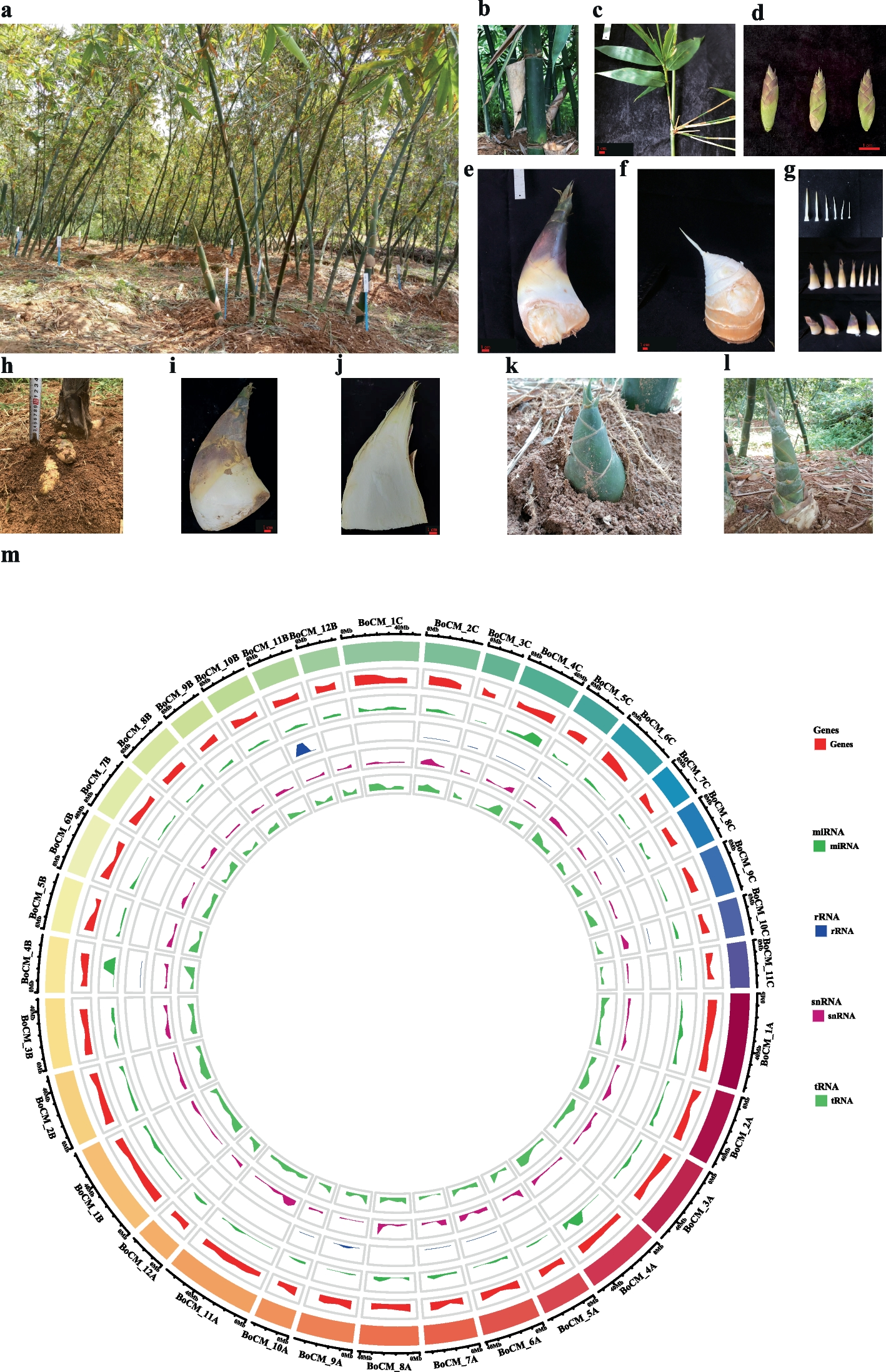
Clumping bamboo has sympodial rhizomes that are characterized by their rhizomes growing outwards from buds on a “mother” rhizome, eventually turning upwards to become new culms (Fig. 1a). The culm height of B. oldhamii ranges from 6 to 12 m, the culm diameter ranges from 3 to 9 cm, depending on the growth environment, the internode length ranges from 20 to 35 cm, and the wall thickness ranges from 4 to 12 mm, depending on growth conditions (Fig. 1b). There are multiple branches in each node of the middle and top culms in B. oldhamii, about 5 leaves in each little branch, and the leaf length ranges from 10–20 cm, and the leaf width ranges from 1.0 to 1.5 cm (Fig. 1c). The flowering period happens between May to November and includes bracts (3–5) and florets (5–9) (Fig. 1d). The shooting period is from May to November, the young edible shoots are sold as fresh vegetables on the market or marked as processed food, and the sheaths of the shoots are deciduous, leathery, dark brown spinous-hairy (Fig. 1e-g). The shoot of B. oldhamii had their best taste and maximum edible portion when they had not erupted out of the soil surface (Fig. 1h-j). When the shoot of B. oldhamii erupted out of the soil surface, the above-ground part of the shoot sheaths would be green with the sunlight and days (Fig. 1k). The old inedible shoots would be degraded or grown into young bamboo culms and then form a new bamboo (Fig. 1l).
Fig. 1
The biological phenotype and genome characteristics of B. oldhamii. a A clumping forest of B. oldhamii; b A culm internode of B. oldhamii; c A leaf branch of B. oldhamii; d The florets of B. oldhamii; e A edible shoot of B. oldhamii; f The shoots of B. oldhamii with shells off; g The off shells of a bamboo shoot; h Phenotype of the shoots under soil surface; i The phenotype of the edible shoot usually sold on market. j The longitudinal section of the edible shoot is usually sold on the market. k–l The shoot has erupted out of the soil surface within several days. m The genome circular graph of B. oldhamii from the inner to outer track represents the density of tRNA, snRNA, rRNA, miRNA, and Genes; the last outer track represents the length of chromosomes in different colors
To comprehend this bamboo species more deeply, the genetic genome information of B. oldhamii was explored. Compared with the internal standard tomato with a genome size of 800 Mb and a peak around 51.03 using flow cytometric fluorescent detection (Fig. S1a), the estimated genome size of B. oldhamii is 1.375 Gb with a peak around 87.77 (Fig. S1b). The Illumina sequencing method was used to estimate the genome size of B. oldhamii around 1.33 Gb based on the 27 K-mer length (Fig. S2). Based on the above genome size estimation results, we processed the whole nuclear genome sequence with PacBio HiFi (40X) and HiC (100X) sequencing projects. After the PacBio HiFi and Hi-C sequencing of B. oldhamii, about 97.23% of contig sequences were anchored on the pseudomolecule chromosomes, and a total of 35 pseudochromosomes were acquired based on synteny analysis with the relative species D. latiflorus. The final Pseudochromosome genome was evaluated with the Chromosome Hi-C signal heatmap, corresponding to the Hi-C assisted genome assembly thesis (Fig. S3), and the final genome BUSCO evaluation is 96.59%.
The final assembled genome size of B. oldhamii is 1.446 Gb, the N50 of all the 1851 scaffolds is 38,936,033 bp, and the GC contents are 45% (Table 1). After the gene structure annotation, a total of 88,140 genes were acquired. All 88,140 genes were annotated in Nr, Swissprot, KEGG, KOG, TrEMBL, Interpro, and GO databases with rates of 76.87%, 50.46%, 49.24%, 47.25%, 76.70%, 62.48%, and 55.74% respectively. Finally, about 78.11% (68,843) of genes acquired the functional annotations (Table 1) (Table S1). The noncoding RNA, including miRNA, tRNA, rRNA, and snRNA, was annotated at the rate of 0.003297%, 0.043679%, 0.356499%, and 0.008666% respectively, on the whole genome (Fig. 1m) (Table S2). In the genome annotation of B. oldhamii, a total of 779,839,625 bp repeat genome sequences were acquired and occupied 53.89% of the genome size (Table S3). Based on the transcription factor annotation, the large number of transcription factor families is bHLH, NAC, MYB, bZIP, C2H2, AP2/ERF-ERF, and AP2/ERF-AP2 (Table S4).
Table 1 Statistics of the assembly genome features and gene annotation in B. oldhamii
The basic genome analysis of B. oldhamii Munro
Through the identification and analysis of genome homologous genes and families can obtain single-copy gene families and multi-copy gene families. We statistics the families and genes in the following species: A. thaliana, A. trichopoda, B. oldhamii (subgenomes A, B, and C), D. latiflorus (subgenomes A, B, and C), O. latifolia, O. sativa, P. edulis (subgenomes C and D), P. trichocarpa, and R. guianensis (Table S5). The results of orthologs and family statistics showed that A. trichopoda has the greatest number of Single-copy orthologs, P. trichocarpa has the greatest number of Multi-copy orthologs, and O. latifolia has the most Unique paralogs (Fig. S4a) (Table S5). 5522 ortholog families exist in both species of B. oldhamii (subgenomes A, B, and C) and O. sativa (Fig. S4b), and 8250 ortholog families that exist in the species of B. oldhamii_A, P. edulis_C, D. latiflorus_A, and O.latifolia (Fig. S4c). As the Phylogenetic tree analysis results show that the species of A. trichopoda is the outer taxa, the other species in the phylogenetic tree are consistent with the phylogenetic positions of the APG IV system. A. thaliana and P. trichocarpa branched together, and the others belong to Poaceae including the bamboos and rice, moreover, the A sub-genome of D. latiflorus_A and B. oldhamii_A branched together, the B sub-genome of D. latiflorus_B and B. oldhamii_B branched together, and the C sub-genome of D. latiflorus_C and B. oldhamii_C branched together (Fig. S5).
Based on the time tree criterion (http://www.timetree.org/), the divergence time between O. sativa and A. thaliana was 130–200 million years ago (Mya), and between P. trichoparca and A. thaliana was 100–120 Mya. As the result shows that the divergence time between O. latifolia and R. guianensis is 47.5 (26.3—69.8) Mya, and the divergence time between O. sativa and bamboos is 100.7 (76.1–126.1) Mya (Fig. 2a). The results of 4DTv analysis showed that the last whole genome duplication of B. oldhamii_A, B. oldhamii_B, and B. oldhamii_C happened after the first divergence of O. sativa. (Fig. 2b). We do the gene family expansion and contraction between genomes and subgenomes (Fig. 2c) (Table S6). The results showed that A. thaliana has the most expansion gene families, and P. trichocarpa has the most contraction gene families, and then is the plant species A. trichopoda (Fig. 2c). The genes in the collinearity fragment have maintained a high degree of conservation throughout species evolution. The genome synteny results showed that there are more blocks between O. sativa and B. oldhamii than between B. oldhamii and D. latiflorus, but the mean block length was larger in B.oldhamii and D. latiflorus than in B. oldhamii and O.sativa (Fig. 2d-f) (Table S7).
Fig. 2
The phenotype and genome characteristics of B. oldhamii. a The estimation of divergence time among species. The number in blue color on the branches means the estimated divergence time (Mya), and the red nodes are tagged as the criteria divergence time (Mya). b The distribution of 4DTv distance (corrected for multiple substitutions). c The gene family expansion and contraction between A. thaliana, A. trichopoda, B. oldhamii (subgenomes A, B, and C), D. latiflorus (subgenomes A, B, and C), O.latifolia, O.sativa, P.edulis (subgenomes C and D), P. trichocarpa, and R. guianensis. d The synteny analysis between O. sativa, B. oldhamii_A, and D. latiflorus_A. e The synteny analysis between O.sativa, B.oldhamii_B, and D.latiflorus_B. (f) The synteny analysis between O. sativa, B. oldhamii_C, and D. latiflorus_C
We acquired the expansion and contraction genes in the three subgenomes of B. oldhamii, in B. oldhamii_A, there were 160 contraction genes and 888 expansion genes (Table S8), and the expansion genes were enriched in the pathway of Plant-pathogen interaction, sulfur relay system, homologous recombination, and valine leucine and isoleucine degradation and others (Fig. S6) (Table S9). In B. oldhamii_B, there are 163 contraction genes belong to 128 families and 669 expansion genes belong to 99 families (Table S10), and the expansion genes were enriched in the pathway of alpha-Linolenic acid metabolism, homologous recombination, glycerophospholipid metabolism, arginine biosynthesis, sesquiterpenoid and triterpenoid biosynthesis, diterpenoid biosynthesis, flavonoid biosynthesis and others (Fig. S7) (Table S11). In B. oldhamii_C, there are 167 contraction genes belonging to 182 families, and 372 expansion genes belong to 46 families (Table S12), and the expansion was enriched in the pathway of homologous recombination and basal transcription factors (Fig. S8) (Table S13).
Data mining of transcriptome and metabolome in multiple shoot developmental phases
Seven developed phases of the shoot in B. oldhamii were picked out searching for the bitter chemicals and key genes contributing to the taste transition from delicious taste to a little bitter taste with transcriptome and metabolome (Fig. 3a). DNBSEQ platforms were used to sequence the 21 shoot samples (7 phases with three biological replicates in each phase). The transcriptome data of each sample is about 6.45 Gb, the average mapping rate of the genome is 91.49%, and the average mapping rate of the gene sets is 65.69%. Based on the reference genome annotation, 88,140 genes were Known Genes, and 3,228 Novel Genes from the RNA-seq data (Table S14). A total of 53,252 genes were detected as expressed genes, 50,129 were known genes (49,365 DEG and 764 Non DEG), and 3,123 genes were novel (3121 DEG and 2 Non DEG) (Fig. 3b) (Table S15). A total of 42,887 new transcripts were detected, 39,659 new alternative splice isoforms distributed in 20,786 known protein-coding genes, and 3,228 were novel gene coding transcripts (Table S16). The FPKM distribution of the transcription factor families in Known Genes was analyzed (Fig. 3c). The results showed that the families of bHLH, MYB-related, bZIP, C3H, HB-, C2H2-, and TUB transcription factors might play crucial roles in shoot growth and development. There are large number of those transcription factors whose expressions were activated in certain shoot developing phases (FPKM > 10).
Fig. 3
Basic data mining of transcriptome and metabolome in multiple shoots developing phases of B. oldhamii. a The sectional shoot morphology of seven developing phases; b The FPKM distribution of expressed Known genes and Novel genes in the transcriptome of seven developing phases. c The FPKM distribution of transcription factors in the genome’s known genes is based on the transcriptome expression data. d The classification of all metabolites detected in the shoot of seven developing phases; e The classification of mined bitter metabolites and relative accumulated patterns in seven developing bamboo shoots of B. oldhamii
A total of 428 qualified metabolites were identified (Table S17). The class of phytochemical compounds includes 37 flavonoids, 29 terpenoids, and others (Fig. 3d). The class of compounds with biological roles includes 31 amino acids, peptides, and analogs, 16 benzene and derivatives, 11 carbohydrates, 11 purines and derivatives, 10 organic acids, and others (Fig. 3d). The class of Lipids includes 15 polyketides, 10 Fatty acyls, and others (Fig. 3d). In order to acquire the bitter metabolites, each metabolite was checked against bitterDB. Finally, 33 bitter metabolites were acquired in this research, major distributed in the families of Amino acids metabolites (L-Isoleucine, L-Leucine, L-Tryptophan, and L-Tyrosine), Flavonoids (Naringenin, Genistein, Genistin, Daidzin, Apigenin, Isorhamnetin, Kaempferol, and Hesperetin), Purines and derivatives (Caffeine, Pentoxifylline, Theobromine, and Adenosine), Terpenoids (Limonin, Andrographolide, Ginkgolide A, L-MENTHONE, Oleuropein, Nerol, Stevioside, Obacunone, Gentiopicrin) and others (Dicumarol, Arbutin, Amarogentin, and others) (Fig. 3e). The taste transition is complex; bitter taste is usually accompanied by astringent taste generation. Tannin is a criterion to measure astringent taste. In the correlation analysis, we added tannin as a measurement criterion for mining the accompanied bitter metabolites. The bitter metabolites positively correlated with the content of tannins were Flavonoids (Genistin, Daidzin, Kaempferol), Terpenoids (Oleuropein, Stevioside, Obacunone), Purine and derivatives (Theobromine), Coumarin and derivatives (Dicumarol), Benzene and derivatives (Salicylic acid), Carbohydrate (Arbutin), and Amarogentin. The negative correlation between bitter metabolites with tannin content was amino acids (L-Isoleucine, L-Leucine, L-Tyrosine), Terpenoids (L-menthone, Nerol, Gentiopicrin), and Sinapic acid (Fig. S9).
The differentially accumulated metabolites between phases were analyzed (Table S18). In the comparison of BoS2 vs. BoS1 (81 DAMs, 46 up DAMs, and 35 down DAMs), all the DAMs were significantly enriched in the pathway of flavone and flavonol biosynthesis, monoterpenoid biosynthesis, and tyrosine metabolism (Fig. S10a). In the comparison of BoS3 vs. BoS2 (49 DAMs, 25 up DAMs, and 24 down DAMs), all the DAMs were significantly enriched in the pathway of phenylalanine, tyrosine, and tryptophan biosynthesis (Fig. S10b). In the comparison of BoS4 vs. BoS3 (61 DAMs, 26 up DAMs, and 35 down DAMs), all the DAMs were significantly enriched in the pathway of Monoterpenoid biosynthesis, Metabolic pathways, and Biosynthesis of secondary metabolites (Fig. S10c). In the comparison of BoS5 vs. BoS4 (172 DAMs, 89 up DAMs, and 83 down DAMs), the total DAMs were significantly enriched in the pathway of aminoacyl-tRNA biosynthesis, metabolic pathway, biosynthesis of secondary metabolites, biosynthesis of amino acids, and phenylalanine metabolism (Fig. S10d). In the comparison of BoS6 vs. BoS5 (45 DAMs, 31 up DAMs, and 14 down DAMs), all the DAMs were enriched in the pathway of Stilbenoid, diarylheptanoid, and gingerol biosynthesis (Fig. S10e). In the comparison of BoS7 vs. BoS6 (23 DAMs, 14 up DAMs, and 9 down DAMs), all the DAMs were enriched in the pathway of flavone and flavonol biosynthesis (Fig. S10f). Finally, a total of 373 non-repeated differentially accumulated metabolites were acquired (Table S19).
Joint analysis between transcriptome and metabolome
From the combined analysis between metabolome and transcriptome, the DAMs and DEGs in each comparison were enriched with KEGG pathways. All the 81 DAMs and 858 DEGs in comparison of BoS2 vs. BoS1 enriched in the pathway of Biosynthesis of secondary metabolites, Glyoxylate and dicarboxylate metabolism, Glycolysis/Gluconeogenesis, and other pathways (Fig. S11) (Table S20). All the 49 DAMs and 439 DEGs in comparison of BoS3 vs. BoS2 enriched in the pathway of Biosynthesis of secondary metabolites, beta-alanine metabolism, Amino sugar and nucleotide sugar metabolism, Alanine, aspartate, and glutamate metabolism, Tryptophan metabolism, and other pathways (Fig. S12) (Table S21). All the 61 DAMs and 388 DEGs in comparison of BoS4 vs. BoS3 enriched in the pathway of Biosynthesis of secondary metabolites, Glutathione metabolism, Flavonoid biosynthesis, Plant hormone signal transduction, and other pathways (Fig. S13) (Table S22). All the 172 DAMs and 516 DEGs in the comparison of BoS5 vs. BoS4 enriched in the pathway of Biosynthesis of secondary metabolites, Isoflavonoid biosynthesis, Plant hormone signal transduction, and other pathways (Fig. S14) (Table S23). All the 45 DAMs and 337 DEGs in comparison of BoS6 vs. BoS5 enriched in the pathway of Biosynthesis of secondary metabolites, Pyrimidine metabolism, and other pathways (Fig. S15) (Table S24). All the 23 DAMs and 410 DEGs in comparison of BoS7 vs. BoS6 enriched in the pathway of Flavone and flavonol biosynthesis and Flavonoid biosynthesis (Fig. S16) (Table S25). From this joint comparison analysis, we find that the pathway of biosynthesis of secondary metabolites, amino acid metabolism, biosynthesis of flavonoid, flavone, flavonol and isoflavonoid, plant hormone signal transduction, and others play important roles in the flavor transition biological process.
The WGCNA analysis between the bitter metabolites and gene expression showed that the bitter metabolites, such as L-Isoleucine, L-Leucine, and L-Tyrosine, were positively correlated with the module of MEturquoise, while Salicylic acid, arbutin, dicumarol, genistin, stevioside, and amarogentin were negatively correlated with the module of MEturquoise (Fig. 4a). The correlated analysis between bitter metabolites and transcription factors showed that the family of bHLH and HB- (including HB-BELL, HB-HD-Zip, HB-KNOX, HB-WOX, and HB-others) has large genes correlated with bitter metabolites, indicating that bHLH and HB- transcription factor families might play pivotal roles in regulating the biosynthesis or metabolism of bitter metabolites (Fig. 4b). To mine out the key genes in bHLH transcription factors, we did the network analysis of the bHLH transcription factor existing in MEturquoise (weight > 0.3) and sorted with degree values, and the results showed that Bo00073447 (bHLH) was a hub gene in the bHLH Co-expression Network (Fig. 4c) (Table S26). To mine out the key genes in HB- transcription factors, we do the network analysis of HB- transcription factors existing in MEturquoise (weight > 0.3) and sorted by degree values, and we find some key genes, Bo00029783(HB-HD-ZIP), Bo00006584(HB-HD-ZIP), Bo00043187(HB-KNOX), and Bo00010600 (HB-WOX) play roles in the HB- Co-expression Network (Fig. 4d) (Table S27).
Fig. 4
Weighted correlation network analysis and gene co-expression network between bitter metabolites and transcription factor families. a Weighted correlation network analysis (WGCNA) between bitter metabolites and expressed genes (FPKM ≥ 5). b Chord Diagram of the correlated relationship between bitter metabolites and transcription factors. c Gene Co-expression Network of bHLH transcription factors existed in the MEturquois module (weight > 0.3). d Gene Co-expression Network of HB- transcription factors existed in the MEturquois module (weight > 0.3)
Model of bitter flavor formation in the shoot of B. oldhamii
From the above joint analysis between bitter metabolites and transcription factors, we found that the expression of bHLH and HB- transcription factors was relevant to the accumulation of bitter metabolites might play roles in the biosynthesis and metabolic pathways of bitter metabolites. In this research, combined with our previous research [15], the mined bitter chemicals were searched against the KEGG pathway database, the bitter metabolites related pathways in each class were simplified and merged with flavonoids, amino acids, terpenoids, purine, solanidine, and hydrogen cyanide pathways (Fig. S17). The flavonoid bitter metabolites (Naringenin, Genistein, Genistin, Daidzin, Apigenin, Kaempferol, and Hesperetin) and other researched metabolites (Salicylate, Salicin, Arbutin, Amygdalin [15], Sinapic acid) are merged in the flavonoid-related pathway originally synthesized from bitter amino acid L-Phenylalanine (Fig. S17a). The bitter amino acids (L-Isoleucine, L-Leucine, L-Tryptophan, and L-Tyrosine) and arginine [15] merged in the amino acid biosynthesis and metabolic pathways (Fig. S17b). The terpenoid metabolites (Limonin, Ginkgolide A, Oleuropein, Nerol, Stevioside, Menthone, and Gentiopicrin) were merged in the terpenoid pathway originating from terpenoid backbone biosynthesis (Fig. S17c). Purine bitter metabolites (Caffeine, Theobromine, and Adenosine) and hypoxanthine [15] were merged in the Purine metabolism pathway (Fig. S17d). Solanidine and Amygdalin were detected in our previous research [15] were merged in the abbreviated biosynthesis of various alkaloids pathway (Fig. S17e) and the abbreviated cyanoamino acid metabolism pathway (Fig. S17f). Amygdalin is a kind of cyanogenic glucoside, but the majority of academics regard taxiphyllin as the major cyanogenic glucoside that exists in the edible bamboo shoot tip [57] and is easily decomposed to hydrogen cyanide (HCN), which causes a bitter flavor. The bitter amino acids (phenylalanine, tyrosine, isoleucine, leucine, and valine) were substances for the biosynthesis of cyanogenic glucosides(Fig. S17f) in plants [12]. We consider that there are several kinds of cyanogenic glucosides that exist in the bamboo shoot, causing the bitter flavor transition, not just one kind of cyanogenic glucoside.
From the above analysis, we hypothesize a simple model about this research (Fig. 5a). In an appropriate environment, when the shoot of B. oldhamii grown out of the soil surface and accepted sunlight signals for days, the expression of bHLH and HB- transcription factors were activated or repressed which influence the genes’ expression in the related pathways (such as, Circadian rhythm; Plant hormone signal transduction; Phenylpropanoid biosynthesis; Flavonoid biosynthesis; Flavone and flavonol biosynthesis; and others) to determine the content accumulation variation of bitter metabolites (such as, flavonoids; amino acids; terpenoids; Purines and derivatives; Cyanogenic glycosides; and others), and finally causing the bitter flavor formation or transition in the tip shoot of B. oldhamii (Fig. 5a). There were exist diversity bamboo species in the world, and the bamboo forests have diverse usages, such as edible shoots collection, bamboo timber collection, city or road ornamental, germplasm preservation, soil improvement, nature reserve, and others. Multiple bamboo species and forests were used to produce edible shoots, which could be sold as fresh shoots or used to make processed food (Picked shoots, Salted shoots, Boiled shoots, Soused shoots, Canned shoots, Dried shoots) (Fig. 5b). The majority of bamboo forests were used for ecosystem balance and bamboo timber, such as bamboo weaving, bamboo charcoal, disposable tableware, bamboo fiber clothes, bamboo timber architecture, bamboo flooring, and bamboo crafts (Fig. 5c). This research was focused on the bamboo species of B. oldhamii, which is especially distributed in the southeast of China, and the shoots that emerged in summer are mainly used as an edible shoot locally. The hypothetical model about the bitter flavor formation or transition in the tip shoot is based on the Multi-omics data in B. oldhamii, which needs to be verified with deep research. Bamboo has a variety of species, and the shoot shape, color, taste, and properties of various bamboo species were different, and the edible portion or part was different depending on the bamboo species. Each bamboo species has its unique flavor and taste; the flavor formation or transition in the edible shoot of other bamboo species still needs further research.
Fig. 5
A hypothetical model of bitter metabolites formation, and the usages of edible bamboo shoot and timber in B. oldhamii. a The hypothetical model of bitter flavor metabolites formation in the shoot tip of B. oldhamii. b The traditional uses of edible bamboo shoots. c The traditional usages of bamboo timber
Xu M, Xu H, Ling YW, Liu JJ, Song P, Fang ZQ, Yue ZS, Duan JL, He F, Wang L. Neutrophil extracellular traps-triggered hepatocellular senescence exacerbates lipotoxicity in non-alcoholic steatohepatitis. J Adv Res. 2025;S2090–1232(25):00175–4. https://doi.org/10.1016/j.jare.2025.03.015. Epub ahead of print. PMID: 40068761.
Article
CAS
Google Scholar
Zhang H, Wang Y, Qu M, Li W, Wu D, Cata JP, et al. Neutrophil, neutrophil extracellular traps and endothelial cell dysfunction in sepsis. Clin Transl Med. 2023;13(1):e1170. https://doi.org/10.1002/ctm2.1170.
Article
CAS
PubMed
PubMed Central
Google Scholar
Ma Y, Wei J, He W, Ren J. Neutrophil extracellular traps in cancer. MedComm. 2024;5(8):e647. https://doi.org/10.1002/mco2.647.
Article
CAS
PubMed
PubMed Central
Google Scholar
Petretto A, Bruschi M, Pratesi F, Croia C, Candiano G, Ghiggeri G, et al. Neutrophil extracellular traps (NET) induced by different stimuli: a comparative proteomic analysis. PLoS ONE. 2019;14(7):e0218946. https://doi.org/10.1371/journal.pone.0218946.
Article
CAS
PubMed
PubMed Central
Google Scholar
Jaillon S, Ponzetta A, Di Mitri D, Santoni A, Bonecchi R, Mantovani A. Neutrophil diversity and plasticity in tumour progression and therapy. Nat Rev Cancer. 2020;20(9):485–503. https://doi.org/10.1038/s41568-020-0281-y.
Article
CAS
PubMed
Google Scholar
Shaul ME, Fridlender ZG. Tumour-associated neutrophils in patients with cancer. Nat Rev Clin Oncol. 2019;16(10):601–20. https://doi.org/10.1038/s41571-019-0222-4.
Article
PubMed
Google Scholar
Ozveren A, Erdogan AP, Ekinci F. The inflammatory prognostic index as a potential predictor of prognosis in metastatic gastric cancer. Sci Rep. 2023;13(1):7755. https://doi.org/10.1038/s41598-023-34778-5. PMID: 37173358; PMCID: PMC10182084.
Article
CAS
PubMed
PubMed Central
Google Scholar
Coffelt SB, Wellenstein MD, de Visser KE. Neutrophils in cancer: neutral no more. Nat Rev Cancer. 2016;16(7):431–46. https://doi.org/10.1038/nrc.2016.52.
Article
CAS
PubMed
Google Scholar
Mantovani A, Cassatella MA, Costantini C, Jaillon S. Neutrophils in the activation and regulation of innate and adaptive immunity. Nat Rev Immunol. 2011;11(8):519–31. https://doi.org/10.1038/nri3024. PMID: 21785456.
Grivennikov SI, Greten FR, Karin M. Immunity, inflammation, and cancer. Cell. 2010;140(6):883–99. https://doi.org/10.1016/j.cell.2010.01.025.
Article
CAS
PubMed
PubMed Central
Google Scholar
Kolaczkowska E, Kubes P. Neutrophil recruitment and function in health and inflammation. Nat Rev Immunol. 2013;13(3):159–75. https://doi.org/10.1038/nri3399.
Article
CAS
PubMed
Google Scholar
Borregaard N. Neutrophils, from marrow to microbes. Immunity. 2010;33(5):657–70. https://doi.org/10.1016/j.immuni.2010.11.011.
Article
CAS
PubMed
Google Scholar
Mantovani A, Cassatella MA, Costantini C, Jaillon S. Neutrophils in the activation and regulation of innate and adaptive immunity. Nat Rev Immunol. 2011;11(8):519–31. https://doi.org/10.1038/nri3024.
Article
CAS
PubMed
Google Scholar
Adrover JM, McDowell SAC, He XY, Quail DF, Egeblad M. NETworking with cancer: the bidirectional interplay between cancer and neutrophil extracellular traps. Cancer Cell. 2023;41(3):505–26. https://doi.org/10.1016/j.ccell.2023.02.001.
Article
CAS
PubMed
PubMed Central
Google Scholar
Papayannopoulos V. Neutrophil extracellular traps in immunity and disease. Nat Rev Immunol. 2018;18(2):134–47. https://doi.org/10.1038/nri.2017.105.
Article
CAS
PubMed
Google Scholar
Brinkmann V, Reichard U, Goosmann C, Fauler B, Uhlemann Y, Weiss DS, et al. Neutrophil extracellular traps kill bacteria. Science. 2004;303(5663):1532–5. https://doi.org/10.1126/science.1092385.
Article
CAS
PubMed
Google Scholar
Fuchs TA, Abed U, Goosmann C, Hurwitz R, Schulze I, Wahn V, et al. Novel cell death program leads to neutrophil extracellular traps. J Cell Biol. 2007;176(2):231–41. https://doi.org/10.1083/jcb.200606027.
Article
CAS
PubMed
PubMed Central
Google Scholar
Herre M, Cedervall J, Mackman N, Olsson AK. Neutrophil extracellular traps in the pathology of cancer and other inflammatory diseases. Physiol Rev. 2023;103(1):277–312. https://doi.org/10.1152/physrev.00062.2021.
Article
CAS
PubMed
Google Scholar
Leung HHL, Perdomo J, Ahmadi Z, Yan F, McKenzie SE, Chong BH. Inhibition of NADPH oxidase blocks NETosis and reduces thrombosis in heparin-induced thrombocytopenia. Blood Adv. 2021;5(23):5439–51. https://doi.org/10.1182/bloodadvances.2020003093.
Chi Z, Chen S, Yang D, Cui W, Lu Y, Wang Z, et al. Gasdermin d-mediated metabolic crosstalk promotes tissue repair. Nature. 2024;634(8036):1168–77. https://doi.org/10.1038/s41586-024-08022-7.
Article
CAS
PubMed
Google Scholar
Zhang J, Yu Q, Jiang D, Yu K, Yu W, Chi Z, Chen S, Li M, Yang D, Wang Z, Xu T, Guo X, Zhang K, Fang H, Ye Q, He Y, Zhang X, Wang D. Epithelial Gasdermin D shapes the host-microbial interface by driving mucus layer formation. Sci Immunol. 2022;7(68):eabk2092. https://doi.org/10.1126/sciimmunol.abk2092. Epub 2022 Feb 4. PMID: 35119941.
Article
CAS
Google Scholar
Fontana P, Du G, Zhang Y, Zhang H, Vora SM, Hu JJ, Shi M, Tufan AB, Healy LB, Xia S, Lee DJ, Li Z, Baldominos P, Ru H, Luo HR, Agudo J, Lieberman J, Wu H. Small-molecule GSDMD agonism in tumors stimulates antitumor immunity without toxicity. Cell. 2024;187(22):6165–e618122. Epub 2024 Sep 6. PMID: 39243763.
Article
CAS
PubMed
PubMed Central
Google Scholar
Bianchi M, Hakkim A, Brinkmann V, Siler U, Seger RA, Zychlinsky A, et al. Restoration of NET formation by gene therapy in CGD controls aspergillosis. Blood. 2009;114(13):2619–22. https://doi.org/10.1182/blood-2009-05-221606.
Article
CAS
PubMed
PubMed Central
Google Scholar
Ermert D, Urban CF, Laube B, Goosmann C, Zychlinsky A, Brinkmann V. Mouse neutrophil extracellular traps in microbial infections. J Innate Immun. 2009;1(3):181–93. https://doi.org/10.1159/000205281.
Article
CAS
PubMed
PubMed Central
Google Scholar
Pilsczek FH, Salina D, Poon KK, Fahey C, Yipp BG, Sibley CD, et al. A novel mechanism of rapid nuclear neutrophil extracellular trap formation in response to Staphylococcus aureus. J Immunol. 2010;185(12):7413–25. https://doi.org/10.4049/jimmunol.1000675.
Article
CAS
PubMed
Google Scholar
Byrd AS, O’Brien XM, Johnson CM, Lavigne LM, Reichner JS. An extracellular matrix-based mechanism of rapid neutrophil extracellular trap formation in response to Candida albicans. J Immunol. 2013;190(8):4136–48. https://doi.org/10.4049/jimmunol.1202671.
Article
CAS
PubMed
Google Scholar
Parker H, Dragunow M, Hampton MB, Kettle AJ, Winterbourn CC. Requirements for NADPH oxidase and myeloperoxidase in neutrophil extracellular trap formation differ depending on the stimulus. J Leukoc Biol. 2012;92(4):841–9. https://doi.org/10.1189/jlb.1211601.
Article
CAS
PubMed
Google Scholar
Douda DN, Khan MA, Grasemann H, Palaniyar N. SK3 channel and mitochondrial ROS mediate NADPH oxidase-independent NETosis induced by calcium influx. Proc Natl Acad Sci U S A. 2015;112(9):2817–22. https://doi.org/10.1073/pnas.1414055112.
Castanheira FVS, Kubes P. Neutrophils and NETs in modulating acute and chronic inflammation. Blood. 2019;133(20):2178–85. https://doi.org/10.1182/blood-2018-11-844530.
Article
CAS
PubMed
Google Scholar
Li M, Lin C, Deng H, Strnad J, Bernabei L, Vogl DT, et al. A novel peptidylarginine deiminase 4 (PAD4) inhibitor BMS-P5 blocks formation of neutrophil extracellular traps and delays progression of multiple myeloma. Mol Cancer Ther. 2020;19(7):1530–8. https://doi.org/10.1158/1535-7163.MCT-19-1020.
Article
CAS
PubMed
PubMed Central
Google Scholar
Papayannopoulos V, Metzler KD, Hakkim A, Zychlinsky A. Neutrophil elastase and myeloperoxidase regulate the formation of neutrophil extracellular traps. J Cell Biol. 2010;191(3):677–91. https://doi.org/10.1083/jcb.201006052.
Article
CAS
PubMed
PubMed Central
Google Scholar
Metzler KD, Fuchs TA, Nauseef WM, Reumaux D, Roesler J, Schulze I, et al. Myeloperoxidase is required for neutrophil extracellular trap formation: implications for innate immunity. Blood. 2011;117(3):953–9. https://doi.org/10.1182/blood-2010-06-290171.
Article
CAS
PubMed
PubMed Central
Google Scholar
Knight JS, Zhao W, Luo W, Subramanian V, O’Dell AA, Yalavarthi S, et al. Peptidylarginine deiminase inhibition is immunomodulatory and vasculoprotective in murine lupus. J Clin Invest. 2013;123(7):2981–93. https://doi.org/10.1172/JCI67390.
Article
CAS
PubMed
PubMed Central
Google Scholar
Guiducci E, Lemberg C, Küng N, Schraner E, Theocharides APA, LeibundGut-Landmann S. Candida albicans-induced NETosis is independent of peptidylarginine deiminase 4. Front Immunol. 2018;9:1573. https://doi.org/10.3389/fimmu.2018.01573.
Article
CAS
PubMed
PubMed Central
Google Scholar
Claushuis TAM, van der Donk LEH, Luitse AL, van Veen HA, van der Wel NN, van Vught LA, Roelofs JJTH, de Boer OJ, Lankelma JM, Boon L, de Vos AF, van ‘t Veer C, van der Poll T. Role of peptidylarginine deiminase 4 in neutrophil extracellular trap formation and host defense during Klebsiella pneumoniae-Induced Pneumonia-Derived sepsis. J Immunol. 2018;201(4):1241–52. https://doi.org/10.4049/jimmunol.1800314. Epub 2018 Jul 9. PMID: 29987161.
Article
CAS
PubMed
Google Scholar
Warnatsch A, Ioannou M, Wang Q, Papayannopoulos V. Inflammation. Neutrophil extracellular traps license macrophages for cytokine production in atherosclerosis. Science. 2015;349(6245):316–20. https://doi.org/10.1126/science.aaa8064. Epub 2015 Jul 16. PMID: 26185250; PMCID: PMC4854322.
Article
CAS
PubMed
PubMed Central
Google Scholar
Branzk N, Lubojemska A, Hardison SE, Wang Q, Gutierrez MG, Brown GD, et al. Neutrophils sense microbe size and selectively release neutrophil extracellular traps in response to large pathogens. Nat Immunol. 2014;15(11):1017–25. https://doi.org/10.1038/ni.2987.
Article
CAS
PubMed
PubMed Central
Google Scholar
Hakkim A, Fuchs TA, Martinez NE, Hess S, Prinz H, Zychlinsky A, Waldmann H. Activation of the Raf-MEK-ERK pathway is required for neutrophil extracellular trap formation. Nat Chem Biol. 2011;7(2):75–7. https://doi.org/10.1038/nchembio.496. Epub 2010 Dec 19. PMID: 21170021.
Article
CAS
PubMed
Google Scholar
Yipp BG, Petri B, Salina D, Jenne CN, Scott BN, Zbytnuik LD, Pittman K, Asaduzzaman M, Wu K, Meijndert HC, Malawista SE, de Boisfleury Chevance A, Zhang K, Conly J, Kubes P. Infection-induced NETosis is a dynamic process involving neutrophil multitasking in vivo. Nat Med. 2012;18(9):1386–93. https://doi.org/10.1038/nm.2847. PMID: 22922410; PMCID: PMC4529131.
Article
CAS
PubMed
PubMed Central
Google Scholar
Yipp BG, Kubes P. NETosis: how vital is it? Blood. 2013;122(16):2784–94. https://doi.org/10.1182/blood-2013-04-457671.
Article
CAS
PubMed
Google Scholar
Wang Y, Li M, Stadler S, Correll S, Li P, Wang D, et al. Histone hypercitrullination mediates chromatin decondensation and neutrophil extracellular trap formation. J Cell Biol. 2009;184(2):205–13. https://doi.org/10.1083/jcb.200806072.
Article
CAS
PubMed
PubMed Central
Google Scholar
McDonald B, Urrutia R, Yipp BG, Jenne CN, Kubes P. Intravascular neutrophil extracellular traps capture bacteria from the bloodstream during sepsis. Cell Host Microbe. 2012;12(3):324–33. https://doi.org/10.1016/j.chom.2012.06.011.
Article
CAS
PubMed
Google Scholar
Jorch SK, Kubes P. An emerging role for neutrophil extracellular traps in noninfectious disease. Nat Med. 2017;23(3):279–87. https://doi.org/10.1038/nm.4294.
Article
CAS
PubMed
Google Scholar
Kim J, Kim HS, Chung JH. Molecular mechanisms of mitochondrial DNA release and activation of the cGAS-STING pathway. Exp Mol Med. 2023;55(3):510–9. https://doi.org/10.1038/s12276-023-00965-7.
Article
CAS
PubMed
PubMed Central
Google Scholar
Yousefi S, Gold JA, Andina N, Lee JJ, Kelly AM, Kozlowski E, et al. Catapult-like release of mitochondrial DNA by eosinophils contributes to antibacterial defense. Nat Med. 2008;14(9):949–53. https://doi.org/10.1038/nm.1855.
Article
CAS
PubMed
Google Scholar
McIlroy DJ, Jarnicki AG, Au GG, Lott N, Smith DW, Hansbro PM, Balogh ZJ. Mitochondrial DNA neutrophil extracellular traps are formed after trauma and subsequent surgery. J Crit Care. 2014;29(6):1133.e1-5. https://doi.org/10.1016/j.jcrc.2014.07.013. Epub 2014 Jul 22. PMID: 25128442.
Article
CAS
PubMed
Google Scholar
Lood C, Blanco LP, Purmalek MM, Carmona-Rivera C, De Ravin SS, Smith CK, et al. Neutrophil extracellular traps enriched in oxidized mitochondrial DNA are interferogenic and contribute to lupus-like disease. Nat Med. 2016;22(2):146–53. https://doi.org/10.1038/nm.4027.
Article
CAS
PubMed
PubMed Central
Google Scholar
Yousefi S, Mihalache C, Kozlowski E, Schmid I, Simon HU. Viable neutrophils release mitochondrial DNA to form neutrophil extracellular traps. Cell Death Differ. 2009;16(11):1438–44. https://doi.org/10.1038/cdd.2009.96.
Article
CAS
PubMed
Google Scholar
Zhang Y, Guo F, Wang Y. Hypoxic tumor microenvironment: destroyer of natural killer cell function. Chin J Cancer Res. 2024;36(2):138–50. https://doi.org/10.21147/j.issn.1000-9604.2024.02.04.
Article
PubMed
PubMed Central
Google Scholar
Guillotin F, Fortier M, Portes M, Demattei C, Mousty E, Nouvellon E, et al. Vital NETosis vs. suicidal NETosis during normal pregnancy and preeclampsia. Front Cell Dev Biol. 2023;10:1099038. https://doi.org/10.3389/fcell.2022.1099038.
Article
PubMed
PubMed Central
Google Scholar
Zhu D, Lu Y, Yang S, Hu T, Tan C, Liang R, et al. PAD4 inhibitor-functionalized layered double hydroxide nanosheets for synergistic sonodynamic therapy/immunotherapy of tumor metastasis. Adv Sci. 2024;11(26):e2401064. https://doi.org/10.1002/advs.202401064.
Article
CAS
Google Scholar
Moiana M, Aranda F, de Larrañaga G. A focus on the roles of histones in health and diseases. Clin Biochem. 2021;94:12–9. https://doi.org/10.1016/j.clinbiochem.2021.04.019.
Article
CAS
PubMed
Google Scholar
Xie W, Yu X, Yang Q, Ke N, Wang P, Kong H, Wu X, Ma P, Chen L, Yang J, Feng X, Wang Y, Shi H, Chen L, Liu YH, Ding BS, Wei Q, Jiang H. An immunomechanical checkpoint PYK2 governs monocyte-to-macrophage differentiation in pancreatic cancer. Cancer Discov. 2025. https://doi.org/10.1158/2159-8290.CD-24-1712. Epub ahead of print. PMID: 40338055.
Yang C, Wang Z, Li L, Zhang Z, Jin X, Wu P, et al. Aged neutrophils form mitochondria-dependent vital NETs to promote breast cancer lung metastasis. J Immunother Cancer. 2021;9(10):e002875. https://doi.org/10.1136/jitc-2021-002875.
Article
PubMed
PubMed Central
Google Scholar
Hao X, Gu H, Chen C, Huang D, Zhao Y, Xie L, Zou Y, Shu HS, Zhang Y, He X, Lai X, Zhang X, Zhou BO, Zhang CC, Chen GQ, Yu Z, Yang Y, Zheng J. Metabolic imaging reveals a unique preference of symmetric cell division and homing of Leukemia-Initiating cells in an endosteal niche. Cell Metab. 2019;29(4):950–e9656. Epub 2018 Dec 20. PMID: 30581117.
Article
CAS
PubMed
Google Scholar
Wu Q, Hu G. A genetic portrait of metastatic seeds in lung adenocarcinoma. Cancer Cell. 2023;41(5):828–30. https://doi.org/10.1016/j.ccell.2023.04.004.
Article
CAS
PubMed
Google Scholar
West AP, Khoury-Hanold W, Staron M, Tal MC, Pineda CM, Lang SM, et al. Mitochondrial DNA stress primes the antiviral innate immune response. Nature. 2015;520(7548):553–7. https://doi.org/10.1038/nature14156.
Article
CAS
PubMed
PubMed Central
Google Scholar
Yamazaki T, Kirchmair A, Sato A, Buqué A, Rybstein M, Petroni G, Bloy N, Finotello F, Stafford L, Navarro Manzano E, de la Ayala F, García-Martínez E, Formenti SC, Trajanoski Z, Galluzzi L. Mitochondrial DNA drives abscopal responses to radiation that are inhibited by autophagy. Nat Immunol. 2020;21(10):1160–71. https://doi.org/10.1038/s41590-020-0751-0. Epub 2020 Aug 3. PMID: 32747819.
Bakhoum SF, Ngo B, Laughney AM, Cavallo JA, Murphy CJ, Ly P, et al. Chromosomal instability drives metastasis through a cytosolic DNA response. Nature. 2018;553(7689):467–72. https://doi.org/10.1038/nature25432.
Article
CAS
PubMed
PubMed Central
Google Scholar
Cheng AN, Cheng LC, Kuo CL, Lo YK, Chou HY, Chen CH, et al. Mitochondrial lon-induced mtDNA leakage contributes to PD-L1-mediated immunoescape via STING-IFN signaling and extracellular vesicles. J Immunother Cancer. 2020;8(2):e001372. https://doi.org/10.1136/jitc-2020-001372.
Article
PubMed
PubMed Central
Google Scholar
Xiong S, Dong L, Cheng L. Neutrophils in cancer carcinogenesis and metastasis. J Hematol Oncol. 2021;14(1):173. https://doi.org/10.1186/s13045-021-01187-y.
Article
CAS
PubMed
PubMed Central
Google Scholar
Yang LY, Luo Q, Lu L, Zhu WW, Sun HT, Wei R, Lin ZF, Wang XY, Wang CQ, Lu M, Jia HL, Chen JH, Zhang JB, Qin LX. Increased neutrophil extracellular traps promote metastasis potential of hepatocellular carcinoma via provoking tumorous inflammatory response. J Hematol Oncol. 2020;13(1):3. https://doi.org/10.1186/s13045-019-0836-0. PMID: 31907001; PMCID: PMC6945602.
Article
CAS
PubMed
PubMed Central
Google Scholar
Demers M, Krause DS, Schatzberg D, Martinod K, Voorhees JR, Fuchs TA, et al. Cancers predispose neutrophils to release extracellular DNA traps that contribute to cancer-associated thrombosis. Proc Natl Acad Sci U S A. 2012;109(32):13076–81. https://doi.org/10.1073/pnas.1200419109.
Article
PubMed
PubMed Central
Google Scholar
Tohme S, Yazdani HO, Al-Khafaji AB, Chidi AP, Loughran P, Mowen K, et al. Neutrophil extracellular traps promote the development and progression of liver metastases after surgical stress. Cancer Res. 2016;76(6):1367–80. https://doi.org/10.1158/0008-5472.CAN-15-1591.
Article
CAS
PubMed
PubMed Central
Google Scholar
Antonio N, Bønnelykke-Behrndtz ML, Ward LC, Collin J, Christensen IJ, Steiniche T, et al. The wound inflammatory response exacerbates growth of pre-neoplastic cells and progression to cancer. EMBO J. 2015;34(17):2219–36. https://doi.org/10.15252/embj.201490147.
Article
CAS
PubMed
PubMed Central
Google Scholar
Hedrick CC, Malanchi I. Neutrophils in cancer: heterogeneous and multifaceted. Nat Rev Immunol. 2022;22(3):173–87. https://doi.org/10.1038/s41577-021-00571-6.
Article
CAS
PubMed
Google Scholar
Liu S, Wu W, Du Y, Yin H, Chen Q, Yu W, et al. The evolution and heterogeneity of neutrophils in cancers: origins, subsets, functions, orchestrations and clinical applications. Mol Cancer. 2023;22(1):148. https://doi.org/10.1186/s12943-023-01843-6.
Article
PubMed
PubMed Central
Google Scholar
Butin-Israeli V, Bui TM, Wiesolek HL, Mascarenhas L, Lee JJ, Mehl LC, Knutson KR, Adam SA, Goldman RD, Beyder A, Wiesmuller L, Hanauer SB, Sumagin R. Neutrophil-induced genomic instability impedes resolution of inflammation and wound healing. J Clin Invest. 2019;129(2):712–26. Epub 2019 Jan 14. PMID: 30640176; PMCID: PMC6355304.
Article
PubMed
PubMed Central
Google Scholar
Hsu YL, Hung JY, Chang WA, Lin YS, Pan YC, Tsai PH, et al. Hypoxic lung cancer-secreted exosomal miR-23a increased angiogenesis and vascular permeability by targeting prolyl hydroxylase and tight junction protein ZO-1. Oncogene. 2017;36(34):4929–42. https://doi.org/10.1038/onc.2017.105.
Article
CAS
PubMed
Google Scholar
Shang M, Weng L, Wu S, Liu B, Yin X, Wang Z, Mao A. HP1BP3 promotes tumor growth and metastasis by upregulating miR-23a to target TRAF5 in esophageal squamous cell carcinoma. Am J Cancer Res. 2021;11(6):2928–43. PMID: 34249436; PMCID: PMC8263663.
Zhou X, Yan T, Huang C, Xu Z, Wang L, Jiang E, Wang H, Chen Y, Liu K, Shao Z, Shang Z. Melanoma cell-secreted Exosomal miR-155-5p induce proangiogenic switch of cancer-associated fibroblasts via SOCS1/JAK2/STAT3 signaling pathway. J Exp Clin Cancer Res. 2018;37(1):242. https://doi.org/10.1186/s13046-018-0911-3. PMID: 30285793; PMCID: PMC6169013.
Article
CAS
PubMed
PubMed Central
Google Scholar
Zheng Z, Sun R, Zhao HJ, Fu D, Zhong HJ, Weng XQ, et al. MiR155 sensitized B-lymphoma cells to anti-PD-L1 antibody via PD-1/PD-L1-mediated lymphoma cell interaction with CD8 + T cells. Mol Cancer. 2019;18(1):54. https://doi.org/10.1186/s12943-019-0977-3.
Article
PubMed
PubMed Central
Google Scholar
Carmody RN, Sarkar A, Reese AT. Gut microbiota through an evolutionary lens. Science. 2021;372(6541):462–3. https://doi.org/10.1126/science.abf0590.
Article
CAS
PubMed
Google Scholar
Ternes D, Tsenkova M, Pozdeev VI, Meyers M, Koncina E, Atatri S, et al. The gut microbial metabolite formate exacerbates colorectal cancer progression. Nat Metab. 2022;4(4):458–75. https://doi.org/10.1038/s42255-022-00558-0.
Article
CAS
PubMed
PubMed Central
Google Scholar
Saitoh T, Komano J, Saitoh Y, Misawa T, Takahama M, Kozaki T, et al. Neutrophil extracellular traps mediate a host defense response to human immunodeficiency virus-1. Cell Host Microbe. 2012;12(1):109–16. https://doi.org/10.1016/j.chom.2012.05.015.
Article
CAS
PubMed
Google Scholar
Ouyang D, Xiang T, Chen Y, Song M, Zhao J, Chen H, Li S, Zhang L, Xu C, Ren Y, Tao Y, Wang Q, He J, Li Y, Xie S, Liu Y, Wang Y, Yang X, You J, Xie S, Li Y, Weng D, Pan Q, Yang Q, Xia J. The CXCL10-CXCR3 axis induces Tumor-Associated neutrophils to interfere with CTLs-Mediated antitumor activity in EBV-Associated epithelial Cancers. Adv sci (Weinh). 2025;e00950. https://doi.org/10.1002/advs.202500950. Epub ahead of print. PMID: 40686457.
Deng Z, Mei S, Ouyang Z, Wang R, Wang L, Zou B, Dai J, Mao K, Li Q, Guo Q, Yi C, Meng F, Xie M, Zhang X, Wang R, Deng T, Wang Z, Li X, Wang Q, Liu B, Tian X. Dysregulation of gut microbiota stimulates NETs-driven HCC intrahepatic metastasis: therapeutic implications of healthy faecal microbiota transplantation. Gut Microbes. 2025;17(1):2476561. Epub 2025 Mar 18. PMID: 40099491; PMCID: PMC11925110.
Article
PubMed
PubMed Central
Google Scholar
Fu A, Yao B, Dong T, Chen Y, Yao J, Liu Y, Li H, Bai H, Liu X, Zhang Y, Wang C, Guo Y, Li N, Cai S. Tumor-resident intracellular microbiota promotes metastatic colonization in breast cancer. Cell. 2022;185(8):1356–e137226. https://doi.org/10.1016/j.cell.2022.02.027. Epub 2022 Apr 7. PMID: 35395179.
Article
CAS
PubMed
Google Scholar
Jin C, Lagoudas GK, Zhao C, Bullman S, Bhutkar A, Hu B, Ameh S, Sandel D, Liang XS, Mazzilli S, Whary MT, Meyerson M, Germain R, Blainey PC, Fox JG, Jacks T. Commensal microbiota promote lung cancer development via γδ T cells. Cell. 2019;176(5):998–e101316. Epub 2019 Jan 31. PMID: 30712876; PMCID: PMC6691977.
Article
CAS
PubMed
PubMed Central
Google Scholar
Branzk N, Papayannopoulos V. Molecular mechanisms regulating NETosis in infection and disease. Semin Immunopathol. 2013;35(4):513–30. https://doi.org/10.1007/s00281-013-0384-6.
Article
CAS
PubMed
PubMed Central
Google Scholar
Liu C, Qi J, Shan B, Gao R, Gao F, Xie H, et al. Pretreatment with cathelicidin-BF ameliorates Pseudomonas aeruginosa pneumonia in mice by enhancing NETosis and the autophagy of recruited neutrophils and macrophages. Int Immunopharmacol. 2018;65:382–91. https://doi.org/10.1016/j.intimp.2018.10.030.
Article
CAS
PubMed
Google Scholar
Garley M, Jabłońska E, Dąbrowska D. NETs in cancer. Tumour Biol. 2016;37(11):14355–61. https://doi.org/10.1007/s13277-016-5328-z.
Article
CAS
PubMed
Google Scholar
Xiao Y, Cong M, Li J, He D, Wu Q, Tian P, Wang Y, Yang S, Liang C, Liang Y, Wen J, Liu Y, Luo W, Lv X, He Y, Cheng DD, Zhou T, Zhao W, Zhang P, Zhang X, Xiao Y, Qian Y, Wang H, Gao Q, Yang QC, Yang Q, Hu G. Cathepsin C promotes breast cancer lung metastasis by modulating neutrophil infiltration and neutrophil extracellular trap formation. Cancer Cell. 2021;39(3):423–e4377. https://doi.org/10.1016/j.ccell.2020.12.012. Epub 2021 Jan 14. PMID: 33450198.
Article
CAS
PubMed
Google Scholar
Qi JL, He JR, Liu CB, Jin SM, Gao RY, Yang X, et al. Pulmonary Staphylococcus aureus infection regulates breast cancer cell metastasis via neutrophil extracellular traps (NETs) formation. MedComm. 2020;1(2):188–201. https://doi.org/10.1002/mco2.22.
Article
PubMed
PubMed Central
Google Scholar
Munir H, Jones JO, Janowitz T, Hoffmann M, Euler M, Martins CP, et al. Stromal-driven and amyloid β-dependent induction of neutrophil extracellular traps modulates tumor growth. Nat Commun. 2021;12(1):683. https://doi.org/10.1038/s41467-021-20982-2.
Article
CAS
PubMed
PubMed Central
Google Scholar
Xie M, Lin Z, Ji X, Luo X, Zhang Z, Sun M, et al. FGF19/FGFR4-mediated elevation of ETV4 facilitates hepatocellular carcinoma metastasis by upregulating PD-L1 and CCL2. J Hepatol. 2023;79(1):109–25. https://doi.org/10.1016/j.jhep.2023.02.036.
Article
CAS
PubMed
Google Scholar
Kim YC, Seok S, Zhang Y, Ma J, Kong B, Guo G, et al. Intestinal FGF15/19 physiologically repress hepatic lipogenesis in the late fed-state by activating SHP and DNMT3A. Nat Commun. 2020;11(1):5969. https://doi.org/10.1038/s41467-020-19803-9.
Article
CAS
PubMed
PubMed Central
Google Scholar
Li C, Chen T, Liu J, Wang Y, Zhang C, Guo L, et al. FGF19-induced inflammatory CAF promoted neutrophil extracellular trap formation in the liver metastasis of colorectal cancer. Adv Sci. 2023;10(24):e2302613. https://doi.org/10.1002/advs.202302613.
Article
CAS
Google Scholar
Cheng Y, Li H, Deng Y, Tai Y, Zeng K, Zhang Y, et al. Cancer-associated fibroblasts induce PDL1 + neutrophils through the IL6-STAT3 pathway that foster immune suppression in hepatocellular carcinoma. Cell Death Dis. 2018;9(4):422. https://doi.org/10.1038/s41419-018-0458-4.
Article
CAS
PubMed
PubMed Central
Google Scholar
Takesue S, Ohuchida K, Shinkawa T, Otsubo Y, Matsumoto S, Sagara A, Yonenaga A, Ando Y, Kibe S, Nakayama H, Iwamoto C, Shindo K, Moriyama T, Nakata K, Miyasaka Y, Ohtsuka T, Toma H, Tominaga Y, Mizumoto K, Hashizume M, Nakamura M. Neutrophil extracellular traps promote liver micrometastasis in pancreatic ductal adenocarcinoma via the activation of cancer–associated fibroblasts. Int J Oncol. 2020;56(2):596–605. https://doi.org/10.3892/ijo.2019.4951. Epub 2019 Dec 24. PMID: 31894273.
Article
CAS
PubMed
Google Scholar
Chen J, Huang Z, Chen Y, Tian H, Chai P, Shen Y, et al. Lactate and lactylation in cancer. Signal Transduct Target Ther. 2025;10(1):38. https://doi.org/10.1038/s41392-024-02082-x.
Article
CAS
PubMed
PubMed Central
Google Scholar
Wise AD, TenBarge EG, Mendonça JDC, Mennen EC, McDaniel SR, Reber CP, Holder BE, Bunch ML, Belevska E, Marshall MG, Vaccaro NM, Blakely CR, Wellawa DH, Ferris J, Sheldon JR, Bieber JD, Johnson JG, Burcham LR, Monteith AJ. Mitochondria sense bacterial lactate and drive release of neutrophil extracellular traps. Cell Host Microbe. 2025;33(3):341–e3579. Epub 2025 Feb 27. PMID: 40020664; PMCID: PMC11955204.
Article
CAS
PubMed
Google Scholar
Cao S, Liu P, Zhu H, Gong H, Yao J, Sun Y, et al. Extracellular acidification acts as a key modulator of neutrophil apoptosis and functions. PLoS One. 2015;10(9):e0139500. https://doi.org/10.1371/journal.pone.0139500.
Article
CAS
PubMed
PubMed Central
Google Scholar
Trevani AS, Andonegui G, Giordano M, López DH, Gamberale R, Minucci F, Geffner JR. Extracellular acidification induces human neutrophil activation. J Immunol. 1999;162(8):4849–57 PMID: 10202029.
Article
CAS
PubMed
Google Scholar
Díaz FE, Dantas E, Cabrera M, Benítez CA, Delpino MV, Duette G, et al. Fever-range hyperthermia improves the anti-apoptotic effect induced by low pH on human neutrophils promoting a proangiogenic profile. Cell Death Dis. 2016;7(10):e2437. https://doi.org/10.1038/cddis.2016.337.
Article
CAS
PubMed
PubMed Central
Google Scholar
Ashby BS. pH studies in human malignant tumours. Lancet. 1966;2(7458):312–5. https://doi.org/10.1016/s0140-6736(66)92598-0.
Article
CAS
PubMed
Google Scholar
Tannock IF, Rotin D. Acid pH in tumors and its potential for therapeutic exploitation. Cancer Res. 1989;49(16):4373–84. PMID: 2545340.
CAS
PubMed
Google Scholar
Estrella V, Chen T, Lloyd M, Wojtkowiak J, Cornnell HH, Ibrahim-Hashim A, Bailey K, Balagurunathan Y, Rothberg JM, Sloane BF, Johnson J, Gatenby RA, Gillies RJ. Acidity generated by the tumor microenvironment drives local invasion. Cancer Res. 2013;73(5):1524–35. https://doi.org/10.1158/0008-5472.CAN-12-2796. Epub 2013 Jan 3. PMID: 23288510; PMCID: PMC3594450.
Article
CAS
PubMed
PubMed Central
Google Scholar
Waldman AD, Fritz JM, Lenardo MJ. A guide to cancer immunotherapy: from T cell basic science to clinical practice. Nat Rev Immunol. 2020;20(11):651–68. https://doi.org/10.1038/s41577-020-0306-5. Epub 2020 May 20. PMID: 32433532; PMCID: PMC7238960.
Article
CAS
PubMed
PubMed Central
Google Scholar
Ramezani-Ali Akbari K, Khaki-Bakhtiarvand V, Mahmoudian J, Asgarian-Omran H, Shokri F, Hojjat-Farsangi M, Jeddi-Tehrani M, Shabani M. Cloning, expression and characterization of a peptibody to deplete myeloid derived suppressor cells in a murine mammary carcinoma model. Protein Expr Purif. 2022;200:106153. https://doi.org/10.1016/j.pep.2022.106153. Epub 2022 Aug 19. PMID: 35995320.
Article
CAS
PubMed
Google Scholar
Qin H, Lerman B, Sakamaki I, Wei G, Cha SC, Rao SS, Qian J, Hailemichael Y, Nurieva R, Dwyer KC, Roth J, Yi Q, Overwijk WW, Kwak LW. Generation of a new therapeutic peptide that depletes myeloid-derived suppressor cells in tumor-bearing mice. Nat Med. 2014;20(6):676–81. https://doi.org/10.1038/nm.3560. Epub 2014 May 25. PMID: 24859530; PMCID: PMC4048321.
Article
CAS
PubMed
PubMed Central
Google Scholar
Tessier-Cloutier B, Twa DD, Marzban M, Kalina J, Chun HE, Pavey N, Tanweer Z, Katz RL, Lum JJ, Salina D. The presence of tumour-infiltrating neutrophils is an independent adverse prognostic feature in clear cell renal cell carcinoma. J Pathol Clin Res. 2021;7(4):385–96. Epub 2021 Mar 4. PMID: 33665979; PMCID: PMC8185362.
Article
CAS
PubMed
PubMed Central
Google Scholar
Xia X, Zhang Z, Zhu C, Ni B, Wang S, Yang S, Yu F, Zhao E, Li Q, Zhao G. Neutrophil extracellular traps promote metastasis in gastric cancer patients with postoperative abdominal infectious complications. Nat Commun. 2022;13(1):1017. https://doi.org/10.1038/s41467-022-28492-5. PMID: 35197446; PMCID: PMC8866499.
Article
CAS
PubMed
PubMed Central
Google Scholar
Guo M, Sheng W, Yuan X, Wang X. Neutrophils as promising therapeutic targets in pancreatic cancer liver metastasis. Int Immunopharmacol. 2024;140:112888. https://doi.org/10.1016/j.intimp.2024.112888. Epub 2024 Aug 11. PMID: 39133956.
Article
CAS
PubMed
Google Scholar
Zhang Y, Guo L, Dai Q, Shang B, Xiao T, Di X, Zhang K, Feng L, Shou J, Wang Y. A signature for pan-cancer prognosis based on neutrophil extracellular traps. J Immunother Cancer. 2022;10(6):e004210. https://doi.org/10.1136/jitc-2021-004210. PMID: 35688556; PMCID: PMC9189842.
Article
PubMed
PubMed Central
Google Scholar
Masucci MT, Minopoli M, Del Vecchio S, Carriero MV. The emerging role of neutrophil extracellular traps (NETs) in tumor progression and metastasis. Front Immunol. 2020;11:1749. https://doi.org/10.3389/fimmu.2020.01749. PMID: 33042107; PMCID: PMC7524869.
Albrengues J, Shields MA, Ng D, Park CG, Ambrico A, Poindexter ME, Upadhyay P, Uyeminami DL, Pommier A, Küttner V, Bružas E, Maiorino L, Bautista C, Carmona EM, Gimotty PA, Fearon DT, Chang K, Lyons SK, Pinkerton KE, Trotman LC, Goldberg MS, Yeh JT, Egeblad M. Neutrophil extracellular traps produced during inflammation awaken dormant cancer cells in mice. Science. 2018;361(6409):eaao4227. https://doi.org/10.1126/science.aao4227. PMID: 30262472; PMCID: PMC6777850.
Article
CAS
PubMed
PubMed Central
Google Scholar
Fang Q, Stehr AM, Naschberger E, Knopf J, Herrmann M, Stürzl M. No NETs no TIME: crosstalk between neutrophil extracellular traps and the tumor immune microenvironment. Front Immunol. 2022;13:1075260. https://doi.org/10.3389/fimmu.2022.1075260. PMID: 36618417; PMCID: PMC9816414.
Article
CAS
PubMed
PubMed Central
Google Scholar
Ganesan R, Bhasin SS, Bakhtiary M, Krishnan U, Cheemarla NR, Thomas BE, Bhasin MK, Sukhatme VP. Taxane chemotherapy induces stromal injury that leads to breast cancer dormancy escape. PLoS Biol. 2023;21(9):e3002275. https://doi.org/10.1371/journal.pbio.3002275. PMID: 37699010; PMCID: PMC10497165.
Article
CAS
PubMed
PubMed Central
Google Scholar
Mousset A, Albrengues J. NETs unleashed: neutrophil extracellular traps boost chemotherapy against colorectal cancer. J Clin Invest. 2024;134(5):e178344. https://doi.org/10.1172/JCI178344. PMID: 38426501; PMCID: PMC10904039.
Article
CAS
PubMed
PubMed Central
Google Scholar
Teijeira Á, Garasa S, Gato M, Alfaro C, Migueliz I, Cirella A, de Andrea C, Ochoa MC, Otano I, Etxeberria I, Andueza MP, Nieto CP, Resano L, Azpilikueta A, Allegretti M, de Pizzol M, Ponz-Sarvisé M, Rouzaut A, Sanmamed MF, Schalper K, Carleton M, Mellado M, Rodriguez-Ruiz ME, Berraondo P, Perez-Gracia JL, Melero I. CXCR1 and CXCR2 chemokine receptor agonists produced by tumors induce neutrophil extracellular traps that interfere with immune cytotoxicity. Immunity. 2020;52(5):856–e8718. https://doi.org/10.1016/j.immuni.2020.03.001. Epub 2020 Apr 13. PMID: 32289253.
Article
CAS
PubMed
Google Scholar
Taifour T, Attalla SS, Zuo D, Gu Y, Sanguin-Gendreau V, Proud H, Solymoss E, Bui T, Kuasne H, Papavasiliou V, Lee CG, Kamle S, Siegel PM, Elias JA, Park M, Muller WJ. The tumor-derived cytokine Chi3l1 induces neutrophil extracellular traps that promote T cell exclusion in triple-negative breast cancer. Immunity. 2023;56(12):2755–e27728. Epub 2023 Nov 30. PMID: 38039967.
Article
CAS
PubMed
Google Scholar
Yang L, Liu Q, Zhang X, Liu X, Zhou B, Chen J, Huang D, Li J, Li H, Chen F, Liu J, Xing Y, Chen X, Su S, Song E. DNA of neutrophil extracellular traps promotes cancer metastasis via CCDC25. Nature. 2020;583(7814):133–8. https://doi.org/10.1038/s41586-020-2394-6. Epub 2020 Jun 11. PMID: 32528174.
Hu C, Long L, Lou J, Leng M, Yang Q, Xu X, Zhou X. CTC-neutrophil interaction: A key driver and therapeutic target of cancer metastasis. Biomed Pharmacother. 2024;180:117474. https://doi.org/10.1016/j.biopha.2024.117474. Epub 2024 Sep 23. PMID: 39316968.
Peinado H, Zhang H, Matei IR, Costa-Silva B, Hoshino A, Rodrigues G, Psaila B, Kaplan RN, Bromberg JF, Kang Y, Bissell MJ, Cox TR, Giaccia AJ, Erler JT, Hiratsuka S, Ghajar CM, Lyden D. Pre-metastatic niches: organ-specific homes for metastases. Nat Rev Cancer. 2017;17(5):302–17. https://doi.org/10.1038/nrc.2017.6. Epub 2017 Mar 17. PMID: 28303905.
Jia J, Wang Y, Li M, Wang F, Peng Y, Hu J, Li Z, Bian Z, Yang S. Neutrophils in the premetastatic niche: key functions and therapeutic directions. Mol Cancer. 2024;23(1):200. https://doi.org/10.1186/s12943-024-02107-7. PMID: 39277750; PMCID: PMC11401288.
Article
PubMed
PubMed Central
Google Scholar
Lee W, Ko SY, Mohamed MS, Kenny HA, Lengyel E, Naora H. Neutrophils facilitate ovarian cancer premetastatic niche formation in the omentum. J Exp Med. 2019;216(1):176–94. https://doi.org/10.1084/jem.20181170. Epub 2018 Dec 19. PMID: 30567719; PMCID: PMC6314534.
Article
CAS
PubMed
PubMed Central
Google Scholar
Lee W, Naora H. Neutrophils fertilize the pre-metastatic niche. Aging (Albany NY). 2019;11(17):6624–5. https://doi.org/10.18632/aging.102258. Epub 2019 Sep 10. PMID: 31509520. PMCID: PMC6756893.
Article
CAS
PubMed
PubMed Central
Google Scholar
Perego M, Tyurin VA, Tyurina YY, Yellets J, Nacarelli T, Lin C, Nefedova Y, Kossenkov A, Liu Q, Sreedhar S, Pass H, Roth J, Vogl T, Feldser D, Zhang R, Kagan VE, Gabrilovich DI. Reactivation of dormant tumor cells by modified lipids derived from stress-activated neutrophils. Sci Transl Med. 2020;12(572):eabb5817. https://doi.org/10.1126/scitranslmed.abb5817. PMID: 33268511; PMCID: PMC8085740.
Article
CAS
PubMed
PubMed Central
Google Scholar
Wculek SK, Malanchi I. Neutrophils support lung colonization of metastasis-initiating breast cancer cells. Nature. 2015;528(7582):413–7. https://doi.org/10.1038/nature16140 Epub 2015 Dec 9. Erratum in: Nature. 2019;571(7763):E2. https://doi.org/10.1038/s41586-019-1328-7 . PMID: 26649828; PMCID: PMC4700594. .
Article
CAS
PubMed
PubMed Central
Google Scholar
Liu Y, Gu Y, Han Y, Zhang Q, Jiang Z, Zhang X, Huang B, Xu X, Zheng J, Cao X. Tumor Exosomal RNAs Promote Lung Pre-metastatic Niche Formation by Activating Alveolar Epithelial TLR3 to Recruit Neutrophils. Cancer Cell. 2016;30(2):243–56. https://doi.org/10.1016/j.ccell.2016.06.021. PMID: 27505671.
Bellomo G, Rainer C, Quaranta V, Astuti Y, Raymant M, Boyd E, Stafferton R, Campbell F, Ghaneh P, Halloran CM, Hammond DE, Morton JP, Palmer D, Vimalachandran D, Jones R, Mielgo A, Schmid MC. Chemotherapy-induced infiltration of neutrophils promotes pancreatic cancer metastasis via Gas6/AXL signalling axis. Gut. 2022;71(11):2284–99. https://doi.org/10.1136/gutjnl-2021-325272. Epub 2022 Jan 12. PMID: 35022267; PMCID: PMC9554050.
Article
CAS
PubMed
Google Scholar
Rys RN, Calcinotto A. Senescent neutrophils: a hidden role in cancer progression. Trends Cell Biol. 2024;S0962–8924(24):00187–9. https://doi.org/10.1016/j.tcb.2024.09.001. Epub ahead of print. PMID: 39362804.
Article
CAS
Google Scholar
Tyagi A, Sharma S, Wu K, Wu SY, Xing F, Liu Y, Zhao D, Deshpande RP, D’Agostino RB Jr, Watabe K. Nicotine promotes breast cancer metastasis by stimulating N2 neutrophils and generating pre-metastatic niche in lung. Nat Commun. 2021;12(1):474. https://doi.org/10.1038/s41467-020-20733-9. PMID: 33473115; PMCID: PMC7817836.
Article
CAS
PubMed
PubMed Central
Google Scholar
Wieland E, Rodriguez-Vita J, Liebler SS, Mogler C, Moll I, Herberich SE, Espinet E, Herpel E, Menuchin A, Chang-Claude J, Hoffmeister M, Gebhardt C, Brenner H, Trumpp A, Siebel CW, Hecker M, Utikal J, Sprinzak D, Fischer A. Endothelial Notch1 activity facilitates metastasis. Cancer Cell. 2017;31(3):355–67. Epub 2017 Feb 23. PMID: 28238683.
Article
CAS
PubMed
Google Scholar
Fang JH, Zhang ZJ, Shang LR, Luo YW, Lin YF, Yuan Y, Zhuang SM. Hepatoma cell-secreted Exosomal microRNA-103 increases vascular permeability and promotes metastasis by targeting junction proteins. Hepatology. 2018;68(4):1459–75. https://doi.org/10.1002/hep.29920. Epub 2018 Jul 25. PMID: 29637568.
Article
CAS
PubMed
Google Scholar
Kuang DM, Zhao Q, Wu Y, Peng C, Wang J, Xu Z, Yin XY, Zheng L. Peritumoral neutrophils link inflammatory response to disease progression by fostering angiogenesis in hepatocellular carcinoma. J Hepatol. 2011;54(5):948–55. Epub 2010 Nov 13. PMID: 21145847.
Article
CAS
PubMed
Google Scholar
Kuang DM, Peng C, Zhao Q, Wu Y, Zhu LY, Wang J, Yin XY, Li L, Zheng L. Tumor-activated monocytes promote expansion of IL-17-producing CD8 + T cells in hepatocellular carcinoma patients. J Immunol. 2010;185(3):1544–9. https://doi.org/10.4049/jimmunol.0904094. Epub 2010 Jun 25. PMID: 20581151.
Article
CAS
PubMed
Google Scholar
Jiang ZZ, Peng ZP, Liu XC, Guo HF, Zhou MM, Jiang D, Ning WR, Huang YF, Zheng L, Wu Y. Neutrophil extracellular traps induce tumor metastasis through dual effects on cancer and endothelial cells. Oncoimmunology. 2022;11(1):2052418. PMID: 35309732; PMCID: PMC8928819.
Article
PubMed
PubMed Central
Google Scholar
Lu K, Xia Y, Cheng P, Li Y, He L, Tao L, Wei Z, Lu Y. Synergistic potentiation of the anti-metastatic effect of a Ginseng-Salvia miltiorrhiza herbal pair and its biological ingredients via the suppression of CD62E-dependent neutrophil infiltration and NETformation. J Adv Res. 2024;S2090–1232(24)00490-9. https://doi.org/10.1016/j.jare.2024.10.036. Epub ahead of print. PMID: 39481643.
Martins-Cardoso K, Almeida VH, Bagri KM, Rossi MID, Mermelstein CS, König S, Monteiro RQ. Neutrophil extracellular traps (NETs) promote Pro-Metastatic phenotype in human breast cancer cells through Epithelial-Mesenchymal transition. Cancers (Basel). 2020;12(6):1542. https://doi.org/10.3390/cancers12061542. PMID: 32545405; PMCID: PMC7352979.
Article
CAS
PubMed
Google Scholar
Kajioka H, Kagawa S, Ito A, Yoshimoto M, Sakamoto S, Kikuchi S, Kuroda S, Yoshida R, Umeda Y, Noma K, Tazawa H, Fujiwara T. Targeting neutrophil extracellular traps with thrombomodulin prevents pancreatic cancer metastasis. Cancer Lett. 2021;497:1–13. Epub 2020 Oct 13. PMID: 33065249.
Article
CAS
PubMed
Google Scholar
Weide LM, Schedel F, Weishaupt C. Neutrophil extracellular traps correlate with tumor necrosis and size in human malignant melanoma metastases. Biology (Basel). 2023;12(6):822. https://doi.org/10.3390/biology12060822. PMID: 37372107; PMCID: PMC10295294.
Article
CAS
PubMed
Google Scholar
Yee PP, Wei Y, Kim SY, Lu T, Chih SY, Lawson C, Tang M, Liu Z, Anderson B, Thamburaj K, Young MM, Aregawi DG, Glantz MJ, Zacharia BE, Specht CS, Wang HG, Li W. Neutrophil-induced ferroptosis promotes tumor necrosis in glioblastoma progression. Nat Commun. 2020;11(1):5424. https://doi.org/10.1038/s41467-020-19193-y. PMID: 33110073; PMCID: PMC7591536.
Article
CAS
PubMed
PubMed Central
Google Scholar
Chen F, Tang H, Cai X, Lin J, Kang R, Tang D, Liu J. DAMPs in Immunosenescence and cancer. Semin Cancer Biol. 2024;106–107:123–42. Epub ahead of print. PMID: 39349230.
Article
PubMed
Google Scholar
Tang D, Kang R, Zeh HJ, Lotze MT. The multifunctional protein HMGB1: 50 years of discovery. Nat Rev Immunol. 2023;23(12):824–41. https://doi.org/10.1038/s41577-023-00894-6. Epub 2023 Jun 15. PMID: 37322174.
Article
CAS
PubMed
Google Scholar
Liu Y, Yan W, Tohme S, Chen M, Fu Y, Tian D, Lotze M, Tang D, Tsung A. Hypoxia induced HMGB1 and mitochondrial DNA interactions mediate tumor growth in hepatocellular carcinoma through Toll-like receptor 9. J Hepatol. 2015;63(1):114–21. Epub 2015 Feb 12. PMID: 25681553; PMCID: PMC4475488.
Article
CAS
PubMed
PubMed Central
Google Scholar
Yang Y, Yang J, Li L, Shao Y, Liu L, Sun B. Neutrophil chemotaxis score and chemotaxis-related genes have the potential for clinical application to prognosticate the survival of patients with tumours. BMC Cancer. 2024;24(1):1244. https://doi.org/10.1186/s12885-024-12993-1. PMID: 39379856; PMCID: PMC11463147.
Article
CAS
PubMed
PubMed Central
Google Scholar
Kamphorst AO, Wieland A, Nasti T, Yang S, Zhang R, Barber DL, Konieczny BT, Daugherty CZ, Koenig L, Yu K, Sica GL, Sharpe AH, Freeman GJ, Blazar BR, Turka LA, Owonikoko TK, Pillai RN, Ramalingam SS, Araki K, Ahmed R. Rescue of exhausted CD8 T cells by PD-1-targeted therapies is CD28-dependent. Science. 2017;355(6332):1423–7. https://doi.org/10.1126/science.aaf0683. Epub 2017 Mar 9. PMID: 28280249; PMCID: PMC5595217.
Article
CAS
PubMed
PubMed Central
Google Scholar
ten Hoeve J, Morris C, Heisterkamp N, Groffen J. Isolation and chromosomal localization of CRKL, a human crk-like gene. Oncogene. 1993;8(9):2469–74. PMID: 8361759.
PubMed
Google Scholar
Zhang J, Gao X, Schmit F, Adelmant G, Eck MJ, Marto JA, Zhao JJ, Roberts TM. CRKL mediates p110β-Dependent PI3K signaling in PTEN-Deficient cancer cells. Cell Rep. 2017;20(3):549–57. PMID: 28723560; PMCID: PMC5704918.
Article
CAS
PubMed
PubMed Central
Google Scholar
Sattler M, Salgia R. Role of the adapter protein CRKL in signal transduction of normal hematopoietic and BCR/ABL-transformed cells. Leukemia. 1998;12(5):637–44. https://doi.org/10.1038/sj.leu.2401010. PMID: 9593259.
Xie P, Yu M, Zhang B, Yu Q, Zhao Y, Wu M, Jin L, Yan J, Zhou B, Liu S, Li X, Zhou C, Zhu X, Huang C, Xu Y, Xiao Y, Zhou J, Fan J, Hung MC, Ye Q, Guo L, Li H. CRKL dictates anti-PD-1 resistance by mediating tumor-associated neutrophil infiltration in hepatocellular carcinoma. J Hepatol. 2024;81(1):93–107. https://doi.org/10.1016/j.jhep.2024.02.009. Epub 2024 Feb 23. PMID: 38403027.
Article
CAS
PubMed
Google Scholar
Lee YH, Martin-Orozco N, Zheng P, Li J, Zhang P, Tan H, Park HJ, Jeong M, Chang SH, Kim BS, Xiong W, Zang W, Guo L, Liu Y, Dong ZJ, Overwijk WW, Hwu P, Yi Q, Kwak L, Yang Z, Mak TW, Li W, Radvanyi LG, Ni L, Liu D, Dong C. Inhibition of the B7-H3 immune checkpoint limits tumor growth by enhancing cytotoxic lymphocyte function. Cell Res. 2017;27(8):1034–45. https://doi.org/10.1038/cr.2017.90. Epub 2017 Jul 7. PMID: 28685773; PMCID: PMC5539354.
Article
CAS
PubMed
PubMed Central
Google Scholar
Suh WK, Gajewska BU, Okada H, Gronski MA, Bertram EM, Dawicki W, Duncan GS, Bukczynski J, Plyte S, Elia A, Wakeham A, Itie A, Chung S, Da Costa J, Arya S, Horan T, Campbell P, Gaida K, Ohashi PS, Watts TH, Yoshinaga SK, Bray MR, Jordana M, Mak TW. The B7 family member B7-H3 preferentially down-regulates T helper type 1-mediated immune responses. Nat Immunol. 2003;4(9):899–906. https://doi.org/10.1038/ni967. Epub 2003 Aug 17. PMID: 12925852.
Article
CAS
PubMed
Google Scholar
Xiong G, Chen Z, Liu Q, Peng F, Zhang C, Cheng M, Ling R, Chen S, Liang Y, Chen D, Zhou Q. CD276 regulates the immune escape of esophageal squamous cell carcinoma through CXCL1-CXCR2 induced NETs. J Immunother Cancer. 2024;12(5):e008662. https://doi.org/10.1136/jitc-2023-008662. PMID: 38724465; PMCID: PMC11086492.
Article
PubMed
PubMed Central
Google Scholar
Song M, Zhang C, Cheng S, Ouyang D, Ping Y, Yang J, Zhang Y, Tang Y, Chen H, Wang QJ, Li YQ, He J, Xiang T, Zhang Y, Xia JC. DNA of Neutrophil Extracellular Traps Binds TMCO6 to Impair CD8 + T-cell Immunity in Hepatocellular Carcinoma. Cancer Res. 2024;84(10):1613–29. https://doi.org/10.1158/0008-5472.CAN-23-2986. PMID: 38381538.
Teijeira A, Garasa S, Ochoa MC, Villalba M, Olivera I, Cirella A, Eguren-Santamaria I, Berraondo P, Schalper KA, de Andrea CE, Sanmamed MF, Melero I. IL8, Neutrophils, and NETs in a collusion against cancer immunity and immunotherapy. Clin Cancer Res. 2021;27(9):2383–93. https://doi.org/10.1158/1078-0432.CCR-20-1319. Epub 2020 Dec 29. PMID: 33376096.
Article
CAS
PubMed
Google Scholar
Zhu X, Heng Y, Ma J, Zhang D, Tang D, Ji Y, He C, Lin H, Ding X, Zhou J, Tao L, Lu L. Prolonged survival of neutrophils induced by Tumor-Derived G-CSF/GM-CSF promotes immunosuppression and progression in laryngeal squamous cell carcinoma. Adv Sci (Weinh). 2024;11(46):e2400836. https://doi.org/10.1002/advs.202400836. Epub 2024 Oct 24. PMID: 39447112; PMCID: PMC11633501.
Article
CAS
PubMed
Google Scholar
Meng Y, Ye F, Nie P, Zhao Q, An L, Wang W, Qu S, Shen Z, Cao Z, Zhang X, Jiao S, Wu D, Zhou Z, Wei L. Immunosuppressive CD10 + ALPL + neutrophils promote resistance to anti-PD-1 therapy in HCC by mediating irreversible exhaustion of T cells. J Hepatol. 2023;79(6):1435–49. Epub 2023 Sep 7. PMID: 37689322.
Article
CAS
PubMed
Google Scholar
Gungabeesoon J, Gort-Freitas NA, Kiss M, Bolli E, Messemaker M, Siwicki M, Hicham M, Bill R, Koch P, Cianciaruso C, Duval F, Pfirschke C, Mazzola M, Peters S, Homicsko K, Garris C, Weissleder R, Klein AM, Pittet MJ. A neutrophil response linked to tumor control in immunotherapy. Cell. 2023;186(7):1448–e146420. https://doi.org/10.1016/j.cell.2023.02.032. PMID: 37001504; PMCID: PMC10132778.
Article
CAS
PubMed
PubMed Central
Google Scholar
Hirschhorn D, Budhu S, Kraehenbuehl L, Gigoux M, Schröder D, Chow A, Ricca JM, Gasmi B, De Henau O, Mangarin LMB, Li Y, Hamadene L, Flamar AL, Choi H, Cortez CA, Liu C, Holland A, Schad S, Schulze I, Betof Warner A, Hollmann TJ, Arora A, Panageas KS, Rizzuto GA, Duhen R, Weinberg AD, Spencer CN, Ng D, He XY, Albrengues J, Redmond D, Egeblad M, Wolchok JD, Merghoub T. T cell immunotherapies engage neutrophils to eliminate tumor antigen escape variants. Cell. 2023;186(7):1432–e144717. https://doi.org/10.1016/j.cell.2023.03.007. PMID: 37001503; PMCID: PMC10994488.
Article
CAS
PubMed
PubMed Central
Google Scholar
Zeng W, Zhang R, Huang P, Chen M, Chen H, Zeng X, Liu J, Zhang J, Huang D, Lao L. Ferroptotic neutrophils induce immunosuppression and chemoresistance in breast cancer. Cancer Res. 2025;85(3):477–96. PMID: 39531510; PMCID: PMC11786957.
Article
CAS
PubMed
Google Scholar
Caronni N, La Terza F, Vittoria FM, Barbiera G, Mezzanzanica L, Cuzzola V, Barresi S, Pellegatta M, Canevazzi P, Dunsmore G, Leonardi C, Montaldo E, Lusito E, Dugnani E, Citro A, Ng MSF, Schiavo Lena M, Drago D, Andolfo A, Brugiapaglia S, Scagliotti A, Mortellaro A, Corbo V, Liu Z, Mondino A, Dellabona P, Piemonti L, Taveggia C, Doglioni C, Cappello P, Novelli F, Iannacone M, Ng LG, Ginhoux F, Crippa S, Falconi M, Bonini C, Naldini L, Genua M, Ostuni R. IL-1β + macrophages fuel pathogenic inflammation in pancreatic cancer. Nature. 2023;623(7986):415–22. https://doi.org/10.1038/s41586-023-06685-2. Epub 2023 Nov 1. PMID: 37914939.
Article
CAS
PubMed
Google Scholar
Kaler P, Augenlicht L, Klampfer L. Macrophage-derived IL-1beta stimulates Wnt signaling and growth of colon cancer cells: a crosstalk interrupted by vitamin D3. Oncogene. 2009;28(44):3892–902. https://doi.org/10.1038/onc.2009.247. Epub 2009 Aug 24. PMID: 19701245; PMCID: PMC2783659.
Article
CAS
PubMed
PubMed Central
Google Scholar
Mousset A, Lecorgne E, Bourget I, Lopez P, Jenovai K, Cherfils-Vicini J, Dominici C, Rios G, Girard-Riboulleau C, Liu B, Spector DL, Ehmsen S, Renault S, Hego C, Mechta-Grigoriou F, Bidard FC, Terp MG, Egeblad M, Gaggioli C, Albrengues J. Neutrophil extracellular traps formed during chemotherapy confer treatment resistance via TGF-β activation. Cancer Cell. 2023;41(4):757–e77510. https://doi.org/10.1016/j.ccell.2023.03.008. PMID: 37037615; PMCID: PMC10228050.
Article
CAS
PubMed
PubMed Central
Google Scholar
Nakazawa D, Kumar SV, Marschner J, Desai J, Holderied A, Rath L, Kraft F, Lei Y, Fukasawa Y, Moeckel GW, Angelotti ML, Liapis H, Anders HJ. Histones and neutrophil extracellular traps enhance tubular necrosis and remote organ injury in ischemic AKI. J Am Soc Nephrol. 2017;28(6):1753–68. Epub 2017 Jan 10. PMID: 28073931; PMCID: PMC5461800.
Article
CAS
PubMed
PubMed Central
Google Scholar
Mousset A, Bellone L, Gaggioli C, Albrengues J. NETscape or nethance: tailoring anti-cancer therapy. Trends Cancer. 2024;10(7):655–67. https://doi.org/10.1016/j.trecan.2024.03.007. Epub 2024 Apr 24. PMID: 38664080.
Article
CAS
PubMed
Google Scholar
Teijeira A, Garasa S, Ochoa MC, Sanchez-Gregorio S, Gomis G, Luri-Rey C, Martinez-Monge R, Pinci B, Valencia K, Palencia B, Barbés B, Bolaños E, Azpilikueta A, García-Cardosa M, Burguete J, Eguren-Santamaría I, Garate-Soraluze E, Berraondo P, Perez-Gracia JL, de Andrea CE, Rodriguez-Ruiz ME, Melero I. Low-Dose ionizing γ-Radiation elicits the extrusion of neutrophil extracellular traps. Clin Cancer Res. 2024;30(18):4131–42. https://doi.org/10.1158/1078-0432.CCR-23-3860. PMID: 38630754; PMCID: PMC11393545.
Article
CAS
PubMed
PubMed Central
Google Scholar
Takeshima T, Pop LM, Laine A, Iyengar P, Vitetta ES, Hannan R. Key role for neutrophils in radiation-induced antitumor immune responses: potentiation with G-CSF. Proc Natl Acad Sci U S A. 2016;113(40):11300–5. https://doi.org/10.1073/pnas.1613187113. Epub 2016 Sep 20. PMID: 27651484; PMCID: PMC5056034.
Article
CAS
PubMed
PubMed Central
Google Scholar
Elvington M, Scheiber M, Yang X, Lyons K, Jacqmin D, Wadsworth C, Marshall D, Vanek K, Tomlinson S. Complement-dependent modulation of antitumor immunity following radiation therapy. Cell Rep. 2014;8(3):818–30. https://doi.org/10.1016/j.celrep.2014.06.051. Epub 2014 Jul 24. PMID: 25066124; PMCID: PMC4137409.
Article
CAS
PubMed
PubMed Central
Google Scholar
Nolan E, Bridgeman VL, Ombrato L, Karoutas A, Rabas N, Sewnath CAN, Vasquez M, Rodrigues FS, Horswell S, Faull P, Carter R, Malanchi I. Radiation exposure elicits a neutrophil-driven response in healthy lung tissue that enhances metastatic colonization. Nat Cancer. 2022;3(2):173–87. https://doi.org/10.1038/s43018-022-00336-7 Epub 2022 Feb 24. Erratum in: Nat Cancer. 2022;3(4):519. https://doi.org/10.1038/s43018-022-00373-2. PMID: 35221334; PMCID: PMC7612918 .
Article
CAS
PubMed
PubMed Central
Google Scholar
de Ruiz-Fernández B, Moreno H, Valencia K, Perurena N, Ruedas P, Walle T, Pezonaga-Torres A, Hinojosa J, Guruceaga E, Pineda-Lucena A, Abengózar-Muela M, Cochonneau D, Zandueta C, Martínez-Canarias S, Teijeira Á, Ajona D, Ortiz-Espinosa S, Morales X, de Ortiz C, Santisteban M, Ramos-García LI, Guembe L, Strnad V, Heymann D, Hervás-Stubbs S, Pío R, Rodríguez-Ruiz ME, de Andrea CE, Vicent S, Melero I, Lecanda F, Martínez-Monge R. Tumor ENPP1 (CD203a)/Haptoglobin axis exploits Myeloid-Derived suppressor cells to promote Post-Radiotherapy local recurrence in breast cancer. Cancer Discov. 2022;12(5):1356–77. https://doi.org/10.1158/2159-8290.CD-21-0932. PMID: 35191482; PMCID: PMC7612709.
Article
Google Scholar
Xia X, Zhang Z, Zhu C, Ni B, Wang S, Yang S, Yu F, Zhao E, Li Q, Zhao G. Neutrophil extracellular traps promote metastasis in gastric cancer patients with postoperative abdominal infectious complications. Nat Commun. 2022;13(1):1017. https://doi.org/10.1038/s41467-022-28492-5. Erratum in: Nat Commun. 2025;16(1):2381. https://doi.org/10.1038/s41467-025-57805-7. PMID: 35197446; PMCID: PMC8866499.
Shinde-Jadhav S, Mansure JJ, Rayes RF, Marcq G, Ayoub M, Skowronski R, Kool R, Bourdeau F, Brimo F, Spicer J, Kassouf W. Role of neutrophil extracellular traps in radiation resistance of invasive bladder cancer. Nat Commun. 2021;12(1):2776. https://doi.org/10.1038/s41467-021-23086-z. PMID: 33986291; PMCID: PMC8119713.
Article
CAS
PubMed
PubMed Central
Google Scholar
Metzler KD, Goosmann C, Lubojemska A, Zychlinsky A, Papayannopoulos V. A myeloperoxidase-containing complex regulates neutrophil elastase release and actin dynamics during NETosis. Cell Rep. 2014;8(3):883–96. https://doi.org/10.1016/j.celrep.2014.06.044. Epub 2014 Jul 24. PMID: 25066128; PMCID: PMC4471680.
Article
CAS
PubMed
PubMed Central
Google Scholar
Kawabata K, Hagio T, Matsuoka S. The role of neutrophil elastase in acute lung injury. Eur J Pharmacol. 2002;451(1):1–10. https://doi.org/10.1016/s0014-2999(02)02182-9. PMID: 12223222.
Hunter MG, Druhan LJ, Massullo PR, Avalos BR. Proteolytic cleavage of granulocyte colony-stimulating factor and its receptor by neutrophil elastase induces growth inhibition and decreased cell surface expression of the granulocyte colony-stimulating factor receptor. Am J Hematol. 2003;74(3):149–55. https://doi.org/10.1002/ajh.10434. PMID: 14587040.
Valenzuela-Fernández A, Planchenault T, Baleux F, Staropoli I, Le-Barillec K, Leduc D, Delaunay T, Lazarini F, Virelizier JL, Chignard M, Pidard D, Arenzana-Seisdedos F. Leukocyte elastase negatively regulates stromal cell-derived factor-1 (SDF-1)/CXCR4 binding and functions by amino-terminal processing of SDF-1 and CXCR4. J Biol Chem. 2002;277(18):15677–89. https://doi.org/10.1074/jbc.M111388200. Epub 2002 Feb 26. PMID: 11867624.
Article
CAS
PubMed
Google Scholar
Shang Y, Jiang YX, Ding ZJ, Shen AL, Xu SP, Yuan SY, Yao SL. Valproic acid attenuates the multiple-organ dysfunction in a rat model of septic shock. Chin Med J (Engl). 2010;123(19):2682–7. PMID: 21034653.
CAS
PubMed
Google Scholar
Aldabbous L, Abdul-Salam V, McKinnon T, Duluc L, Pepke-Zaba J, Southwood M, Ainscough AJ, Hadinnapola C, Wilkins MR, Toshner M, Wojciak-Stothard B. Neutrophil extracellular traps promote angiogenesis: evidence from vascular pathology in pulmonary hypertension. Arterioscler Thromb Vasc Biol. 2016;36(10):2078–87. https://doi.org/10.1161/ATVBAHA.116.307634. Epub 2016 Jul 28. PMID: 27470511.
Article
CAS
PubMed
Google Scholar
Houghton AM, Rzymkiewicz DM, Ji H, Gregory AD, Egea EE, Metz HE, Stolz DB, Land SR, Marconcini LA, Kliment CR, Jenkins KM, Beaulieu KA, Mouded M, Frank SJ, Wong KK, Shapiro SD. Neutrophil elastase-mediated degradation of IRS-1 accelerates lung tumor growth. Nat Med. 2010;16(2):219–23. Epub 2010 Jan 17. PMID: 20081861; PMCID: PMC2821801.
Article
CAS
PubMed
PubMed Central
Google Scholar
Caruso JA, Akli S, Pageon L, Hunt KK, Keyomarsi K. The Serine protease inhibitor Elafin maintains normal growth control by opposing the mitogenic effects of neutrophil elastase. Oncogene. 2015;34(27):3556–67. https://doi.org/10.1038/onc.2014.284. Epub 2014 Sep 8. PMID: 25195861; PMCID: PMC4362782.
Article
CAS
PubMed
Google Scholar
Ho AS, Chen CH, Cheng CC, Wang CC, Lin HC, Luo TY, Lien GS, Chang J. Neutrophil elastase as a diagnostic marker and therapeutic target in colorectal cancers. Oncotarget. 2014;5(2):473–80. https://doi.org/10.18632/oncotarget.1631. PMID: 24457622; PMCID: PMC3964222.
Article
PubMed
PubMed Central
Google Scholar
Foekens JA, Ries C, Look MP, Gippner-Steppert C, Klijn JG, Jochum M. The prognostic value of polymorphonuclear leukocyte elastase in patients with primary breast cancer. Cancer Res. 2003;63(2):337–41. PMID: 12543785.
CAS
PubMed
Google Scholar
Lerman I, Garcia-Hernandez ML, Rangel-Moreno J, Chiriboga L, Pan C, Nastiuk KL, Krolewski JJ, Sen A, Hammes SR. Infiltrating myeloid cells exert protumorigenic actions via neutrophil elastase. Mol Cancer Res. 2017;15(9):1138–52. https://doi.org/10.1158/1541-7786.MCR-17-0003. Epub 2017 May 16. PMID: 28512253; PMCID: PMC5581693.
Article
CAS
PubMed
PubMed Central
Google Scholar
Zeng W, Song Y, Wang R, He R, Wang T. Neutrophil elastase: from mechanisms to therapeutic potential. J Pharm Anal. 2023;13(4):355–66. https://doi.org/10.1016/j.jpha.2022.12.003. Epub 2023 Jan 7. PMID: 37181292; PMCID: PMC10173178.
Article
CAS
PubMed
PubMed Central
Google Scholar
Wada Y, Yoshida K, Tsutani Y, Shigematsu H, Oeda M, Sanada Y, Suzuki T, Mizuiri H, Hamai Y, Tanabe K, Ukon K, Hihara J. Neutrophil elastase induces cell proliferation and migration by the release of TGF-alpha, PDGF and VEGF in esophageal cell lines. Oncol Rep. 2007;17(1):161–7. PMID: 17143494.
CAS
PubMed
Google Scholar
Wada Y, Yoshida K, Hihara J, Konishi K, Tanabe K, Ukon K, Taomoto J, Suzuki T, Mizuiri H. Sivelestat, a specific neutrophil elastase inhibitor, suppresses the growth of gastric carcinoma cells by preventing the release of transforming growth factor-alpha. Cancer Sci. 2006;97(10):1037–43. https://doi.org/10.1111/j.1349-7006.2006.00278.x. Epub 2006 Aug 17. PMID: 16918998; PMCID: PMC11158772.
Article
CAS
PubMed
PubMed Central
Google Scholar
Lerman I, Hammes SR. Neutrophil elastase in the tumor microenvironment. Steroids. 2018;133:96–101. https://doi.org/10.1016/j.steroids.2017.11.006. Epub 2017 Nov 16. PMID: 29155217; PMCID: PMC5870895.
Article
CAS
PubMed
Google Scholar
Pivetta E, Danussi C, Wassermann B, Modica TM, Del Bel Belluz L, Canzonieri V, Colombatti A, Spessotto P. Neutrophil elastase-dependent cleavage compromises the tumor suppressor role of EMILIN1. Matrix Biol. 2014;34:22–32. Epub 2014 Feb 7. PMID: 24513040.
Article
CAS
PubMed
Google Scholar
Maiorani O, Pivetta E, Capuano A, Modica TM, Wassermann B, Bucciotti F, Colombatti A, Doliana R, Spessotto P. Neutrophil elastase cleavage of the gC1q domain impairs the EMILIN1-α4β1 integrin interaction, cell adhesion and anti-proliferative activity. Sci Rep. 2017;7:39974. https://doi.org/10.1038/srep39974. PMID: 28074935; PMCID: PMC5225433.
Article
CAS
PubMed
PubMed Central
Google Scholar
El Rayes T, Catena R, Lee S, Stawowczyk M, Joshi N, Fischbach C, Powell CA, Dannenberg AJ, Altorki NK, Gao D, Mittal V. Lung inflammation promotes metastasis through neutrophil protease-mediated degradation of Tsp-1. Proc Natl Acad Sci U S A. 2015;112(52):16000–5. https://doi.org/10.1073/pnas.1507294112. Epub 2015 Dec 14. PMID: 26668367; PMCID: PMC4703007.
Article
CAS
PubMed
PubMed Central
Google Scholar
Tamakuma S, Ogawa M, Aikawa N, Kubota T, Hirasawa H, Ishizaka A, Taenaka N, Hamada C, Matsuoka S, Abiru T. Relationship between neutrophil elastase and acute lung injury in humans. Pulm Pharmacol Ther. 2004;17(5):271–9. https://doi.org/10.1016/j.pupt.2004.05.003. PMID: 15477122.
Matsuzaki K, Hiramatsu Y, Homma S, Sato S, Shigeta O, Sakakibara Y. Sivelestat reduces inflammatory mediators and preserves neutrophil deformability during simulated extracorporeal circulation. Ann Thorac Surg. 2005;80(2):611–7. https://doi.org/10.1016/j.athoracsur.2005.02.038. PMID: 16039215.
Aikawa N, Kawasaki Y. Clinical utility of the neutrophil elastase inhibitor Sivelestat for the treatment of acute respiratory distress syndrome. Ther Clin Risk Manag. 2014;10:621–9. https://doi.org/10.2147/TCRM.S65066. PMID: 25120368; PMCID: PMC4130327.
Article
CAS
PubMed
PubMed Central
Google Scholar
Okamoto M, Mizuno R, Kawada K, Itatani Y, Kiyasu Y, Hanada K, Hirata W, Nishikawa Y, Masui H, Sugimoto N, Tamura T, Inamoto S, Sakai Y, Obama K. Neutrophil extracellular traps promote metastases of colorectal cancers through activation of ERK signaling by releasing neutrophil elastase. Int J Mol Sci. 2023;24(2):1118. https://doi.org/10.3390/ijms24021118. PMID: 36674635; PMCID: PMC9867023.
Silva CMS, Wanderley CWS, Veras FP, Sonego F, Nascimento DC, Gonçalves AV, Martins TV, Cólon DF, Borges VF, Brauer VS, Damasceno LEA, Silva KP, Toller-Kawahisa JE, Batah SS, Souza ALJ, Monteiro VS, Oliveira AER, Donate PB, Zoppi D, Borges MC, Almeida F, Nakaya HI, Fabro AT, Cunha TM, Alves-Filho JC, Zamboni DS, Cunha FQ. Gasdermin D Inhibition prevents multiple organ dysfunction during sepsis by blocking NET formation. Blood. 2021;138(25):2702–13. https://doi.org/10.1182/blood.2021011525. PMID: 34407544; PMCID: PMC8703366.
Article
CAS
PubMed
PubMed Central
Google Scholar
Jia W, Mao Y, Luo Q, Wu J, Guan Q. Targeting neutrophil elastase is a promising direction for future cancer treatment. Discov Oncol. 2024;15(1):167. https://doi.org/10.1007/s12672-024-01010-3. PMID: 38750338; PMCID: PMC11096153.
Article
CAS
PubMed
PubMed Central
Google Scholar
Cui C, Chakraborty K, Tang XA, Zhou G, Schoenfelt KQ, Becker KM, Hoffman A, Chang YF, Blank A, Reardon CA, Kenny HA, Vaisar T, Lengyel E, Greene G, Becker L. Neutrophil elastase selectively kills cancer cells and attenuates tumorigenesis. Cell. 2021;184(12):3163–e317721. Epub 2021 May 7. PMID: 33964209; PMCID: PMC10712736.
Article
CAS
PubMed
PubMed Central
Google Scholar
Eruslanov EB, Singhal S, Albelda SM. Mouse versus human neutrophils in cancer: A major knowledge gap. Trends Cancer. 2017;3(2):149–60. https://doi.org/10.1016/j.trecan.2016.12.006. Epub 2017 Jan 19. PMID: 28718445; PMCID: PMC5518602.
Article
CAS
PubMed
PubMed Central
Google Scholar
Kessenbrock K, Plaks V, Werb Z. Matrix metalloproteinases: regulators of the tumor microenvironment. Cell. 2010;141(1):52–67. https://doi.org/10.1016/j.cell.2010.03.015. PMID: 20371345; PMCID: PMC2862057.
Article
CAS
PubMed
PubMed Central
Google Scholar
Mondal S, Adhikari N, Banerjee S, Amin SA, Jha T. Matrix metalloproteinase-9 (MMP-9) and its inhibitors in cancer: A minireview. Eur J Med Chem. 2020;194:112260. https://doi.org/10.1016/j.ejmech.2020.112260. Epub 2020 Mar 21. Erratum in: Eur J Med Chem. 2020;205:112642. https://doi.org/10.1016/j.ejmech.2020.112642. PMID: 32224379.
Cai N, Cheng K, Ma Y, Liu S, Tao R, Li Y, Li D, Guo B, Jia W, Liang H, Zhao J, Xia L, Ding ZY, Chen J, Zhang W. Targeting MMP9 in CTNNB1 mutant hepatocellular carcinoma restores CD8 + T cell-mediated antitumour immunity and improves anti-PD-1 efficacy. Gut. 2024;73(6):985–99. https://doi.org/10.1136/gutjnl-2023-331342. PMID: 38123979; PMCID: PMC11103337.
Article
CAS
PubMed
Google Scholar
Lee CJ, Jang TY, Jeon SE, Yun HJ, Cho YH, Lim DY, Nam JS. The dysadherin/MMP9 axis modifies the extracellular matrix to accelerate colorectal cancer progression. Nat Commun. 2024;15(1):10422. https://doi.org/10.1038/s41467-024-54920-9. PMID: 39613801; PMCID: PMC11607440.
Article
CAS
PubMed
PubMed Central
Google Scholar
Bories D, Raynal MC, Solomon DH, Darzynkiewicz Z, Cayre YE. Down-regulation of a serine protease, myeloblastin, causes growth arrest and differentiation of promyelocytic leukemia cells. Cell. 1989;59(6):959–68. https://doi.org/10.1016/0092-8674(89)90752-6. PMID: 2598267.
Article
CAS
PubMed
Google Scholar
Campanelli D, Detmers PA, Nathan CF, Gabay JE. Azurocidin and a homologous Serine protease from neutrophils. Differential antimicrobial and proteolytic properties. J Clin Invest. 1990;85(3):904–15. PMID: 2312733; PMCID: PMC296509.
Article
CAS
PubMed
PubMed Central
Google Scholar
Coeshott C, Ohnemus C, Pilyavskaya A, Ross S, Wieczorek M, Kroona H, Leimer AH, Cheronis J. Converting enzyme-independent release of tumor necrosis factor alpha and IL-1beta from a stimulated human monocytic cell line in the presence of activated neutrophils or purified proteinase 3. Proc Natl Acad Sci U S A. 1999;96(11):6261–6. https://doi.org/10.1073/pnas.96.11.6261. PMID: 10339575; PMCID: PMC26869.
Article
CAS
PubMed
PubMed Central
Google Scholar
Mehlen P, Puisieux A. Metastasis: a question of life or death. Nat Rev Cancer. 2006;6(6):449–58. https://doi.org/10.1038/nrc1886. PMID: 16723991.
Article
CAS
PubMed
Google Scholar
Schneider C, Fehr MK, Steiner RA, Hagen D, Haller U, Fink D. Frequency and distribution pattern of distant metastases in breast cancer patients at the time of primary presentation. Arch Gynecol Obstet. 2003;269(1):9–12. https://doi.org/10.1007/s00404-002-0445-x. Epub 2002 Nov 15. PMID: 14605816.
Article
PubMed
Google Scholar
Baggiolini M, Bretz U, Dewald B, Feigenson ME. The polymorphonuclear leukocyte. Agents Actions. 1978;8(1–2):3–10. https://doi.org/10.1007/BF01972395. PMID: 345782.
Article
CAS
PubMed
Google Scholar
Pham CT. Neutrophil serine proteases: specific regulators of inflammation. Nat Rev Immunol. 2006;6(7):541–50. https://doi.org/10.1038/nri1841. PMID: 16799473.
Cox TR. The matrix in cancer. Nat Rev Cancer. 2021;21(4):217–38. https://doi.org/10.1038/s41568-020-00329-7. Epub 2021 Feb 15. PMID: 33589810.
Article
CAS
PubMed
Google Scholar
Guan X, Lu Y, Zhu H, Yu S, Zhao W, Chi X, Xie C, Yin Z. The crosstalk between cancer cells and neutrophils enhances hepatocellular carcinoma metastasis via neutrophil extracellular Traps-Associated cathepsin G component: A potential therapeutic target. J Hepatocell Carcinoma. 2021;8:451–65. PMID: 34046369; PMCID: PMC8144903.
Article
PubMed
PubMed Central
Google Scholar
Fu Z, Akula S, Thorpe M, Hellman L. Potent and broad but not unselective cleavage of cytokines and chemokines by human neutrophil elastase and proteinase 3. Int J Mol Sci. 2020;21(2):651. https://doi.org/10.3390/ijms21020651. PMID: 31963828; PMCID: PMC7014372.
Article
CAS
PubMed
PubMed Central
Google Scholar
Burster T, Knippschild U, Molnár F, Zhanapiya A. Cathepsin G and its Dichotomous Role in Modulating Levels of MHC Class I Molecules. Arch Immunol Ther Exp (Warsz). 2020;68(4):25. https://doi.org/10.1007/s00005-020-00585-3. PMID: 32815043.
Kudo T, Kigoshi H, Hagiwara T, Takino T, Yamazaki M, Yui S, Cathepsin G. A neutrophil protease, induces compact cell-cell adhesion in MCF-7 human breast cancer cells. Mediators Inflamm. 2009;2009:850940. https://doi.org/10.1155/2009/850940. Epub 2009 Nov 10. PMID: 19920860; PMCID: PMC2775934.
Article
CAS
PubMed
PubMed Central
Google Scholar
Song Y, Jin D, Chen J, Luo Z, Chen G, Yang Y, Liu X. Identification of an immune-related long non-coding RNA signature and nomogram as prognostic target for muscle-invasive bladder cancer. Aging. 2020;12(12):12051–73. https://doi.org/10.18632/aging.103369. Epub 2020 Jun 24. PMID: 32579540; PMCID: PMC7343518.
Article
CAS
PubMed
PubMed Central
Google Scholar
Chan S, Wang X, Wang Z, Du Y, Zuo X, Chen J, Sun R, Zhang Q, Lin L, Yang Y, Yu Z, Zhao H, Zhang H, Chen W. CTSG suppresses colorectal cancer progression through negative regulation of Akt/mTOR/Bcl2 signaling pathway. Int J Biol Sci. 2023;19(7):2220–33. https://doi.org/10.7150/ijbs.82000. PMID: 37151875; PMCID: PMC10158020.
Article
CAS
PubMed
PubMed Central
Google Scholar
Shi L, Yao H, Liu Z, Xu M, Tsung A, Wang Y. Endogenous PAD4 in breast cancer cells mediates cancer extracellular chromatin network formation and promotes lung metastasis. Mol Cancer Res. 2020;18(5):735–47. https://doi.org/10.1158/1541-7786.MCR-19-0018. Epub 2020 Mar 19. PMID: 32193354; PMCID: PMC7668292.
Article
CAS
PubMed
PubMed Central
Google Scholar
Chang X, Han J. Expression of peptidylarginine deiminase type 4 (PAD4) in various tumors. Mol Carcinog. 2006;45(3):183 – 96. https://doi.org/10.1002/mc.20169. PMID: 16355400.
Chang X, Han J, Pang L, Zhao Y, Yang Y, Shen Z. Increased PADI4 expression in blood and tissues of patients with malignant tumors. BMC Cancer. 2009;9:40. https://doi.org/10.1186/1471-2407-9-40. PMID: 19183436; PMCID: PMC2637889.
Yuzhalin AE, Gordon-Weeks AN, Tognoli ML, Jones K, Markelc B, Konietzny R, Fischer R, Muth A, O’Neill E, Thompson PR, Venables PJ, Kessler BM, Lim SY, Muschel RJ. Colorectal cancer liver metastatic growth depends on PAD4-driven citrullination of the extracellular matrix. Nat Commun. 2018;9(1):4783. https://doi.org/10.1038/s41467-018-07306-7. PMID: 30429478; PMCID: PMC6235861.
Article
CAS
PubMed
PubMed Central
Google Scholar
Wang Y, Lyu Y, Tu K, Xu Q, Yang Y, Salman S, Le N, Lu H, Chen C, Zhu Y, Wang R, Liu Q, Semenza GL. Histone citrullination by PADI4 is required for HIF-dependent transcriptional responses to hypoxia and tumor vascularization. Sci Adv. 2021;7(35):eabe3771. https://doi.org/10.1126/sciadv.abe3771. PMID: 34452909; PMCID: PMC8397272.
Article
CAS
PubMed
PubMed Central
Google Scholar
Liu K, Zhang Y, Du G, Chen X, Xiao L, Jiang L, Jing N, Xu P, Zhao C, Liu Y, Zhao H, Sun Y, Wang J, Cheng C, Wang D, Pan J, Xue W, Zhang P, Zhang ZG, Gao WQ, Jiang SH, Zhang K, Zhu HH. 5-HT orchestrates histone serotonylation and citrullination to drive neutrophil extracellular traps and liver metastasis. J Clin Invest. 2025;135(8):e183544. https://doi.org/10.1172/JCI183544. PMID: 39903533; PMCID: PMC11996869.
Article
CAS
PubMed
PubMed Central
Google Scholar
Zhu D, Lu Y, Yan Z, Deng Q, Hu B, Wang Y, Wang W, Wang Y, Wang Y. A β-Carboline derivate PAD4 inhibitor reshapes neutrophil phenotype and improves the tumor immune microenvironment against Triple-Negative breast cancer. J Med Chem. 2024;67(10):7973–94. https://doi.org/10.1021/acs.jmedchem.4c00030. Epub 2024 May 10. PMID: 38728549.
Article
CAS
PubMed
Google Scholar
Tan H, Jiang Y, Shen L, Nuerhashi G, Wen C, Gu L, Wang Y, Qi H, Cao F, Huang T, Liu Y, Xie W, Deng W, Fan W. Cryoablation-induced neutrophil Ca2 + elevation and NET formation exacerbate immune escape in colorectal cancer liver metastasis. J Exp Clin Cancer Res. 2024;43(1):319. https://doi.org/10.1186/s13046-024-03244-z. PMID: 39648199; PMCID: PMC11626751.
Article
CAS
PubMed
PubMed Central
Google Scholar
Luo Y, Arita K, Bhatia M, Knuckley B, Lee YH, Stallcup MR, Sato M, Thompson PR. Inhibitors and inactivators of protein arginine deiminase 4: functional and structural characterization. Biochemistry. 2006;45(39):11727–36. https://doi.org/10.1021/bi061180d. PMID: 17002273; PMCID: PMC1808342.
Article
CAS
PubMed
Google Scholar
Cedervall J, Dragomir A, Saupe F, Zhang Y, Ärnlöv J, Larsson E, Dimberg A, Larsson A, Olsson AK. Pharmacological targeting of peptidylarginine deiminase 4 prevents cancer-associated kidney injury in mice. Oncoimmunology. 2017;6(8):e1320009. PMID: 28919990; PMCID: PMC5593702.
Article
PubMed
PubMed Central
Google Scholar
Zhou Y, An LL, Chaerkady R, Mittereder N, Clarke L, Cohen TS, Chen B, Hess S, Sims GP, Mustelin T. Evidence for a direct link between PAD4-mediated citrullination and the oxidative burst in human neutrophils. Sci Rep. 2018;8(1):15228. https://doi.org/10.1038/s41598-018-33385-z. PMID: 30323221; PMCID: PMC6189209.
Article
CAS
PubMed
PubMed Central
Google Scholar
Mollica Poeta V, Massara M, Capucetti A, Bonecchi R. Chemokines and chemokine receptors: new targets for cancer immunotherapy. Front Immunol. 2019;10:379. https://doi.org/10.3389/fimmu.2019.00379. PMID: 30894861; PMCID: PMC6414456.
Article
CAS
PubMed
PubMed Central
Google Scholar
Nie M, Yang L, Bi X, Wang Y, Sun P, Yang H, Liu P, Li Z, Xia Y, Jiang W. Neutrophil extracellular traps induced by IL8 promote diffuse large B-cell lymphoma progression via the TLR9 signaling. Clin Cancer Res. 2019;25(6):1867–79. https://doi.org/10.1158/1078-0432.CCR-18-1226. Epub 2018 Nov 16. PMID: 30446590.
Article
CAS
PubMed
Google Scholar
Bullock K, Richmond A. Suppressing MDSC recruitment to the tumor microenvironment by antagonizing CXCR2 to enhance the efficacy of immunotherapy. Cancers (Basel). 2021;13(24):6293. https://doi.org/10.3390/cancers13246293. PMID: 34944914; PMCID: PMC8699249.
Article
CAS
PubMed
Google Scholar
Sano M, Ijichi H, Takahashi R, Miyabayashi K, Fujiwara H, Yamada T, Kato H, Nakatsuka T, Tanaka Y, Tateishi K, Morishita Y, Moses HL, Isayama H, Koike K. Blocking CXCLs-CXCR2 axis in tumor-stromal interactions contributes to survival in a mouse model of pancreatic ductal adenocarcinoma through reduced cell invasion/migration and a shift of immune-inflammatory microenvironment. Oncogenesis. 2019;8(2):8. https://doi.org/10.1038/s41389-018-0117-8. PMID: 30659170; PMCID: PMC6338726.
Article
CAS
PubMed
PubMed Central
Google Scholar
Gulhati P, Schalck A, Jiang S, Shang X, Wu CJ, Hou P, Ruiz SH, Soto LS, Parra E, Ying H, Han J, Dey P, Li J, Deng P, Sei E, Maeda DY, Zebala JA, Spring DJ, Kim M, Wang H, Maitra A, Moore D, Clise-Dwyer K, Wang YA, Navin NE, DePinho RA. Targeting T cell checkpoints 41BB and LAG3 and myeloid cell CXCR1/CXCR2 results in antitumor immunity and durable response in pancreatic cancer. Nat Cancer. 2023;4(1):62–80. https://doi.org/10.1038/s43018-022-00500-z. Epub 2022 Dec 30. PMID: 36585453; PMCID: PMC9925045.
Article
CAS
PubMed
Google Scholar
Nywening TM, Belt BA, Cullinan DR, Panni RZ, Han BJ, Sanford DE, Jacobs RC, Ye J, Patel AA, Gillanders WE, Fields RC, DeNardo DG, Hawkins WG, Goedegebuure P, Linehan DC. Targeting both tumour-associated CXCR2 + neutrophils and CCR2 + macrophages disrupts myeloid recruitment and improves chemotherapeutic responses in pancreatic ductal adenocarcinoma. Gut. 2018;67(6):1112–23. https://doi.org/10.1136/gutjnl-2017-313738. Epub 2017 Dec 1. PMID: 29196437; PMCID: PMC5969359.
Article
CAS
PubMed
Google Scholar
Cheng Y, Mo F, Li Q, Han X, Shi H, Chen S, Wei Y, Wei X. Targeting CXCR2 inhibits the progression of lung cancer and promotes therapeutic effect of cisplatin. Mol Cancer. 2021;20(1):62. https://doi.org/10.1186/s12943-021-01355-1. PMID: 33814009; PMCID: PMC8019513.
Article
CAS
PubMed
PubMed Central
Google Scholar
Pant A, Hwa-Lin Bergsneider B, Srivastava S, Kim T, Jain A, Bom S, Shah P, Kannapadi N, Patel K, Choi J, Cho KB, Verma R, Yu-Ju Wu C, Brem H, Tyler B, Pardoll DM, Jackson C, Lim M. CCR2 and CCR5 co-inhibition modulates immunosuppressive myeloid milieu in glioma and synergizes with anti-PD-1 therapy. Oncoimmunology. 2024;13(1):2338965. PMID: 38590799; PMCID: PMC11000615.
Article
PubMed
PubMed Central
Google Scholar
Chao CC, Lee CW, Chang TM, Chen PC, Liu JF. CXCL1/CXCR2 paracrine axis contributes to lung metastasis in osteosarcoma. Cancers (Basel). 2020;12(2):459. https://doi.org/10.3390/cancers12020459. PMID: 32079335; PMCID: PMC7072404.
Article
CAS
PubMed
Google Scholar
Li YM, Liu ZY, Wang JC, Yu JM, Li ZC, Yang HJ, Tang J, Chen ZN. Receptor-Interacting protein kinase 3 deficiency recruits Myeloid-Derived suppressor cells to hepatocellular carcinoma through the chemokine (C-X-C Motif) ligand 1-Chemokine (C-X-C Motif) receptor 2 axis. Hepatology. 2019;70(5):1564–81. Epub 2019 Jul 17. PMID: 31021443; PMCID: PMC6900048.
Article
CAS
PubMed
Google Scholar
Dahmani A, Delisle JS. TGF-β in T cell biology: implications for cancer immunotherapy. Cancers (Basel). 2018;10(6):194. https://doi.org/10.3390/cancers10060194. PMID: 29891791; PMCID: PMC6025055.
Article
CAS
PubMed
Google Scholar
Suzuki E, Kim S, Cheung HK, Corbley MJ, Zhang X, Sun L, Shan F, Singh J, Lee WC, Albelda SM, Ling LE. A novel small-molecule inhibitor of transforming growth factor beta type I receptor kinase (SM16) inhibits murine mesothelioma tumor growth in vivo and prevents tumor recurrence after surgical resection. Cancer Res. 2007;67(5):2351–9. https://doi.org/10.1158/0008-5472.CAN-06-2389. PMID: 17332368.
Article
CAS
PubMed
Google Scholar
Fridlender ZG, Sun J, Kim S, Kapoor V, Cheng G, Ling L, Worthen GS, Albelda SM. Polarization of tumor-associated neutrophil phenotype by TGF-beta: N1 versus N2 TAN. Cancer Cell. 2009;16(3):183–94. PMID: 19732719; PMCID: PMC2754404.
Article
CAS
PubMed
PubMed Central
Google Scholar
Chung JY, Chan MK, Li JS, Chan AS, Tang PC, Leung KT, To KF, Lan HY, Tang PM. TGF-β signaling: from tissue fibrosis to tumor microenvironment. Int J Mol Sci. 2021;22(14):7575. https://doi.org/10.3390/ijms22147575. PMID: 34299192; PMCID: PMC8303588.
Article
CAS
PubMed
PubMed Central
Google Scholar
Chan MK, Chung JY, Tang PC, Chan AS, Ho JY, Lin TP, Chen J, Leung KT, To KF, Lan HY, Tang PM. TGF-β signaling networks in the tumor microenvironment. Cancer Lett. 2022;550:215925. https://doi.org/10.1016/j.canlet.2022.215925. Epub 2022 Sep 29. PMID: 36183857.
Article
CAS
PubMed
Google Scholar
Chung JY, Tang PC, Chan MK, Xue VW, Huang XR, Ng CS, Zhang D, Leung KT, Wong CK, Lee TL, Lam EW, Nikolic-Paterson DJ, To KF, Lan HY, Tang PM. Smad3 is essential for polarization of tumor-associated neutrophils in non-small cell lung carcinoma. Nat Commun. 2023;14(1):1794. https://doi.org/10.1038/s41467-023-37515-8. PMID: 37002229; PMCID: PMC10066366.
Article
CAS
PubMed
PubMed Central
Google Scholar
Tang PM, Zhou S, Meng XM, Wang QM, Li CJ, Lian GY, Huang XR, Tang YJ, Guan XY, Yan BP, To KF, Lan HY. Smad3 promotes cancer progression by inhibiting E4BP4-mediated NK cell development. Nat Commun. 2017;8:14677. https://doi.org/10.1038/ncomms14677. PMID: 28262747; PMCID: PMC5343519.
Article
PubMed
PubMed Central
Google Scholar
Peng H, Shen J, Long X, Zhou X, Zhang J, Xu X, Huang T, Xu H, Sun S, Li C, Lei P, Wu H, Zhao J. Local release of TGF-β inhibitor modulates Tumor-Associated neutrophils and enhances pancreatic cancer response to combined irreversible electroporation and immunotherapy. Adv Sci (Weinh). 2022;9(10):e2105240. https://doi.org/10.1002/advs.202105240. Epub 2022 Feb 7. PMID: 35128843; PMCID: PMC8981446.
Article
CAS
PubMed
Google Scholar
Pei Y, Chen L, Huang Y, Wang J, Feng J, Xu M, Chen Y, Song Q, Jiang G, Gu X, Zhang Q, Gao X, Chen J. Sequential targeting TGF-β signaling and KRAS mutation increases therapeutic efficacy in pancreatic cancer. Small. 2019;15(24):e1900631. https://doi.org/10.1002/smll.201900631. Epub 2019 Apr 29. PMID: 31033217.
Article
CAS
PubMed
Google Scholar
Chen Z, Zhang H, Qu M, Nan K, Cao H, Cata JP, Chen W, Miao C. Review: the emerging role of neutrophil extracellular traps in sepsis and sepsis-Associated thrombosis. Front Cell Infect Microbiol. 2021;11:653228. PMID: 33816356; PMCID: PMC8010653.
Article
CAS
PubMed
PubMed Central
Google Scholar
Hisada Y, Grover SP, Maqsood A, Houston R, Ay C, Noubouossie DF, Cooley BC, Wallén H, Key NS, Thålin C, Farkas ÁZ, Farkas VJ, Tenekedjiev K, Kolev K, Mackman N. Neutrophils and neutrophil extracellular traps enhance venous thrombosis in mice bearing human pancreatic tumors. Haematologica. 2020;105(1):218–25. Epub 2019 May 2. PMID: 31048354; PMCID: PMC6939515.
Article
CAS
PubMed
PubMed Central
Google Scholar
Kim SW, Lee JK. Role of HMGB1 in the interplay between NETosis and thrombosis in ischemic stroke: A review. Cells. 2020;9(8):1794. https://doi.org/10.3390/cells9081794. PMID: 32731558; PMCID: PMC7464684.
Article
CAS
PubMed
PubMed Central
Google Scholar
Spicer JD, McDonald B, Cools-Lartigue JJ, Chow SC, Giannias B, Kubes P, Ferri LE. Neutrophils promote liver metastasis via Mac-1-mediated interactions with Circulating tumor cells. Cancer Res. 2012;72(16):3919–27. https://doi.org/10.1158/0008-5472.CAN-11-2393. Epub 2012 Jul 2. PMID: 22751466.
Article
CAS
PubMed
Google Scholar
Szczerba BM, Castro-Giner F, Vetter M, Krol I, Gkountela S, Landin J, Scheidmann MC, Donato C, Scherrer R, Singer J, Beisel C, Kurzeder C, Heinzelmann-Schwarz V, Rochlitz C, Weber WP, Beerenwinkel N, Aceto N. Neutrophils escort Circulating tumour cells to enable cell cycle progression. Nature. 2019;566(7745):553–7. https://doi.org/10.1038/s41586-019-0915-y. Epub 2019 Feb 6. PMID: 30728496.
Article
CAS
PubMed
Google Scholar
Bianchi M, Sun M, Jeldres C, Shariat SF, Trinh QD, Briganti A, Tian Z, Schmitges J, Graefen M, Perrotte P, Menon M, Montorsi F, Karakiewicz PI. Distribution of metastatic sites in renal cell carcinoma: a population-based analysis. Ann Oncol. 2012;23(4):973–80. https://doi.org/10.1093/annonc/mdr362. Epub 2011 Sep 2. PMID: 21890909.
Premier portfolio across the highest-return U.S. shale basins drives significant free cash flow and enhanced stockholder value
Pro forma second quarter of 2025 production totaled 526 MBoe/d
Pro forma full-year 2025 consensus free cash flow of more than $1.4 billion
Step-change in free cash flow supports sustained return of capital
Value-Driven Synergies
Proven management and a world-class technical team positioned to deliver identified and achievable annual synergies of approximately $200 million with upside potential
Synergies create potential for accelerated debt repayment and improved through-cycle returns
Value-Accretive Substance
Significant accretion on key per share financial metrics, before synergies
Free cash flow to be prioritized for debt reduction and sustainable quarterly fixed dividend of $0.20 per share
Committed to leading in sustainability and environmental stewardship while expanding our positive impact in the communities where we operate
Companies to host a live Q&A call today at 8:00 a.m. Mountain time/10:00 a.m. Eastern time
DENVER, Nov. 3, 2025 /PRNewswire/ — SM Energy Company (“SM Energy”) (NYSE: SM) and Civitas Resources, Inc. (“Civitas”) (NYSE: CIVI) today announced they have entered into a definitive merger agreement involving an all-stock transaction (the “Transaction”).
Under the terms of the Transaction, each common share of Civitas will be exchanged for 1.45 shares of SM Energy common stock. The combined company’s enterprise value of approximately $12.8 billion is inclusive of each company’s net debt.
The combined company will have a premier portfolio of approximately 823,000 net acres, with the Permian position being the cornerstone. Pro forma full-year 2025 consensus free cash flow generation of more than $1.4 billion enables sustained capital returns, and increased market capitalization enhances trading liquidity with broader investment appeal.
Transformational Combination Delivering Superior Value
Value-Enhancing Scale. The combined company will operate a premier asset portfolio consisting of approximately 823,000 net acres across the highest-return U.S. shale basins, immediately transformed into a top-10 U.S. independent oil-focused producer. We expect that this premier portfolio will deliver a step-change in free cash flow enabling sustained capital returns.
Synergy-Enhanced Free Cash Flow. Identified and achievable annual synergies totaling $200 million, with upside potential to $300 million, is expected to enhance stockholder value. Identified synergies include opportunities across the combined organization consisting of overhead and G&A, drilling and completion and operational costs, and cost of capital. These synergies are expected to accelerate deleveraging and support a sustainable returns strategy.
Proven Management. A trusted leadership team, supported by a combined world-class technical team, equipped with the processes and infrastructure to deliver a successful integration.
Significant Accretion on Key Financial Per Share Metrics, Before Synergies. The combination is expected to be immediately accretive to key per share financial metrics, including operating cash flow, debt-adjusted cash flow, free cash flow, and net asset value.
Financial Discipline. Free cash flow will be prioritized for debt reduction with path to 1.0x net leverage by YE 2027 at $65/Bbl WTI and $3.50/MMBtu Henry Hub with substantial liquidity and an improved credit profile.
Sustainable Quarterly Fixed Dividend Maintained at $0.20/Share. The combined company will deliver sustainable dividends, a program that SM Energy has grown on a per share basis by 33% since the program was introduced in 2022.
Advancing Our Collective Commitment to Sustainability and Stewardship. The combined company will uphold its long-standing focus on responsible operations, safety, and environmental excellence, while integrating best practices.
SM Energy Chief Executive Officer Herb Vogel comments: “This strategic combination creates a leading oil and gas company with enhanced scale, numerous value-adding synergies, and significant free cash flow, driving superior value to stockholders. Congratulations to the Civitas team on building a leading sustainable energy company in the Permian and DJ basins since its inception in 2021. Their operational excellence and talent are reflected in today’s transaction. Together, we look forward to unlocking stockholder value as a unified organization.”
SM Energy President and Chief Operating Officer Beth McDonald comments: “This merger combines two premier operators and establishes a company with transformative scale in the highest-return U.S. shale basins. By combining two complementary portfolios, we expect to unlock significant free cash flow to strengthen our balance sheet, accelerate stockholder returns, and position us for sustainable growth through every cycle.”
Civitas Interim Chief Executive Officer Wouter van Kempen comments: “Today marks a pivotal moment for Civitas and SM Energy as we announce a merger that unlocks new potential to deliver enhanced stockholder value and achieve outcomes beyond the reach of either company alone. By combining our strong technical teams and complementary assets, we gain scale, sharpen our competitive edge, and strengthen our ability to responsibly produce energy that contributes to energy security and prosperity. This merger positions us to lead with operational and environmental excellence, generate meaningful synergies, and accelerate value creation.”
“This transformative transaction will immediately create a leading independent E&P company, with a strong asset position across the premium oil oriented basins in the U.S.,” said Ben Dell from Kimmeridge. “The step-change in scale coupled with identified operational synergies should enhance long-term value to all shareholders for years to come.”
TRANSACTION DETAILS
Under the terms of the agreement, Civitas stockholders will receive 1.45 shares of SM Energy common stock at closing. After closing, the company will continue to trade as SM Energy (NYSE: SM). Upon completion of the Transaction, SM Energy stockholders will own approximately 48% of the combined company and Civitas stockholders will own approximately 52% on a fully diluted basis. At this exchange ratio, and the respective companies’ closing share prices on October 31, 2025, inclusive of net debt, the combined company would have an enterprise value of approximately $12.8 billion. SM Energy will issue approximately 126.3 million shares of common stock as consideration to the holders of Civitas common shares in accordance with the terms of the merger agreement.
GOVERNANCE AND LEADERSHIP
Following the merger, the Board of Directors will total 11 members and will be comprised of 6 representatives from SM Energy and 5 representatives from Civitas. Julio Quintana will serve as Non-Executive Chairman. The combined company will be headquartered in Denver, Colorado.
Herb Vogel will serve as Chief Executive Officer of the combined company, and the previously announced expected CEO transition to Beth McDonald remains on-track.
TIMING AND APPROVALS
The combination has been unanimously approved by the boards of directors of both companies. The Transaction is expected to close in the first quarter of 2026. The Transaction is subject to customary closing conditions, including approvals by SM Energy and Civitas stockholders and regulatory clearances.
ADVISORS
Evercore is serving as financial advisor and Gibson, Dunn & Crutcher LLP as legal advisor to SM Energy.
J.P. Morgan is serving as financial advisor and Kirkland & Ellis LLP as legal advisor to Civitas Resources.
CONFERENCE CALL AND ADDITIONAL MATERIALS
November 3, 2025 – Please join SM Energy and Civitas management at 8:00 a.m. Mountain time/10:00 a.m. Eastern time today for a joint conference call to discuss the Transaction.
The discussion will be accessible via:
An investor presentation regarding the Transaction can also be found at www.sm-energy.com and www.civitasresources.com.
SM Energy’s third quarter 2025 earnings pre-recorded webcast originally scheduled for November 4, 2025, and the live Q&A session originally scheduled for November 5, 2025, have been cancelled and replaced with today’s joint conference call.
ABOUT SM ENERGY
SM Energy Company is an independent energy company engaged in the acquisition, exploration, development, and production of crude oil, natural gas, and NGLs in the states of Texas and Utah. SM Energy routinely posts important information about the Company on its website. For more information about SM Energy, please visit its website at www.sm-energy.com.
ABOUT CIVITAS
Civitas Resources, Inc. is an independent exploration and production company focused on the acquisition, development, and production of crude oil and liquids-rich natural gas from its premier assets in the Permian Basin in Texas and New Mexico and the DJ Basin in Colorado. Civitas’ proven business model to maximize shareholder returns is focused on four key strategic pillars: generating significant free cash flow, maintaining a premier balance sheet, returning capital to shareholders, and demonstrating ESG leadership. For more information about Civitas, please visit www.civitasresources.com.
NOTICE REGARDING INFORMATION CONTAINED IN THIS RELEASE
FORWARD LOOKING STATEMENTS
This press release contains “forward-looking statements” within the meaning of Section 27A of the Securities Act of 1933, as amended, and Section 21E of the Securities Exchange Act of 1934, as amended. All statements, other than statements of historical fact, included in this press release that address events, or developments that SM Energy and Civitas expect, believe, or anticipate will or may occur in the future are forward-looking statements. The words “intend,” “expect,” and similar expressions are intended to identify forward-looking statements. Forward-looking statements in this press release include, but are not limited to, statements regarding the Transaction, pro forma descriptions of the combined company and its operations, integration and transition plans, synergies, opportunities and anticipated future performance. There are a number of risks and uncertainties that could cause actual results to differ materially from the forward-looking statements included in this communication. These include the expected timing and likelihood of completion of the Transaction, including the timing, receipt and terms and conditions of any required governmental and regulatory approvals of the Transaction that could reduce anticipated benefits or cause the parties to abandon the Transaction, the ability to successfully integrate the businesses, the occurrence of any event, change or other circumstances that could give rise to the termination of the merger agreement, the possibility that stockholders of SM Energy or Civitas may not approve the Transaction, the risk that the parties may not be able to satisfy the conditions to the Transaction in a timely manner or at all, risks related to disruption of management time from ongoing business operations due to the Transaction, the risk that any announcements relating to the Transaction could have adverse effects on the market price of SM Energy’s common stock or Civitas common stock, the risk that the Transaction and its announcement could have an adverse effect on the ability of SM Energy and Civitas to retain customers and retain and hire key personnel and maintain relationships with their suppliers and customers and on their operating results and businesses generally, the risk the pending Transaction could distract management of both entities and they will incur substantial costs, the risk that problems may arise in successfully integrating the businesses of the companies, which may result in the combined company not operating as effectively and efficiently as expected, the risk that the combined company may be unable to achieve synergies or it may take longer than expected to achieve those synergies and other important factors that could cause actual results to differ materially from those projected. All such factors are difficult to predict and are beyond SM Energy’s or Civitas’ control, including those detailed in SM Energy’s annual reports on Form 10-K, quarterly reports on Form 10-Q and current reports on Form 8-K that are available on its website at www.sm-energy.com/investors and on the SEC’s website at www.sec.gov, and those detailed in Civitas’ annual reports on Form 10-K, quarterly reports on Form 10-Q and current reports on Form 8-K that are available on Civitas’ website at ir.civitasresources.com/investor-relations and on the SEC’s website at www.sec.gov. All forward-looking statements are based on assumptions that SM Energy or Civitas believe to be reasonable but that may not prove to be accurate. Such forward-looking statements are based on assumptions and analyses made by SM Energy and Civitas in light of their perceptions of current conditions, expected future developments, and other factors that SM Energy and Civitas believe are appropriate under the circumstances. These statements are subject to a number of known and unknown risks and uncertainties. Forward-looking statements are not guarantees of future performance and actual events may be materially different from those expressed or implied in the forward-looking statements. The forward-looking statements in this press release speak as of the date of this press release.
SM ENERGY INVESTOR CONTACT
Patrick Lytle, [email protected], 303-864-2502
CIVITAS INVESTOR CONTACT
Brad Whitmarsh, [email protected], 832-736-8909
NO OFFER OR SOLICITATION
This communication is for informational purposes only and is not intended to, and shall not, constitute an offer to buy or sell or the solicitation of an offer to buy or sell any securities, or a solicitation of any vote or approval, nor shall there be any sale of securities in any jurisdiction in which such offer, solicitation or sale would be unlawful prior to registration or qualification under the securities laws of any such jurisdiction. No offering of securities shall be made, except by means of a prospectus meeting the requirements of Section 10 of the Securities Act of 1933, as amended.
ADDITIONAL INFORMATION AND WHERE TO FIND IT
In connection with the proposed Transaction, SM Energy intends to file with the SEC a registration statement on Form S-4 (the “Registration Statement”) that will include a joint proxy statement of SM Energy and Civitas and a prospectus of SM Energy (the “Joint Proxy Statement/Prospectus”). Each of SM Energy and Civitas may also file other relevant documents with the SEC regarding the proposed Transaction. This communication is not a substitute for the Joint Proxy Statement/Prospectus or Registration Statement or any other document that SM Energy or Civitas, as applicable, may file with the SEC in connection with the proposed Transaction. After the Registration Statement has been declared effective by the SEC, a definitive Joint Proxy Statement/Prospectus will be mailed to the stockholders of each of SM Energy and Civitas. BEFORE MAKING ANY VOTING OR INVESTMENT DECISION, INVESTORS AND SECURITY HOLDERS OF SM ENERGY AND CIVITAS ARE URGED TO READ THE REGISTRATION STATEMENT, THE JOINT PROXY STATEMENT/PROSPECTUS AND ANY OTHER RELEVANT DOCUMENTS THAT MAY BE FILED WITH THE SEC, AS WELL AS ANY AMENDMENTS OR SUPPLEMENTS TO THESE DOCUMENTS, CAREFULLY AND IN THEIR ENTIRETY IF AND WHEN THEY BECOME AVAILABLE BECAUSE THEY CONTAIN OR WILL CONTAIN IMPORTANT INFORMATION ABOUT SM ENERGY, CIVITAS, THE PROPOSED TRANSACTION AND RELATED MATTERS. Investors and security holders will be able to obtain free copies of the Registration Statement and the Joint Proxy Statement/Prospectus, as well as other filings containing important information about SM Energy, Civitas and the proposed Transaction, once such documents are filed with the SEC through the website maintained by the SEC at www.sec.gov. Copies of the documents filed with the SEC by SM Energy will be available free of charge on SM Energy’s website at www.sm-energy.com/investors. Copies of the documents filed with the SEC by Civitas will be available free of charge on Civitas’ website at ir.civitasresources.com/investor-relations. The information included on, or accessible through, SM Energy’s or Civitas’ website is not incorporated by reference into this communication.
PARTICIPANTS IN THE SOLICITATION
SM Energy, Civitas and certain of their respective directors and executive officers may be deemed to be participants in the solicitation of proxies in respect of the proposed Transaction. Information about the directors and executive officers of SM Energy, including a description of their direct or indirect interests, by security holdings or otherwise, is set forth in SM Energy’s proxy statement for its 2025 Annual Meeting of Stockholders, which was filed with the SEC on April 7, 2025 (and which is available at www.sec.gov/Archives/edgar/data/893538/000089353825000032/sm-20250404.htm) and a Form 8-K filed by SM Energy on September 8, 2025 (and which is available at www.sec.gov/Archives/edgar/data/893538/000089353825000116/sm-20250904.htm). Information about the directors and executive officers of Civitas, including a description of their direct or indirect interests, by security holdings or otherwise, is set forth in a Form 8-K filed by Civitas on August 6, 2025 (and which is available at www.sec.gov/Archives/edgar/data/1509589/000110465925074774/tm2522747d1_8k.htm), a Form 8-K filed by Civitas on May 7, 2025 (and which is available at www.sec.gov/Archives/edgar/data/1509589/000110465925045550/tm2514090d1_8k.htm), and Civitas’ proxy statement for its 2025 Annual Meeting of Stockholders, which was filed with the SEC on April 21, 2025 (and which is available at www.sec.gov/Archives/edgar/data/1509589/000155837025005077/civi-20241231xdef14a.htm). Other information regarding the participants in the proxy solicitations and a description of their direct and indirect interests, by security holdings or otherwise, will be contained in the Joint Proxy Statement/Prospectus and other relevant materials to be filed with the SEC regarding the proposed Transaction when such materials become available. Investors should read the Joint Proxy Statement/Prospectus carefully when it becomes available before making any voting or investment decisions. You may obtain free copies of these documents from SM Energy and Civitas using the sources indicated above.
Fastmarkets’ MB-MNO-0001 manganese ore high grade index tracks the spot prices of high-grade manganese ore in the CIF China market, with manganese content and other chemical specifications set to match the prevailing brands.
Fastmarkets’ MB-MNO-0005 manganese ore high grade port index tracks the spot prices of high-grade manganese ore in the FOT China market, with manganese content and other chemical specifications set to match the prevailing brands.
After a period of observation of spot market trends and developments, Fastmarkets has gathered sufficient data and background to recalibrate the base brands to reflect the higher liquidity and diversity of material in these markets.
This adjustment has been kept within the current parameters of the methodology, using the same chemical specifications, to maintain stability of the indices.
This adjustment in base brands has been applied from November 1. This change was previously announced on October 13.
Disclaimer: Fastmarkets does not endorse or vet base brands as superior to others. They are used solely as representative anchors for normalization purposes. Fastmarkets reserves the right to amend the base brand(s) and index specifications in response to fundamental changes in products and market behavior.
Fastmarkets specifications define only chemical and physical properties. Soft factors and base brands are not disclosed.
To provide feedback on this decision on the manganese ore high grade indices, please send feedback to pricing@fastmarkets.com and ores_alloys@fastmarkets.com. Please add the subject heading “FAO: Paul Lim/Janie Davies, re: manganese ore high grade indices.”
Please indicate if comments are confidential. Fastmarkets will consider all comments received and will make comments not marked as confidential available upon request.
To see all Fastmarkets pricing methodology and specification documents, go to the Fastmarkets methodology page.
Coronary heart disease is the leading cause of death worldwide, accounting for approximately 30% of all deaths.1 Percutaneous coronary intervention (PCI) can directly implant stents in the most severely narrowed coronary arteries to improve the degree of coronary artery stenosis. It has the characteristics of rapidly restoring myocardial blood and oxygen supply and thereby improving the clinical symptoms of patients, and has become the most effective means of treating coronary heart disease.2,3 However, in-stent restenosis (ISR) reduces the overall effectiveness of PCI. Although the incidence of ISR in bare-metal stents is 30% at 6 months, and that in drug-eluting stents (DES) drops to 7% at 4 years,4 ISR remains the main cause of long-term failure after PCI.5 A 10-year data from a DES randomized trial showed that ISR led to a target lesion revascularization rate of approximately 20% at 10 years.6 A study by Seiler et al demonstrated that at 1-year follow-up, the MACE rate after ISR treatment was 19.7%; 15.4% of patients experienced TVF (target vessel failure), with myocardial infarction (MI) and stent thrombosis occurring in 5.9% and 2.1% of patients, respectively.7 Another study has demonstrated that the rate of target vessel revascularization due to in-stent restenosis three years after drug-eluting stent implantation reached 5.2%.8 Therefore, early identification and control of related risk factors are key approaches to reducing the incidence of ISR and improving patient prognosis.
Fractional Flow Reserve (FFR) is an invasive diagnostic technique widely regarded as the “gold standard” for assessing the hemodynamic significance of coronary artery lesions. It effectively quantifies the extent of myocardial ischemia, offers clinical guidance on the necessity of stent implantation during PCI, and aids in predicting patient outcomes following PCI.9 Previous studies have shown that FFR has certain value in the diagnosis and prognosis assessment of ISR.10 However, its invasive nature, high cost, large radiation dose and adverse reactions have limited its clinical application. Therefore, a non-invasive CT-based fractional flow reserve (CT-FFR) has emerged. CT-FFR is obtained through computational fluid dynamics (CFD) simulation,11 and in recent years, CT-FFR software based on machine learning (ML) algorithms has gradually matured.12 Existing studies have demonstrated that CT-FFR has a good correlation with invasive FFR in identifying ISR (OR = 0.84).13
Inflammation plays a significant role in the occurrence and development of ISR. Pericoronary adipose tissue (PCAT) refers to the adipose tissue that surrounds the coronary arteries and exhibits a significant bidirectional interaction with the vascular wall.14 Fat attenuation index (FAI), a novel and non-invasive imaging biomarker based on coronary computed tomography angiography (CCTA), visualizes and quantifies the inflammation around coronary arteries by mapping the attenuation gradient of PCAT and tracking the changes in the size of local adipocytes and lipid content around coronary arteries.15 Previous studies have shown that FAI is associated with high-risk and vulnerable plaques and has high diagnostic value for coronary artery stenosis and myocardial ischemia, and is significantly superior to CCTA alone.16–18 Further studies have confirmed that FAI is significantly associated with adverse cardiac events and ISR, and has high predictive value.19,20 However, the predictive value of CT-FFR combined with FAI based on deep learning for ISR after PCI and its prognostic assessment have not been systematically verified.
To explore how the fractional flow reserve and fat attenuation index around the stent affect ISR, this study proposes the following verifiable hypothesis: The CT-FFR and FAI values derived from the CCTA, are correlated with the occurrence of ISR. Therefore, this study aims to investigate the correlation between CT-FFR and FAI values derived from the CCTA deep learning method and ISR following percutaneous coronary intervention, as well as to evaluate their predictive value for ISR by integrating the functional and inflammatory parameters around the stent.
Materials and Methods
Patient Population
This was a single-center retrospective study. A retrospective collection was made of patients with coronary heart disease who underwent coronary stent implantation at Linyi Central Hospital from January 2019 to December 2024 and were readmitted for treatment due to recurrent angina pectoris and other symptoms. All patients underwent coronary CT angiography (CCTA) and invasive coronary angiography (CAG) simultaneously. Inclusion criteria were: (1) Patients who underwent both CCTA and CAG after coronary stent implantation, with an interval of ≥ 90 days, The interval between CCTA and CAG is less than two weeks; (2) Good image quality, suitable for image analysis and measurement of FAI and CT-FFR values. Exclusion criteria were: (1) Patients with incomplete clinical data; (2) Patients who did not undergo CAG or the time interval between CAG and CCTA < 90 days; (3) Patients with stents located only in the left main trunk or branches, except for the three main arteries, or stents located within 10 mm of the proximal right coronary artery; (4) Patients with congenital coronary artery anomalies or congenital heart disease; (5) Patients who underwent coronary artery bypass grafting (CABG) surgery; (6) Patients with poor image quality due to various reasons, making assessment impossible. The clinical data of patients were collected from the hospital’s electronic medical record system, outpatient records, and telephone follow-up interviews. Record the patient’s age, gender, height, weight, BMI, hypertension, hyperlipidemia, diabetes, smoking, drinking and laboratory test data during the CCTA examination. Laboratory tests included lipid analysis, myocardial enzyme spectrum analysis, C-reactive protein and erythrocyte sedimentation rate analysis. If a patient had multiple laboratory tests, the results closest to CCTA were selected. Record whether the patient has achieved complete revascularization, the pharmacological therapy following revascularization, and the clinical presentation of patients, LV functions and comorbidities. Record the characteristics of the stent, including the vessel and segment where the stent is located, the diameter of the stent, the total length of the stent, the number of stents, and the overlapping situation.
CT Acquisition and Reconstruction
CCTA was performed with a third-generation dual-source CT device (Siemens SOMATOM Force). All patients signed informed consent forms. Scanning range: The upper boundary starts 2cm below the tracheal bifurcation, and the lower boundary ends above the diaphragm. The contrast dose tracking and triggering technique was used to select the ROI at the ascending aorta root, set the triggering threshold to 100 Hu, and delay 5 seconds for automatic scanning. Scanning parameters: Tube current modulation technology was adopted, and the tube voltage ranged from 70–120 kV. The interval is 0.5 mm, and the layer thickness is 0.75 mm. During the examination, the patient’s electrocardiogram, heart rate, blood pressure, clinical symptoms, and other relevant parameters are continuously monitored. The procedure will be terminated if any of the following abnormalities occur: arrhythmia, a sustained decrease in heart rate, a blood pressure drop exceeding 40 mmHg compared to baseline, acute chest pain, or new-onset ST-segment elevation or depression.
CCTA Image Analysis
All CCTA images were uploaded to the post-processing workstation (Syngo.Via, Siemens), and the best diastolic images were selected for image processing. Two radiologists with more than five years of experience in coronary CTA interpretation analyzed the CCTA measurements. The CCTA measurement parameters encompass the normal lumen diameter at the proximal stent segment, the minimum lumen diameter (MLD) within the stent, and the minimum lumen area (MLA) of the stent, all derived through standardized image post-processing techniques such as multiplanar reconstruction (MPR), curved planar reconstruction (CPR), maximum intensity projection (MIP), and volume rendering technique (VRT).
CT-FFR measurement: All patients’ CCTA data were uploaded to the artificial intelligence analysis software (Coronary Doc. Shukun Technology, China) in DICOM format. The software leverages artificial intelligence technology based on neural network models to learn the relationship between computational fluid dynamics (CFD) and anatomical structures, enabling the calculation of CT-FFR values for blood vessels with diameters exceeding 2 millimeters.21,22 CT-FFR measurement includes the CT-FFR value at the proximal edge of the stent (CT-FFRpro), the CT-FFR value at the minimum area of the stent (CT-FFRmin), the CT-FFR value at the distal edge of the stent (CT-FFRdis), the CT-FFR value 2 cm from the distal edge of the stent (CT-FFR2cm). Additionally, the ΔCT-FFR value was calculated as the difference between CT-FFRpro and CT-FFRdis. The study also recorded the rate of change of CT-FFR values relative to stent length, expressed as ΔCT-FFR per unit length (ΔCT-FFR/length).
FAI measurement: The fat area value range is from −190HU to −30HU. The radial distance of the outer wall of the coronary artery is manually modified to the length of the target vessel diameter. Based on the vessel level, the software can automatically calculate the FAI value within 40 mm of the proximal end of the three main coronary arteries of the patient. To minimize the influence of the aortic wall, the stent area placed within the 10 mm segment at the distal end of the right coronary artery was excluded in this study.15 Based on the vessel level, the FAI value of the target vessel where the stent is located is recorded. Based on the lesion level, the length range of the stent from the proximal to the distal end (including 5 mm within the proximal and distal edges) is manually defined, and the radial distance is manually adjusted to make the diameter equal to the diameter of the stent, for the measurement of lesion-specific FAI (Lesion-specific FAI, FAIlesion) around the stent.23 Two senior radiologists, each with more than five years of experience in cardiovascular imaging diagnosis, independently performed CT-FFR and FAI analyses in a double-blind fashion, without access to clinical data or ICA results. In the event of discrepancies, a senior physician was consulted to resolve differences through consensus evaluation.
ICA Acquisition
The examination was conducted using the Philips Azurion 7 M20 angiography machine. Conventional angiography of the left and right coronary arteries was performed in various positions. Two senior cardiologists made the diagnosis of in-stent restenosis (ISR) without knowledge of the CCTA results, and the results were recorded and stored in the hospital’s electronic medical record system. ISR was defined as a lumen diameter stenosis of ≥ 50% in the stent segment or its proximal or distal edge (a segment adjacent to the stent with a length of 5 mm).24 According to ICA measurements, all patients who underwent coronary stenting were divided into two groups: vessels with ISR (ISR group) and vessels without ISR (non-ISR group).
Statistical Analysis
SPSS 26.0 and R (4.2.1) software were used for statistical analysis. Continuous data conforming to a normal distribution are presented as the means ± standard deviations and were compared between groups with the independent sample t test. Continuous data that were not normally distributed are expressed as medians (interquartile intervals) and were compared between groups with the Wilcoxon rank-sum test. Categorical variables are expressed as frequencies and percentages and were compared with the chi-square test. Spearman correlation analysis was employed to explore the relationships between various risk factors and ISR. Following the collinearity analysis of the research parameters, both univariate and multivariate logistic regression analyses were performed to identify independent risk factors for ISR. Subsequently, nomogram models and various clinical models were developed. The predictive performance of these models was assessed using the area under the curve (AUC) of the receiver operating characteristic curve and the concordance index (C-index). Additionally, calibration curves were employed to evaluate the agreement between predicted probabilities and observed outcomes, while decision curve analysis was conducted to determine the clinical utility of the models. A P value <0.05 was considered statistically significant.
Results
Patient Characteristics and Clinical Outcomes
A total of 378 patients were ultimately included in this study. Among them, 120 cases were included in the ISR group (31.7%), 88 were male (73.3%), and the age was 65.05 ± 8.58 (years). A total of 258 cases were included in the non-ISR group (68.3%), with 176 males (68.2%) and an age of 63.36 ± 8.71 (years). The flow chart is shown in Figure 1. There were statistically significant differences between the two patient groups with respect to hyperlipidemia, lipoprotein(a), hydroxybutyrate dehydrogenase, troponin, NT-proBNP levels and ACEI/ARB. The results of the blood vessels where the stents were located showed that there were 218 cases (57.7%) of LAD, 69 cases (18.2%) of LCX, and 91 cases (24.1%) of RCA. The results of the stent segments showed that there were 143 cases (37.8%) in the proximal segment, 90 cases (23.8%) in the proximal and middle segments, 82 cases (21.7%) in the middle segment, and 63 cases (16.7%) in the distal segment. There were statistically significant differences in stent length, number of stents, stent diameter, minimum stent lumen area, and minimum lumen diameter between the two groups (P < 0.05). There was no statistically significant difference in the diameter of normal blood vessels in the proximal segment of the stent and the overlapping stents. There were statistically significant differences in CT-FFRpro, CT-FFRmin, CT-FFRdis, CT-FFR2cm, ΔCT-FFR, and ΔCT-FFR/length between the two groups (P < 0.05). The FAI values of the target vessels and the FAI values (FAIlesion) around the stents showed statistically significant differences between the two groups (P < 0.05). The patient and stent characteristics as well as relevant results of CCTA, CT-FFR and FAI measurements are displayed in Table 1.
Table 1 Comparison of General Data Between the Two Groups
Figure 1 Flow chart of inclusion and exclusion criteria for the study population.
Correlation Analysis of CT-FFR, FAI and ISR
According to the Spearman correlation analysis, CT-FFR2cm showed a moderate negative correlation with ISR (R = −0.60, P < 0.001). ΔCT-FFR demonstrated a moderate positive correlation with ISR (R = 0.684, P < 0.001). FAIlesion also exhibited a moderate positive association with ISR (R = 0.576, P < 0.001). Furthermore, ΔCT-FFR/length was moderately positively correlated with ISR (R = 0.635, P < 0.001) (Figure 2).
Figure 2 The correlation between various factors and in-stent restenosis after PCI.
Univariate and Multivariate Analyses for ISR
Univariate logistic regression analysis demonstrated that CT-FFRpro, CT-FFRmin, CT-FFRdis, CT-FFR2cm, ΔCT-FFR, FAI, FAIlesion, stent length, ΔCT-FFR/length, stents number, MLD and MLA, brain natriuretic peptide precursor, hyperlipidemia, ACEI/ARB and lipoprotein(a) were all associated with ISR. Following collinearity assessment, CT-FFRpro and CT-FFRdis were excluded, due to multicollinearity. The remaining variables were entered into a multivariate logistic regression model. The results showed that CT-FFR2cm, ΔCT-FFR, FAIlesion, ΔCT-FFR/length, hyperlipidemia and lipoprotein a were independent predictors of ISR (Table 2).
Table 2 Univariate and Multivariate Logistic Regression Analysis for ISR
Nomogram Construction and Prediction Performance of ISR Test Parameters
A nomogram model was developed based on the aforementioned independent influencing factors to predict the occurrence of in-stent restenosis (ISR) in patients who underwent percutaneous coronary intervention (PCI) (Figure 3). The model demonstrated high predictive accuracy, as evidenced by a C-index of 0.966. Receiver operating characteristic (ROC) curve analysis was performed to evaluate the performance of the predictive model. The results indicated that ΔCT-FFR exhibited the highest predictive value for ISR, with an AUC of 0.923 (95% CI: 0.889–0.957), outperforming both FAIlesion (AUC=0.857, 95% CI: 0.817–0.896) and clinical data (AUC=0.688, 95% CI: 0.628–0.748). This difference was statistically significant (P < 0.05) (Figure 4). Several clinical models were constructed, and calibration and decision curve analyses were conducted to further validate the ISR prediction model. Model 1 only includes ΔCT-FFR; Model 2 includes ΔCT-FFR and FAIlesion; Model 3 includes ΔCT-FFR, FAIlesion and clinical data. The ROC results showed that the area under the curve (AUC), cut-off value, sensitivity, specificity, accuracy, positive predictive value (PPV), and negative predictive value (NPV) of Model 1 for predicting ISR were 0.923 (95% CI: 0.889–0.957), 0.08, 0.965, 0.783, 0.907, 0.905, and 0.913, respectively. For Model 2, the corresponding values were 0.955 (95% CI: 0.930–0.981), 0.439, 0.957, 0.858, 0.926, 0.936, and 0.904, respectively. For Model 3, these values were 0.958 (95% CI: 0.932–0.984), 0.337, 0.942, 0.892, 0.926, 0.949, and 0.877, respectively. Model 3 had the highest predictive value (AUC: 0.958, 95% CI, 0.932–0.984). There was a statistically significant difference between Model 1 and Model 2 (AUC: 0.923 vs 0.955, P < 0.05). The predictive value of ΔCT-FFR in combination with FAI is significantly greater than that of either parameter alone. There was no statistically significant difference between Model 2 and Model 3 (AUC: 0.955 vs 0.958, P > 0.05) (Figure 5 and Table 3). The calibration curve results show that most of the data points in the ISR prediction model are close to the ideal line, indicating that the model has a high degree of calibration (Figure 6). The results of the decision curve analysis show that in ISR prediction, the net gain of the model in most threshold probability ranges is higher than that under the assumption that all patients are positive or negative, indicating that the model has high clinical practicability (Figure 7).
Table 3 The Results of the Work Characteristic Curves of ISR Predicted by Each Model
Figure 3 A nomogram model was developed to predict the occurrence of in-stent restenosis.
Figure 4 The receiver operating characteristic curves of each factor for predicting in-stent restenosis (ISR).
Figure 5 The receiver operating characteristic curves of each model for predicting in-stent restenosis.
Figure 6 Calibration curves of each model for the prediction of in-stent restenosis.
Figure 7 Decision curves of each model for the prediction of in-stem restenosis.
Discussion
To the best of our knowledge, this is the first study to investigate the correlation between CT-FFR combined with FAI and its potential in predicting in-stent restenosis (ISR) in patients with coronary heart disease following percutaneous coronary intervention (PCI). Our findings demonstrate that ΔCT-FFR, CT-FFR2cm, and peri-stent FAI derived from CCTA are moderately associated with ISR and may act as independent predictors for ISR after stent implantation. Notably, while the integration of imaging features and clinical data enhances the identification of ISR, imaging features exhibit greater predictive value compared to clinical data alone.
Percutaneous coronary intervention initiates two distinct pathological processes that ultimately lead to ISR. In the early phase, ISR is primarily attributed to excessive neointimal hyperplasia, whereas in the later phase, it is predominantly caused by neoatherosclerosis.25,26 Plaque rupture may further precipitate acute coronary syndrome and potentially result in major adverse cardiovascular events (MACE), including sudden cardiac death, thereby posing a significant threat to patient safety.27 The fractional flow reserve (FFR) measured after PCI reflects the hemodynamic alterations in the target vessel where the stent is deployed. A lower FFR value indicates a reduced maximum blood flow capacity in the stented vessel under hyperemic conditions compared to a normal vessel, which may result in impaired blood flow, promote platelet aggregation and vascular occlusion, and consequently increase the risk and severity of ISR.28 Conversely, a higher FFR level suggests favorable myocardial perfusion, adequate subendocardial oxygen supply, and diminished ischemia-induced inflammatory responses. These factors contribute to stable cellular metabolism, protection against ischemia-reperfusion injury, promotion of collateral circulation development, mitigation of adverse effects associated with vascular stenosis, and inhibition of ISR progression.29,30 Previous studies have indicated that the FFR can serve as a valuable tool for assessing prognosis following PCI, with a significant post-PCI improvement in FFR being linked to greater symptom relief and a reduced incidence of adverse cardiovascular events.9,31 Onuma et al32 were the first to demonstrate the feasibility of using computational fluid dynamics (CFD)-based CT-derived FFR in patients undergoing PCI with bioabsorbable stents. Andreini et al33 reported a case of severe ISR that was missed by coronary CT angiography but accurately detected by CT-FFR. In this case, CCTA did not detect significant stenosis within the RCA stent of the patient. On the contrary, CT-FFR analysis showed obvious distal stenosis of the RCA stent segment. Invasive coronary angiography confirmed severe ISR in RCA. Wang et al evaluated the predictive value of CT-FFR prior to PCI for target vessel failure (TVF) following stent implantation and found that CT-FFR, as an independent predictor of TVF, significantly improved risk reclassification compared with a clinical risk factor model.34 Tang et al13 were the first to investigate the predictive performance of machine learning (ML)-based CT-FFR for ISR. Their results indicated that CT-FFR achieved an accuracy rate of 85% in identifying ISR. During the follow-up period, statistically significant differences were observed between the ISR and non-ISR groups in terms of ΔCT-FFR and ΔCT-FFR/length. Moreover, ΔCT-FFR/length was identified as an independent predictor of ISR. In this study, statistically significant differences in CT-FFR2cm, ΔCT-FFR, and ΔCT-FFR/length were found at each measured location between the two groups (P < 0.05), and CT-FFR2cm, ΔCT-FFR, and ΔCT-FFR/length were all independently associated with ISR. These findings are largely consistent with those of previous studies. Furthermore, this study revealed that the predictive values of CT-FFR2cm, ΔCT-FFR, and ΔCT-FFR/length for ISR were significantly higher than those of traditional risk factors such as hyperlipidemia and lipoprotein(a) [Lp(a)]. Therefore, we believe that for patients undergoing follow-up after PCI, it is essential to measure CT-FFR values at multiple locations within the stent, with particular emphasis on the difference in CT-FFR between the proximal and distal ends. As an independent predictor of ISR, ΔCT-FFR demonstrates high predictive accuracy for ISR assessment, significantly outperforming conventional clinical data.
Previous studies have confirmed that the development of ISR is closely associated with inflammatory processes.35 Following stent implantation, vascular endothelial cells are damaged, and mechanical injury to the vascular wall triggers an inflammatory response. This inflammation promotes the formation of neointimal hyperplasia (NIH) and contributes to the development of neoatherosclerosis. Peri-coronary adipose tissue (PCAT) is capable of directly releasing substantial amounts of pro-inflammatory adipokines, cytokines, and chemokines, which contribute to endothelial dysfunction, inflammatory cell infiltration, and smooth muscle cell migration.36,37 Additionally, mediators secreted from the inflamed vascular wall, such as interleukin-6 (IL-6), tumor necrosis factor-alpha (TNF-α), and plasminogen activator inhibitor-1 (PAI-1), can exert paracrine effects on PCAT. These mediators inhibit the proliferation and differentiation of human preadipocytes within PCAT, thereby suppressing lipid accumulation, reducing adipocyte numbers, and lowering overall lipid content.38,39 Nogic et al initially examined the association between the mean attenuation of lesion-specific pericoronary adipose tissue (PCATlesion) prior to stent implantation and the occurrence of stent failure following PCI. Their findings indicated that PCATlesion values were significantly elevated in patients who experienced stent failure compared to those who did not, and an increased PCATlesion was identified as an independent predictor of stent failure.23 The fat attenuation index, which is derived from CCTA, quantitatively assesses the CT attenuation gradient of PCAT, thereby reflecting the inflammatory activity of the coronary arteries and enabling non-invasive detection of coronary vascular inflammation. Adolf et al investigated the predictive significance of lesion-specific FAI (FAIlesion) prior to stent placement with respect to in-stent restenosis (ISR). They found a significant correlation between FAIlesion and ISR, with elevated FAIlesion levels serving as an independent predictor of stent restenosis.20 Qin et al first investigated the predictive value of peristent FAI for ISR and demonstrated that peristent FAI serves as an independent predictor of ISR, potentially functioning as a non-invasive biomarker for assessing the risk and severity of ISR following stent implantation.40 These findings align with our research results. In our study, the lesion-specific peristent FAI (FAIlesion) was significantly higher in the ISR group compared to the non-ISR group. As an independent predictor of ISR, FAIlesion exhibited a moderate correlation with the occurrence of ISR. Moreover, the predictive value of FAIlesion for ISR was found to be significantly greater than that of hyperlipidemia and lipoprotein(a) (Lp(a)). This study demonstrates that the FAI surrounding the stent, as a novel imaging biomarker of inflammation, holds significant clinical potential for the non-invasive assessment of ISR and may serve as a predictive tool for evaluating ISR risk following stent implantation. However, findings regarding the application value of FAI in ISR exhibit inconsistency. Another study evaluating the diagnostic value of radiomic features of pericoronary adipose tissue for in-stent restenosis reported no significant difference in peristent FAI between the ISR and non-ISR groups.41 Therefore, the clinical utility of FAI in ISR requires comprehensive validation through large-scale clinical trials.
Limitations of this study include the following: (1) This was a single-center retrospective study, and selection bias may have occurred during patient enrollment. (2) The subjective editing of the stent vessel lumen profile could potentially influence subsequent CT-FFR and FAI calculations. Therefore, larger-scale and more comprehensive studies are required to further validate the clinical application of these parameters in the context of ISR. (3) Previous research has indicated that early and late in-stent restenosis involve distinct pathophysiological mechanisms.42,43 However, this study did not differentiate between early and late ISR.
In conclusion, ΔCT-FFR and peri-stent FAI are independent predictors of in-stent restenosis following percutaneous coronary intervention, and demonstrate superior predictive performance for ISR compared to clinical characteristics. The combined application of these two parameters further enhances the predictive performance for ISR. The integration of CT-FFR and FAI techniques derived from CCTA enables a comprehensive and systematic evaluation of ISR through a “one-stop” assessment encompassing functional and inflammatory data. This non-invasive approach provides additional diagnostic value for ISR risk assessment: it not only helps reduce unnecessary invasive examinations in some patients and optimize the clinical management pathway for post-PCI ISR, but also lays a crucial foundation for individualized treatment decisions.
Data Sharing Statement
The datasets used or analyzed during the current study are available from the corresponding author on reasonable request.
Ethics Statement
This study was approved by the ethic committee of Linyi Central Hospital (LCH-LW-2025115) and it was carried out following the guidelines of the Helsinki Declaration (World Medical Association Declaration of Helsinki). Written informed consent was obtained from the patients for their participation in this study.
Disclosure
The authors declare that the research was conducted in the absence of any commercial or financial relationships that could be construed as a potential conflict of interest.
2. Sulava EF, Johnson JC. Management of Coronary Artery Disease. Surg Clin North Am. 2022;102(3):449–464. doi:10.1016/j.suc.2022.01.005
3. Lawton JS, Tamis-Holland JE, Bangalore S, et al. Writing Committee Members. 2021 ACC/AHA/SCAI guideline for coronary artery revascularization: a report of the American college of cardiology/american heart association joint committee on clinical practice guidelines. J Am Coll Cardiol. 2022;79(2):e21–e129. doi:10.1016/j.jacc.2021.09.006
4. Madhavan MV, Kirtane AJ, Redfors B, et al. Stent-related adverse events >1 year after percutaneous coronary intervention. J Am Coll Cardiol. 2020;75(6):590–604. doi:10.1016/j.jacc.2019.11.058
5. Kawai K, Virmani R, Finn AV. In-Stent Restenosis. Interv Cardiol Clin. 2022;11(4):429–443. doi:10.1016/j.iccl.2022.02.005
6. Kufner S, Joner M, Thannheimer A, et al. Ten-year clinical outcomes from a trial of three limus-eluting stents with different polymer coatings in patients with coronary artery disease. Circulation. 2019;139:325–333. doi:10.1161/CIRCULATIONAHA.118.038065
7. Seiler T, Attinger-Toller A, Cioffi GM, et al. Treatment of in-stent restenosis using a dedicated super high-pressure balloon. Cardiovasc Revasc Med. 2023;46:29–35. doi:10.1016/j.carrev.2022.08.018
8. Cuesta J, Pérez-Vizcayno MJ, García Del Blanco B, et al. Long-term results of bioresorbable vascular scaffolds in patients with in-stent restenosis: the RIBS VI study. JACC Cardiovasc Interv. 2024;17(15):1825–1836.
9. Frøbert O, Götberg M. Fractional flow reserve-dichotomous decisions in myocardial ischemia. Circ Cardiovasc Interv. 2022;15(2):e011787. doi:10.1161/CIRCINTERVENTIONS.122.011787
10. BKrüger S, Koch KC, Kaumanns I, et al. Use of fractional flow reserve versus stress perfusion scintigraphy in stent restenosis. Eur J Intern Med. 2005;16(6):429–431. doi:10.1016/j.ejim.2005.01.022
11. Nørgaard BL, Leipsic J, Gaur S, et al. NXT Trial Study Group. Diagnostic performance of noninvasive fractional flow reserve derived from coronary computed tomography angiography in suspected coronary artery disease: the NXT trial (Analysis of coronary blood flow using CT angiography: next steps). J Am Coll Cardiol. 2014;63(12):1145–1155. doi:10.1016/j.jacc.2013.11.043
12. Kawase Y, Matsuo H, Kuramitsu S, et al. Clinical use of physiological lesion assessment using pressure guidewires: an expert consensus document of the Japanese association of cardiovascular intervention and therapeutics-update 2022. Cardiovasc Interv Ther. 2022;37(3):425–439. doi:10.1007/s12928-022-00863-1
13. Tang CX, Guo BJ, Schoepf JU, et al. Feasibility and prognostic role of machine learning-based FFRCT in patients with stent implantation. Eur Radiol. 2021;31(9):6592–6604. doi:10.1007/s00330-021-07922-w
14. Farag SI, Mostafa SA, El-Rabbat KE, El-Kaffas SM, Awara DM. The relation between pericoronary fat thickness and density quantified by coronary computed tomography angiography with coronary artery disease severity. Indian Heart J. 2023;75(1):53–58. doi:10.1016/j.ihj.2023.01.006
15. Oikonomou EK, Marwan M, Desai MY, et al. Non-invasive detection of coronary inflammation using computed tomography and prediction of residual cardiovascular risk (the CRISP CT study): a post-hoc analysis of prospective outcome data. Lancet. 2018;392(10151):929–939. doi:10.1016/S0140-6736(18)31114-0
16. Guo B, Jiang M, Guo X, et al. Diagnostic and prognostic performance of artificial intelligence-based fully-automated on-site CT-FFR in patients with CAD. Sci Bull. 2024;69(10):1472–1485. doi:10.1016/j.scib.2024.03.053
17. Von Knebel Doeberitz PL, De Cecco CN, Schoepf UJ, et al. Coronary CT angiography-derived plaque quantification with artificial intelligence CT fractional flow reserve for the identification of lesion-specific ischemia. Eur Radiol. 2019;29(5):2378–2387. doi:10.1007/s00330-018-5834-z
18. Zhang R, Ju Z, Li Y, Gao Y, Gu H, Wang X. Pericoronary fat attenuation index is associated with plaque parameters and stenosis severity in patients with acute coronary syndrome: a cross-sectional study. J Thorac Dis. 2022;14(12):4865–4876. doi:10.21037/jtd-22-1536
19. Yu Y, Shan D, Wang X, et al. Predictive value of the perivascular fat attenuation index for MACE in young people suspected of CAD. BMC Cardiovasc Disord. 2025;25(1):80. doi:10.1186/s12872-024-04401-0
20. Adolf R, Krinke I, Datz J, et al. Specific calcium deposition on pre-procedural CCTA at the time of percutaneous coronary intervention predicts in-stent restenosis in symptomatic patients. J Cardiovasc Comput Tomogr. 2025;19(1):9–16. doi:10.1016/j.jcct.2024.09.010
21. Jensen JM, Bøtker HE, Mathiassen ON, et al. Computed tomography derived fractional flow reserve testing in stable patients with typical angina pectoris: influence on downstream rate of invasive coronary angiography. Eur Heart J Cardiovasc Imaging. 2018;19(4):405–414. doi:10.1093/ehjci/jex068
22. Kueh SH, Mooney J, Ohana M, et al. Fractional flow reserve derived from coronary computed tomography angiography reclassification rate using value distal to lesion compared to lowest value. J Cardiovasc Comput Tomogr. 2017;11(6):462–467. doi:10.1016/j.jcct.2017.09.009
23. Nogic J, Kim J, Layland J, et al. Peri-coronary adipose tissue is a predictor of stent failure in patients undergoing percutaneous coronary intervention. Cardiovasc Revasc Med. 2023;53:61–66. doi:10.1016/j.carrev.2023.02.022
24. Kuntz RE, Baim DS. Defining coronary restenosis. Newer clinical and angiographic paradigms. Circulation. 1993;88(3):1310–1323. doi:10.1161/01.cir.88.3.1310
25. Otsuka F, Byrne RA, Yahagi K, et al. Neoatherosclerosis: overview of histopathologic findings and implications for intravascular imaging assessment. Eur Heart J. 2015;36(32):2147–2159. doi:10.1093/eurheartj/ehv205
26. Pelliccia F, Zimarino M, Niccoli G, et al. In-stent restenosis after percutaneous coronary intervention: emerging knowledge on biological pathways. Eur Heart J Open. 2023;3(5):oead083. PMID: 37808526; PMCID: PMC10558044. doi:10.1093/ehjopen/oead083
27. Sakamoto A, Sato Y, Kawakami R, et al. Risk prediction of in-stent restenosis among patients with coronary drug-eluting stents: current clinical approaches and challenges. Expert Rev Cardiovasc Ther. 2021;19(9):801–816. doi:10.1080/14779072.2021.1856657
28. McInerney A, Travieso Gonzalez A, Castro Mejía A, et al. Long-term outcomes after deferral of revascularization of in-stent restenosis using fractional flow reserve. Catheter Cardiovasc Interv. 2022;99(3):723–729. doi:10.1002/ccd.29823
29. Erdoğan M, Erdöl MA, Öztürk S, Durmaz T. Systemic immune-inflammation index is a novel marker to predict functionally significant coronary artery stenosis. Biomarker Med. 2020;14(16):1553–1561. doi:10.2217/bmm-2020-0274
30. le Noble F, Kupatt C. Interdependence of angiogenesis and arteriogenesis in development and disease. Int J Mol Sci. 2022;23(7):3879. doi:10.3390/ijms23073879
31. Hirai K, Kawasaki T, Sakakura K, et al. Determinants of insufficient improvement in fractional flow reserve following percutaneous coronary intervention. Heart Vessels. 2020;35(12):1650–1656. doi:10.1007/s00380-020-01645-6
32. Onuma Y, Collet C, van Geuns RJ, et al. ABSORB investigators. Long-term serial non-invasive multislice computed tomography angiography with functional evaluation after coronary implantation of a bioresorbable everolimus-eluting scaffold: the ABSORB cohort B MSCT substudy. Eur Heart J Cardiovasc Imaging. 2017;18(8):870–879. doi:10.1093/ehjci/jex022
33. Andreini D, Mushtaq S, Pontone G, Rogers C, Pepi M, Bartorelli AL. Severe in-stent restenosis missed by coronary CT angiography and accurately detected with FFRCT. Int J Cardiovasc Imaging. 2017;33(1):119–120. doi:10.1007/s10554-016-0971-4
34. Wang Z, Tang C, Zuo R, et al. Pre-PCI CT-FFR predicts target vessel failure after stent implantation. J Thorac Imaging. 2024;39(4):232–240. doi:10.1097/RTI.0000000000000791
35. Pepe M, Napoli G, Carulli E, et al. Autoimmune diseases in patients undergoing percutaneous coronary intervention: a risk factor for in-stent restenosis? Atherosclerosis. 2021;333:24–31. doi:10.1016/j.atherosclerosis.2021.08.007
36. Fernández-Alfonso MS, Gil-Ortega M, García-Prieto CF, Aranguez I, Ruiz-Gayo M, Somoza B. Mechanisms of perivascular adipose tissue dysfunction in obesity. Int J Endocrinol. 2013;2013:402053. doi:10.1155/2013/402053
37. Oguz M, Akbulut T, Saylik F, Sipal A, Erdal E. Association of coronary artery severity and late in-stent restenosis: an angiographic imaging study. Angiology. 2024;75(2):122–130. doi:10.1177/00033197221150953
38. Antonopoulos AS, Simantiris S. Detecting the vulnerable patient: toward preventive imaging by coronary computed tomography angiography. Circ Cardiovasc Imaging. 2023;16(2):e015135. doi:10.1161/CIRCIMAGING.122.015135
39. Antonopoulos AS, Sanna F, Sabharwal N, et al. Detecting human coronary inflammation by imaging perivascular fat. Sci Transl Med. 2017;9(398):eaal2658. doi:10.1126/scitranslmed.aal2658
40. Qin B, Li Z, Zhou H, Liu Y, Wu H, Wang Z. The predictive value of the perivascular adipose tissue CT fat attenuation index for coronary in-stent restenosis. Front Cardiovasc Med. 2022;9:822308. doi:10.3389/fcvm.2022.822308
41. Cui K, Liang S, Hua M, et al. Diagnostic performance of machine learning-derived radiomics signature of pericoronary adipose tissue in coronary computed tomography angiography for coronary artery in-stent restenosis. Acad Radiol. 2023;30(12):2834–2843. doi:10.1016/j.acra.2023.04.006
42. Jinnouchi H, Kuramitsu S, Shinozaki T, et al. Difference of tissue characteristics between early and late restenosis after second-generation drug-eluting stents implantation- an optical coherence tomography study. Circ J. 2017;81(4):450–457. doi:10.1253/circj.CJ-16-1069
43. Feng C, Zhang P, Han B, et al. Optical coherence tomographic analysis of drug-eluting in-stent restenosis at different times: a STROBE compliant study. Medicine. 2018;97(34):e12117. PMID: 30142870; PMCID: PMC6372013. doi:10.1097/MD.0000000000012117
Miller KD, Nogueira L, Devasia T, Mariotto AB, Yabroff KR, Jemal A, et al. Cancer treatment and survivorship statistics, 2022. CA Cancer J Clin. 2022;72:409–36.
PubMed
Google Scholar
Bray F, Laversanne M, Sung H, Ferlay J, Siegel RL, Soerjomataram I, et al. Global cancer statistics 2022: GLOBOCAN estimates of incidence and mortality worldwide for 36 cancers in 185 countries. CA Cancer J Clin. 2024;74:229–63.
PubMed
Google Scholar
Jokhadze N, Das A, Dizon DS. Global cancer statistics: A healthy population relies on population health. CA Cancer J Clin. 2024;74:224–6.
PubMed
Google Scholar
Chabner BA, Roberts TG Jr. Timeline: Chemotherapy and the war on cancer. Nat Rev Cancer. 2005;5:65–72.
CAS
PubMed
Google Scholar
DeVita VT Jr, Chu E. A history of cancer chemotherapy. Cancer Res. 2008;68:8643–53.
CAS
PubMed
Google Scholar
Jang JY, Kim D, Kim ND. Recent Developments in Combination Chemotherapy for Colorectal and Breast Cancers with Topoisomerase Inhibitors. Int J Mol Sci. 2023;24:8457.
Tilsed CM, Fisher SA, Nowak AK, Lake RA, Lesterhuis WJ. Cancer chemotherapy: insights into cellular and tumor microenvironmental mechanisms of action. Front Oncol. 2022;12:960317.
CAS
PubMed
PubMed Central
Google Scholar
Liu D, Auguste DT. Cancer targeted therapeutics: From molecules to drug delivery vehicles. J Control Release. 2015;219:632–43.
CAS
PubMed
PubMed Central
Google Scholar
Tibau A, Hwang TJ, Molto C, Avorn J, Kesselheim AS. Clinical value of molecular targets and FDA-approved genome-targeted cancer therapies. JAMA Oncol. 2024;10:634–41.
PubMed
PubMed Central
Google Scholar
Longley DB, Harkin DP, Johnston PG. 5-fluorouracil: mechanisms of action and clinical strategies. Nat Rev Cancer. 2003;3:330–8.
CAS
PubMed
Google Scholar
Finn RS. Development of molecularly targeted therapies in hepatocellular carcinoma: where do we go now? Clin Cancer Res. 2010;16:390–7.
CAS
PubMed
Google Scholar
Bedard PL, Hyman DM, Davids MS, Siu LL. Small molecules, big impact: 20 years of targeted therapy in oncology. Lancet. 2020;395:1078–88.
CAS
PubMed
Google Scholar
Vanneman M, Dranoff G. Combining immunotherapy and targeted therapies in cancer treatment. Nat Rev Cancer. 2012;12:237–51.
CAS
PubMed
PubMed Central
Google Scholar
Tabish TA, Narayan RJ. Mitochondria-targeted graphene for advanced cancer therapeutics. Acta Biomater. 2021;129:43–56.
CAS
PubMed
Google Scholar
Fu Z, Li S, Han S, Shi C, Zhang Y. Antibody drug conjugate: the “biological missile” for targeted cancer therapy. Signal Transduct Target Ther. 2022;7:93.
CAS
PubMed
PubMed Central
Google Scholar
Tsuchikama K, Anami Y, Ha SYY, Yamazaki CM. Exploring the next generation of antibody-drug conjugates. Nat Rev Clin Oncol. 2024;21:203–23.
CAS
PubMed
Google Scholar
Tolcher AW. Antibody drug conjugates: lessons from 20 years of clinical experience. Ann Oncol. 2016;27:2168–72.
CAS
PubMed
Google Scholar
Tsuchikama K, An Z. Antibody-drug conjugates: recent advances in conjugation and linker chemistries. Protein Cell. 2018;9:33–46.
CAS
PubMed
Google Scholar
Theocharopoulos C, Lialios PP, Samarkos M, Gogas H, Ziogas DC. Antibody-Drug Conjugates: Functional Principles and Applications in Oncology and Beyond. Vaccines (Basel). 2021;9:1111.
Beck A, Goetsch L, Dumontet C, Corvaïa N. Strategies and challenges for the next generation of antibody-drug conjugates. Nat Rev Drug Discov. 2017;16:315–37.
CAS
PubMed
Google Scholar
Peters C, Brown S. Antibody-drug conjugates as novel anti-cancer chemotherapeutics. Biosci Rep. 2015;35:e00225.
Matikonda SS, McLaughlin R, Shrestha P, Lipshultz C, Schnermann MJ. Structure-Activity Relationships of Antibody-Drug Conjugates: A Systematic Review of Chemistry on the Trastuzumab Scaffold. Bioconjug Chem. 2022;33:1241–53.
CAS
PubMed
Google Scholar
Zhao P, Zhang Y, Li W, Jeanty C, Xiang G, Dong Y. Recent advances of antibody drug conjugates for clinical applications. Acta Pharm Sin B. 2020;10:1589–600.
CAS
PubMed
PubMed Central
Google Scholar
Gogia P, Ashraf H, Bhasin S, Xu Y. Antibody-Drug Conjugates: A Review of Approved Drugs and Their Clinical Level of Evidence. Cancers (Basel). 2023;15:3886.
Gauzy-Lazo L, Sassoon I, Brun MP. Advances in Antibody-Drug Conjugate Design: Current Clinical Landscape and Future Innovations. SLAS Discov. 2020;25:843–68.
CAS
PubMed
Google Scholar
Ruan DY, Wu HX, Meng Q, Xu RH. Development of antibody-drug conjugates in cancer: Overview and prospects. Cancer Commun (Lond). 2024;44:3–22.
PubMed
Google Scholar
Alrosan AZ, Dmour I, Rataan AO, Heilat GB, Alrosan K. Comparative pharmacological analysis of fam-trastuzumab deruxtecan-nxki and sacituzumab govitecan-hziy: Two recently developed chemotherapies in the crucial battle against breast cancer. Toxicol Rep. 2025;14:102054.
CAS
PubMed
PubMed Central
Google Scholar
Gandullo-Sánchez L, Ocaña A, Pandiella A. Generation of Antibody-Drug Conjugate Resistant Models. Cancers (Basel). 2021;13:4631.
Khoury R, Saleh K, Khalife N, Saleh M, Chahine C, Ibrahim R, et al. Mechanisms of Resistance to Antibody-Drug Conjugates. Int J Mol Sci. 2023;24:9674.
Hunter FW, Barker HR, Lipert B, Rothé F, Gebhart G, Piccart-Gebhart MJ, et al. Mechanisms of resistance to trastuzumab emtansine (T-DM1) in HER2-positive breast cancer. Br J Cancer. 2020;122:603–12.
CAS
PubMed
Google Scholar
Ma J, Chan JJ, Toh CH, Yap YS. Emerging systemic therapy options beyond CDK4/6 inhibitors for hormone receptor-positive HER2-negative advanced breast cancer. NPJ Breast Cancer. 2023;9:74.
CAS
PubMed
PubMed Central
Google Scholar
Birnboim-Perach R, Benhar I. Using combination therapy to overcome diverse challenges of immune checkpoint inhibitors treatment. Int J Biol Sci. 2024;20:3911–22.
PubMed
PubMed Central
Google Scholar
Teicher BA, Morris J. Antibody-drug conjugate targets, drugs, and linkers. Curr Cancer Drug Targets. 2022;22:463–529.
CAS
PubMed
Google Scholar
Larrosa C, Mora J, Cheung NK. Global Impact of Monoclonal Antibodies (mAbs) in Children: A Focus on Anti-GD2. Cancers (Basel). 2023;15:3729.
Zhang X, Huang AC, Chen F, Chen H, Li L, Kong N, et al. Novel development strategies and challenges for anti-Her2 antibody-drug conjugates. Antib Ther. 2022;5:18–29.
CAS
PubMed
PubMed Central
Google Scholar
Huang H, Zhou Y, Shang C, Zhang Y, Shen Y. A novel anti-HER2/EGFR bispecific antibody-drug conjugate demonstrates promising antitumor efficacy and overcomes resistance to HER2- or EGFR-targeted ADCs. Invest New Drugs. 2025;43:262–75.
CAS
PubMed
Google Scholar
Yamazaki CM, Yamaguchi A, Anami Y, Xiong W, Otani Y, Lee J, et al. Antibody-drug conjugates with dual payloads for combating breast tumor heterogeneity and drug resistance. Nat Commun. 2021;12:3528.
CAS
PubMed
PubMed Central
Google Scholar
Yue H, Xu H, Ma L, Li X, Yang B, Wang X, et al. A DXd/TLR7-Agonist Dual-Conjugate Anti-HER2 ADC Exerts Robust Antitumor Activity Through Tumor Cell Killing and Immune Activation. Mol Cancer Ther. 2024;23:1639–51.
CAS
PubMed
Google Scholar
Zhao RJ, Fan XX. Advances in Antibody-Based Immune-Stimulating Drugs: Driving Innovation in Cancer Therapy. Int J Mol Sci. 2025;26:1440.
Zhang C, Liu Y, Li G, Yang Z, Han C, Sun X, et al. Targeting the undruggables-the power of protein degraders. Sci Bull (Beijing). 2024;69:1776–97.
CAS
PubMed
Google Scholar
Kang DH, Lee J, Im S, Chung C. Navigating the Complexity of Resistance in Lung Cancer Therapy: Mechanisms, Organoid Models, and Strategies for Overcoming Treatment Failure. Cancers (Basel). 2024;16:3996.
Bandara S, Raveendran S. Current Landscape and Future Directions in Cancer Immunotherapy: Therapies, Trials, and Challenges. Cancers (Basel). 2025;17:821.
Tarantino P, Carmagnani Pestana R, Corti C, Modi S, Bardia A, Tolaney SM, et al. Antibody-drug conjugates: Smart chemotherapy delivery across tumor histologies. CA Cancer J Clin. 2022;72:165–82.
PubMed
Google Scholar
Fuentes-Antrás J, Genta S, Vijenthira A, Siu LL. Antibody-drug conjugates: in search of partners of choice. Trends Cancer. 2023;9:339–54.
PubMed
Google Scholar
Conilh L, Sadilkova L, Viricel W, Dumontet C. Payload diversification: a key step in the development of antibody-drug conjugates. J Hematol Oncol. 2023;16:3.
CAS
PubMed
PubMed Central
Google Scholar
Shen BQ, Xu K, Liu L, Raab H, Bhakta S, Kenrick M, et al. Conjugation site modulates the in vivo stability and therapeutic activity of antibody-drug conjugates. Nat Biotechnol. 2012;30:184–9.
CAS
PubMed
Google Scholar
Drago JZ, Modi S, Chandarlapaty S. Unlocking the potential of antibody-drug conjugates for cancer therapy. Nat Rev Clin Oncol. 2021;18:327–44.
Ponziani S, Di Vittorio G, Pitari G, Cimini AM, Ardini M, Gentile R, et al. Antibody-Drug Conjugates: The New Frontier of Chemotherapy. Int J Mol Sci. 2020;21:5510.
Khera E, Thurber GM. Pharmacokinetic and Immunological Considerations for Expanding the Therapeutic Window of Next-Generation Antibody-Drug Conjugates. BioDrugs. 2018;32:465–80.
PubMed
Google Scholar
Swain SM, Shastry M, Hamilton E. Targeting HER2-positive breast cancer: advances and future directions. Nat Rev Drug Discov. 2023;22:101–26.
CAS
PubMed
Google Scholar
Indini A, Rijavec E, Grossi F. Trastuzumab Deruxtecan: Changing the Destiny of HER2 Expressing Solid Tumors. Int J Mol Sci. 2021;22:4774.
Okeley NM, Miyamoto JB, Zhang X, Sanderson RJ, Benjamin DR, Sievers EL, et al. Intracellular activation of SGN-35, a potent anti-CD30 antibody-drug conjugate. Clin Cancer Res. 2010;16:888–97.
CAS
PubMed
Google Scholar
Francisco JA, Cerveny CG, Meyer DL, Mixan BJ, Klussman K, Chace DF, et al. cAC10-vcMMAE, an anti-CD30-monomethyl auristatin E conjugate with potent and selective antitumor activity. Blood. 2003;102:1458–65.
CAS
PubMed
Google Scholar
Shi F, Liu Y, Zhou X, Shen P, Xue R, Zhang M. Disitamab vedotin: a novel antibody-drug conjugates for cancer therapy. Drug Deliv. 2022;29:1335–44.
CAS
PubMed
PubMed Central
Google Scholar
Liu K, Li M, Li Y, Li Y, Chen Z, Tang Y, et al. A review of the clinical efficacy of FDA-approved antibody-drug conjugates in human cancers. Mol Cancer. 2024;23:62.
CAS
PubMed
PubMed Central
Google Scholar
Rugo HS, Bardia A, Marmé F, Cortés J, Schmid P, Loirat D, et al. Overall survival with sacituzumab govitecan in hormone receptor-positive and human epidermal growth factor receptor 2-negative metastatic breast cancer (TROPiCS-02): a randomised, open-label, multicentre, phase 3 trial. Lancet. 2023;402:1423–33.
CAS
PubMed
Google Scholar
Shastry M, Jacob S, Rugo HS, Hamilton E. Antibody-drug conjugates targeting TROP-2: clinical development in metastatic breast cancer. Breast. 2022;66:169–77.
PubMed
PubMed Central
Google Scholar
Li JY, Perry SR, Muniz-Medina V, Wang X, Wetzel LK, Rebelatto MC, et al. A Biparatopic HER2-Targeting Antibody-Drug Conjugate Induces Tumor Regression in Primary Models Refractory to or Ineligible for HER2-Targeted Therapy. Cancer Cell. 2016;29:117–29.
CAS
PubMed
Google Scholar
Perez Bay AE, Faulkner D, DaSilva JO, Young TM, Yang K, Giurleo JT, et al. A Bispecific METxMET Antibody-Drug Conjugate with Cleavable Linker Is Processed in Recycling and Late Endosomes. Mol Cancer Ther. 2023;22:357–70.
PubMed
PubMed Central
Google Scholar
Evers A, Krah S, Demir D, Gaa R, Elter D, Schroeter C, et al. Engineering hydrophobicity and manufacturability for optimized biparatopic antibody-drug conjugates targeting c-MET. MAbs. 2024;16:2302386.
PubMed
PubMed Central
Google Scholar
Junutula JR, Raab H, Clark S, Bhakta S, Leipold DD, Weir S, et al. Site-specific conjugation of a cytotoxic drug to an antibody improves the therapeutic index. Nat Biotechnol. 2008;26:925–32.
Gu Y, Zhao Q. Clinical Progresses and Challenges of Bispecific Antibodies for the Treatment of Solid Tumors. Mol Diagn Ther. 2024;28:669–702.
PubMed
PubMed Central
Google Scholar
Gökbuget N, Dombret H, Bonifacio M, Reichle A, Graux C, Faul C, et al. Blinatumomab for minimal residual disease in adults with B-cell precursor acute lymphoblastic leukemia. Blood. 2018;131:1522–31.
PubMed
PubMed Central
Google Scholar
Qiao G, Kone LB, Phillips EH, Lee SS, Brown GE, Khetani SR, et al. LIGHT enhanced bispecific antibody armed T-cells to treat immunotherapy resistant colon cancer. Oncogene. 2022;41:2054–68.
CAS
PubMed
PubMed Central
Google Scholar
Singh A, Dees S, Grewal IS. Overcoming the challenges associated with CD3+ T-cell redirection in cancer. Br J Cancer. 2021;124:1037–48.
CAS
PubMed
PubMed Central
Google Scholar
Gu Y, Wang Z, Wang Y. Bispecific antibody drug conjugates: Making 1+1>2. Acta Pharm Sin B. 2024;14:1965–86.
CAS
PubMed
PubMed Central
Google Scholar
Wei B, Gunzner-Toste J, Yao H, Wang T, Wang J, Xu Z, et al. Discovery of Peptidomimetic Antibody-Drug Conjugate Linkers with Enhanced Protease Specificity. J Med Chem. 2018;61:989–1000.
CAS
PubMed
Google Scholar
Mahmood I. Clinical Pharmacology of Antibody-Drug Conjugates. Antibodies (Basel). 2021;10:20.
Paci A, Desnoyer A, Delahousse J, Blondel L, Maritaz C, Chaput N, et al. Pharmacokinetic/pharmacodynamic relationship of therapeutic monoclonal antibodies used in oncology: Part 1, monoclonal antibodies, antibody-drug conjugates and bispecific T-cell engagers. Eur J Cancer. 2020;128:107–18.
CAS
PubMed
Google Scholar
Bargh JD, Isidro-Llobet A, Parker JS, Spring DR. Cleavable linkers in antibody-drug conjugates. Chem Soc Rev. 2019;48:4361–74.
CAS
PubMed
Google Scholar
Khongorzul P, Ling CJ, Khan FU, Ihsan AU, Zhang J. Antibody-Drug Conjugates: A Comprehensive Review. Mol Cancer Res. 2020;18:3–19.
CAS
PubMed
Google Scholar
Su Z, Xiao D, Xie F, Liu L, Wang Y, Fan S, et al. Antibody-drug conjugates: recent advances in linker chemistry. Acta Pharm Sin B. 2021;11:3889–907.
CAS
PubMed
PubMed Central
Google Scholar
Lewis Phillips GD, Li G, Dugger DL, Crocker LM, Parsons KL, Mai E, et al. Targeting HER2-positive breast cancer with trastuzumab-DM1, an antibody-cytotoxic drug conjugate. Cancer Res. 2008;68:9280–90.
CAS
PubMed
Google Scholar
Dorywalska M, Strop P, Melton-Witt JA, Hasa-Moreno A, Farias SE, Galindo Casas M, et al. Effect of attachment site on stability of cleavable antibody drug conjugates. Bioconjug Chem. 2015;26:650–9.
CAS
PubMed
Google Scholar
Ducry L, Stump B. Antibody-drug conjugates: linking cytotoxic payloads to monoclonal antibodies. Bioconjug Chem. 2010;21:5–13.
CAS
PubMed
Google Scholar
Chari RV, Miller ML, Widdison WC. Antibody-drug conjugates: an emerging concept in cancer therapy. Angew Chem Int Ed Engl. 2014;53:3796–827.
CAS
PubMed
Google Scholar
Agarwal P, Bertozzi CR. Site-specific antibody-drug conjugates: the nexus of bioorthogonal chemistry, protein engineering, and drug development. Bioconjug Chem. 2015;26:176–92.
CAS
PubMed
Google Scholar
Pillow TH, Sadowsky JD, Zhang D, Yu SF, Del Rosario G, Xu K, et al. Decoupling stability and release in disulfide bonds with antibody-small molecule conjugates. Chem Sci. 2017;8:366–70.
CAS
PubMed
Google Scholar
Kellogg BA, Garrett L, Kovtun Y, Lai KC, Leece B, Miller M, et al. Disulfide-linked antibody-maytansinoid conjugates: optimization of in vivo activity by varying the steric hindrance at carbon atoms adjacent to the disulfide linkage. Bioconjug Chem. 2011;22:717–27.
CAS
PubMed
Google Scholar
Erickson HK, Widdison WC, Mayo MF, Whiteman K, Audette C, Wilhelm SD, et al. Tumor delivery and in vivo processing of disulfide-linked and thioether-linked antibody-maytansinoid conjugates. Bioconjug Chem. 2010;21:84–92.
CAS
PubMed
Google Scholar
Hamann PR, Hinman LM, Hollander I, Beyer CF, Lindh D, Holcomb R, et al. Gemtuzumab ozogamicin, a potent and selective anti-CD33 antibody-calicheamicin conjugate for treatment of acute myeloid leukemia. Bioconjug Chem. 2002;13:47–58.
CAS
PubMed
Google Scholar
Feld J, Barta SK, Schinke C, Braunschweig I, Zhou Y, Verma AK. Linked-in: design and efficacy of antibody drug conjugates in oncology. Oncotarget. 2013;4:397–412.
PubMed
PubMed Central
Google Scholar
Hafeez U, Parakh S, Gan HK, Scott AM. Antibody-Drug Conjugates for Cancer Therapy. Molecules. 2020;25:4764.
Hinman LM, Hamann PR, Wallace R, Menendez AT, Durr FE, Upeslacis J. Preparation and characterization of monoclonal antibody conjugates of the calicheamicins: a novel and potent family of antitumor antibiotics. Cancer Res. 1993;53:3336–42.
CAS
PubMed
Google Scholar
Lambert JM, Chari RV. Ado-trastuzumab emtansine (T-DM1): an antibody-drug conjugate (ADC) for HER2-positive breast cancer. J Med Chem. 2014;57:6949–64.
CAS
PubMed
Google Scholar
Sanchez-Moreno P, Ortega-Vinuesa JL, Peula-Garcia JM, Marchal JA, Boulaiz H. Smart drug-delivery systems for cancer nanotherapy. Curr Drug Targets. 2018;19:339–59.
CAS
PubMed
Google Scholar
Martínez-Carmona M, Lozano D, Colilla M, Vallet-Regí M. Lectin-conjugated pH-responsive mesoporous silica nanoparticles for targeted bone cancer treatment. Acta Biomater. 2018;65:393–404.
PubMed
Google Scholar
Teicher BA, Chari RV. Antibody conjugate therapeutics: challenges and potential. Clin Cancer Res. 2011;17:6389–97.
CAS
PubMed
Google Scholar
Hobson AD. Antibody drug conjugates beyond cytotoxic payloads. Prog Med Chem. 2023;62:1–59.
PubMed
Google Scholar
Gromek SM, Balunas MJ. Natural products as exquisitely potent cytotoxic payloads for antibody- drug conjugates. Curr Top Med Chem. 2015;14:2822–34.
PubMed
Google Scholar
Shefet-Carasso L, Benhar I. Antibody-targeted drugs and drug resistance–challenges and solutions. Drug Resist Updat. 2015;18:36–46.
PubMed
Google Scholar
Singh D, Dheer D, Samykutty A, Shankar R. Antibody drug conjugates in gastrointestinal cancer: from lab to clinical development. J Control Release. 2021;340:1–34.
CAS
PubMed
Google Scholar
Birrer MJ, Moore KN, Betella I, Bates RC. Antibody-drug conjugate-based therapeutics: state of the science. J Natl Cancer Inst. 2019;111:538–49.
PubMed
Google Scholar
Jiang J, Li S, Tang N, Wang L, Xin W, Li S. Preclinical safety profile of RC88-ADC: a novel mesothelin-targeted antibody conjugated with Monomethyl auristatin E. Drug Chem Toxicol. 2023;46:24–34.
PubMed
Google Scholar
Bianchi G, Anderson KC. Understanding biology to tackle the disease: Multiple myeloma from bench to bedside, and back. CA Cancer J Clin. 2014;64:422–44.
PubMed
Google Scholar
Almodovar Diaz AA, Alouch SS, Chawla Y, Gonsalves WI. The antibody drug conjugate, belantamab-mafodotin, in the treatment of multiple myeloma: a comprehensive review. Blood Lymphat Cancer. 2024;14:71–87.
PubMed
PubMed Central
Google Scholar
Scribner JA, Hicks SW, Sinkevicius KW, Yoder NC, Diedrich G, Brown JG, et al. Preclinical Evaluation of IMGC936, a Next-Generation Maytansinoid-based Antibody-drug Conjugate Targeting ADAM9-expressing Tumors. Mol Cancer Ther. 2022;21:1047–59.
CAS
PubMed
Google Scholar
Marei HE, Cenciarelli C, Hasan A. Potential of antibody-drug conjugates (ADCs) for cancer therapy. Cancer Cell Int. 2022;22:255.
CAS
PubMed
PubMed Central
Google Scholar
Wiedemeyer WR, Gavrilyuk J, Schammel A, Zhao X, Sarvaiya H, Pysz M, et al. ABBV-011, A Novel, Calicheamicin-Based Antibody-Drug Conjugate, Targets SEZ6 to Eradicate Small Cell Lung Cancer Tumors. Mol Cancer Ther. 2022;21:986–98.
CAS
PubMed
PubMed Central
Google Scholar
Yao HP, Zhao H, Hudson R, Tong XM, Wang MH. Duocarmycin-based antibody-drug conjugates as an emerging biotherapeutic entity for targeted cancer therapy: pharmaceutical strategy and clinical progress. Drug Discov Today. 2021;26:1857–74.
CAS
PubMed
Google Scholar
Joseph AM, Nahar K, Daw S, Hasan MM, Lo R, Le TBK, et al. Mechanistic insight into the repair of C8-linked pyrrolobenzodiazepine monomer-mediated DNA damage. RSC Med Chem. 2022;13:1621–33.
CAS
PubMed
PubMed Central
Google Scholar
Anderson MG, Zhang Q, Rodriguez LE, Hecquet CM, Donawho CK, Ansell PJ, et al. ABBV-176, a PRLR antibody drug conjugate with a potent DNA-damaging PBD cytotoxin and enhanced activity with PARP inhibition. BMC Cancer. 2021;21:681.
CAS
PubMed
PubMed Central
Google Scholar
Perez HL, Cardarelli PM, Deshpande S, Gangwar S, Schroeder GM, Vite GD, et al. Antibody-drug conjugates: current status and future directions. Drug Discov Today. 2014;19:869–81.
CAS
PubMed
Google Scholar
Khera E, Dong S, Huang H, de Bever L, van Delft FL, Thurber GM. Cellular-Resolution Imaging of Bystander Payload Tissue Penetration from Antibody-Drug Conjugates. Mol Cancer Ther. 2022;21:310–21.
CAS
PubMed
Google Scholar
Wang R, Hu B, Pan Z, Mo C, Zhao X, Liu G, et al. Antibody-Drug Conjugates (ADCs): current and future biopharmaceuticals. J Hematol Oncol. 2025;18:51.
PubMed
PubMed Central
Google Scholar
Panowski S, Bhakta S, Raab H, Polakis P, Junutula JR. Site-specific antibody drug conjugates for cancer therapy. MAbs. 2014;6:34–45.
PubMed
Google Scholar
Izzo D, Ascione L, Guidi L, Marsicano RM, Koukoutzeli C, Trapani D, et al. Innovative payloads for ADCs in cancer treatment: moving beyond the selective delivery of chemotherapy. Ther Adv Med Oncol. 2025;17:17588359241309460.
PubMed
PubMed Central
Google Scholar
Modi S, Jacot W, Yamashita T, Sohn J, Vidal M, Tokunaga E, et al. Trastuzumab Deruxtecan in Previously Treated HER2-Low Advanced Breast Cancer. N Engl J Med. 2022;387:9–20.
CAS
PubMed
PubMed Central
Google Scholar
Rosellini M, Santoni M, Mollica V, Rizzo A, Cimadamore A, Scarpelli M, et al. Treating Prostate Cancer by Antibody-Drug Conjugates. Int J Mol Sci. 2021;22:1551.
Bardia A, Sun S, Thimmiah N, Coates JT, Wu B, Abelman RO, et al. Antibody-drug conjugate sacituzumab govitecan enables a sequential TOP1/PARP inhibitor therapy strategy in patients with breast cancer. Clin Cancer Res. 2024;30:2917–24.
CAS
PubMed
PubMed Central
Google Scholar
Najdi T, Awad L, Chartouni A, Soueidy C, Kourie H. Navigating antibody‒drug conjugates (ADCs): from metastatic to early breast cancer treatment strategies. Invest New Drugs. 2025;43:466-503.
Cheung A, Chenoweth AM, Johansson A, Laddach R, Guppy N, Trendell J, et al. Anti-EGFR antibody-drug conjugate carrying an inhibitor targeting CDK restricts triple-negative breast cancer growth. Clin Cancer Res. 2024;30:3298–315.
CAS
PubMed
PubMed Central
Google Scholar
Boni V, Sharma MR, Patnaik A. The Resurgence of Antibody Drug Conjugates in Cancer Therapeutics: Novel Targets and Payloads. Am Soc Clin Oncol Educ Book. 2020;40:1–17.
PubMed
Google Scholar
Umotoy JC, de Taeye SW. Antibody Conjugates for Targeted Therapy Against HIV-1 as an Emerging Tool for HIV-1 Cure. Front Immunol. 2021;12:708806.
CAS
PubMed
PubMed Central
Google Scholar
Choi J, Jang H, Choi J, Choi Y, Yang Y, Shim MK, et al. Immune checkpoint-targeted drug conjugates: a promising tool for remodeling tumor immune microenvironment. J Control Release. 2023;359:85–96.
CAS
PubMed
Google Scholar
Li C, Shi K, Zhao S, Liu J, Zhai Q, Hou X, et al. Natural-source payloads used in the conjugated drugs architecture for cancer therapy: Recent advances and future directions. Pharmacol Res. 2024;207:107341.
CAS
PubMed
Google Scholar
Xi M, Zhu J, Zhang F, Shen H, Chen J, Xiao Z, et al. Antibody-drug conjugates for targeted cancer therapy: recent advances in potential payloads. Eur J Med Chem. 2024;276:116709.
CAS
PubMed
Google Scholar
Riccardi F, Dal Bo M, Macor P, Toffoli G. A comprehensive overview on antibody-drug conjugates: from the conceptualization to cancer therapy. Front Pharmacol. 2023;14:1274088.
CAS
PubMed
PubMed Central
Google Scholar
Kovtun YV, Goldmacher VS. Cell killing by antibody-drug conjugates. Cancer Lett. 2007;255:232–40.
CAS
PubMed
Google Scholar
Nguyen TD, Bordeau BM, Balthasar JP. Mechanisms of ADC Toxicity and Strategies to Increase ADC Tolerability. Cancers (Basel). 2023;15:713.
Dumontet C, Reichert JM, Senter PD, Lambert JM, Beck A. Antibody-drug conjugates come of age in oncology. Nat Rev Drug Discov. 2023;22:641–61.
CAS
PubMed
Google Scholar
Colombo R, Tarantino P, Rich JR, LoRusso PM, de Vries EGE. The Journey of Antibody-Drug Conjugates: Lessons Learned from 40 Years of Development. Cancer Discov. 2024;14:2089–108.
CAS
PubMed
Google Scholar
Okada R, Asakage T. Near-infrared photoimmunotherapy: basics and clinical application. Jpn J Clin Oncol. 2025;55:843-851.
Buyukgolcigezli I, Tenekeci AK, Sahin IH. Opportunities and Challenges in Antibody-Drug Conjugates for Cancer Therapy: A New Era for Cancer Treatment. Cancers (Basel). 2025;17:958.
Lv Y, Cui X, Li T, Liu C, Wang A, Wang T, et al. Mechanism of action and future perspectives of ADCs in combination with immune checkpoint inhibitors for solid tumors. Clin Exp Med. 2025;25:139.
PubMed
PubMed Central
Google Scholar
Jen EY, Ko CW, Lee JE, Del Valle PL, Aydanian A, Jewell C, et al. FDA Approval: Gemtuzumab Ozogamicin for the Treatment of Adults with Newly Diagnosed CD33-Positive Acute Myeloid Leukemia. Clin Cancer Res. 2018;24:3242–6.
CAS
PubMed
Google Scholar
Bross PF, Beitz J, Chen G, Chen XH, Duffy E, Kieffer L, et al. Approval summary: gemtuzumab ozogamicin in relapsed acute myeloid leukemia. Clin Cancer Res. 2001;7:1490–6.
CAS
PubMed
Google Scholar
Chia CSB. A Patent Review on FDA-Approved Antibody-Drug Conjugates. Their Linkers and Drug Payloads ChemMedChem. 2022;17:e202200032.
CAS
PubMed
Google Scholar
Jiang X, Nik Nabil WN, Ze Y, Dai R, Xi Z, Xu H. Unlocking natural potential: antibody-drug conjugates with naturally derived payloads for cancer therapy. Phytother Res. 2025;39:789–874.
CAS
PubMed
Google Scholar
Salinas Y, Chauhan SC, Bandyopadhyay D. Small-Molecule Mitotic Inhibitors as Anticancer Agents: Discovery, Classification, Mechanisms of Action, and Clinical Trials. Int J Mol Sci. 2025;26:3279.
Narayan P, Dilawari A, Osgood C, Feng Z, Bloomquist E, Pierce WF, et al. US food and drug administration approval summary: Fam-trastuzumab deruxtecan-nxki for human epidermal growth factor receptor 2-low unresectable or metastatic breast cancer. J Clin Oncol. 2023;41:2108–16.
CAS
PubMed
Google Scholar
Spring LM, Nakajima E, Hutchinson J, Viscosi E, Blouin G, Weekes C, et al. Sacituzumab govitecan for metastatic triple-negative breast cancer: clinical overview and management of potential toxicities. Oncologist. 2021;26:827–34.
CAS
PubMed
PubMed Central
Google Scholar
Bardia A, Hurvitz SA, Tolaney SM, Loirat D, Punie K, Oliveira M, et al. Sacituzumab Govitecan in metastatic triple-negative breast cancer. N Engl J Med. 2021;384:1529–41.
CAS
PubMed
Google Scholar
Samantasinghar A, Sunildutt NP, Ahmed F, Soomro AM, Salih ARC, Parihar P, et al. A comprehensive review of key factors affecting the efficacy of antibody drug conjugate. Biomed Pharmacother. 2023;161:114408.
CAS
PubMed
Google Scholar
Li BT, Smit EF, Goto Y, Nakagawa K, Udagawa H, Mazières J, et al. Trastuzumab deruxtecan in HER2-mutant non-small-cell lung cancer. N Engl J Med. 2022;386:241–51.
CAS
PubMed
Google Scholar
Shitara K, Bang YJ, Iwasa S, Sugimoto N, Ryu MH, Sakai D, et al. Trastuzumab deruxtecan in previously treated HER2-positive gastric cancer. N Engl J Med. 2020;382:2419–30.
CAS
PubMed
Google Scholar
Powles T, Rosenberg JE, Sonpavde GP, Loriot Y, Durán I, Lee JL, et al. Enfortumab vedotin in previously treated advanced urothelial carcinoma. N Engl J Med. 2021;384:1125–35.
CAS
PubMed
PubMed Central
Google Scholar
Johns AC, Campbell MT. Toxicities From Antibody-Drug Conjugates. Cancer J. 2022;28:469–78.
CAS
PubMed
Google Scholar
D’Arienzo A, Verrazzo A, Pagliuca M, Napolitano F, Parola S, Viggiani M, et al. Toxicity profile of antibody-drug conjugates in breast cancer: practical considerations. EClinicalMedicine. 2023;62:102113.
PubMed
PubMed Central
Google Scholar
Pedersini R, Buffoni M, Petrelli F, Ghidini A, di Mauro P, Amoroso V, et al. Gastrointestinal toxicity of antibody drug conjugates (ADCs) in metastatic breast cancer: a pooled analysis. Clin Breast Cancer. 2024;24:411–20.
CAS
PubMed
Google Scholar
Atiq S, Hirshman N, Shariff A, Zhang T. The management of toxicities from immune, targeted and ADCs treatments in patients with urothelial cancer. Urol Oncol. 2023;41:410–9.
CAS
PubMed
Google Scholar
Shi Y, Yao K, Zhao J, Yue Y, Wu H. Gastrointestinal toxicity of antibody-drug conjugates: a pharmacovigilance study using the FAERS database. BMC Pharmacol Toxicol. 2025;26:50.
CAS
PubMed
PubMed Central
Google Scholar
Baba T, Kusumoto M, Kato T, Kurihara Y, Sasaki S, Oikado K, et al. Clinical and imaging features of interstitial lung disease in cancer patients treated with trastuzumab deruxtecan. Int J Clin Oncol. 2023;28:1585–96.
CAS
PubMed
PubMed Central
Google Scholar
Liao D, Zhang J, Yan T, Chen Y, Fu Y, Xie N, et al. A systematic review of mechanisms, incidence, and management of Trastuzumab deruxtecan induced ILD/pneumonitis in solid tumors. Drug Des Devel Ther. 2025;19:1655–68.
CAS
PubMed
PubMed Central
Google Scholar
Tsao LC, Wang JS, Ma X, Sodhi S, Ragusa JV, Liu B, et al. Effective extracellular payload release and immunomodulatory interactions govern the therapeutic effect of trastuzumab deruxtecan (T-DXd). Nat Commun. 2025;16:3167.
CAS
PubMed
PubMed Central
Google Scholar
Rugo HS, Tolaney SM, Loirat D, Punie K, Bardia A, Hurvitz SA, et al. Safety analyses from the phase 3 ASCENT trial of sacituzumab govitecan in metastatic triple-negative breast cancer. NPJ Breast Cancer. 2022;8:98.
CAS
PubMed
PubMed Central
Google Scholar
Best RL, LaPointe NE, Azarenko O, Miller H, Genualdi C, Chih S, et al. Microtubule and tubulin binding and regulation of microtubule dynamics by the antibody drug conjugate (ADC) payload, monomethyl auristatin E (MMAE): mechanistic insights into MMAE ADC peripheral neuropathy. Toxicol Appl Pharmacol. 2021;421:115534.
CAS
PubMed
Google Scholar
Lacouture ME, Patel AB, Rosenberg JE, O’Donnell PH. Management of dermatologic events associated with the Nectin-4-directed antibody-drug conjugate Enfortumab Vedotin. Oncologist. 2022;27:e223–32.
PubMed
PubMed Central
Google Scholar
Bouleftour W, Guillot A, Magne N. The anti-Nectin 4: a promising tumor cells target. A systematic review. Mol Cancer Ther. 2022;21:493–501.
CAS
PubMed
Google Scholar
Metrangolo V, Engelholm LH. Antibody-Drug Conjugates: The Dynamic Evolution from Conventional to Next-Generation Constructs. Cancers (Basel). 2024;16:447.
Sasso JM, Tenchov R, Bird R, Iyer KA, Ralhan K, Rodriguez Y, et al. The evolving landscape of antibody-drug conjugates. In depth analysis of recent research progress. Bioconjug Chem. 2023;34:1951–2000.
CAS
PubMed
PubMed Central
Google Scholar
Bogani G, Coleman RL, Vergote I, van Gorp T, Ray-Coquard I, Oaknin A, et al. Mirvetuximab soravtansine-gynx: first antibody/antigen-drug conjugate (ADC) in advanced or recurrent ovarian cancer. Int J Gynecol Cancer. 2024;34:469–77.
PubMed
Google Scholar
Yao P, Zhang Y, Zhang S, Wei X, Liu Y, Du C, et al. Knowledge atlas of antibody-drug conjugates on CiteSpace and clinical trial visualization analysis. Front Oncol. 2022;12:1039882.
CAS
PubMed
Google Scholar
Krop IE, Masuda N, Mukohara T, Takahashi S, Nakayama T, Inoue K, et al. Patritumab deruxtecan (HER3-DXd), a human epidermal growth factor receptor 3-directed antibody-drug conjugate, in patients with previously treated human epidermal growth factor receptor 3-expressing metastatic breast cancer: a multicenter, phase I/II trial. J Clin Oncol. 2023;41:5550–60.
CAS
PubMed
PubMed Central
Google Scholar
Feustel K, Martin J, Falchook GS. B7–H3 inhibitors in oncology clinical trials: a review. J Immunother Precis Oncol. 2024;7:53–66.
PubMed
PubMed Central
Google Scholar
Brignole C, Calarco E, Bensa V, Giusto E, Perri P, Ciampi E, et al. Antitumor activity of the investigational B7-H3 antibody-drug conjugate, vobramitamab duocarmazine, in preclinical models of neuroblastoma. J Immunother Cancer. 2023;11:e007174.
Decary S, Berne PF, Nicolazzi C, Lefebvre AM, Dabdoubi T, Cameron B, et al. Preclinical activity of SAR408701: a novel Anti-CEACAM5-maytansinoid antibody-drug conjugate for the treatment of CEACAM5-positive epithelial tumors. Clin Cancer Res. 2020;26:6589–99.
CAS
PubMed
Google Scholar
Cardenas KCA, Enos CW, Spear MR, Austin DE, Almofeez R, Kortchak S, et al. CT109-SN-38, a novel antibody-drug conjugate with dual specificity for CEACAM5 and 6, elicits potent killing of pancreatic cancer cells. Curr Cancer Drug Targets. 2024;24:720–32.
PubMed
Google Scholar
Xie M, Li P, Lu H, Dong S, Wilski N, Pandey P, et al. Abstract 2880: TM4SF1 as a novel target for different modalities (mAb, bispecific ADC, targeted siRNA) for the treatment of different solid tumor cancers. Cancer Res. 2025;85:2880–2880.
Google Scholar
Visintin A, Knowlton K, Tyminski E, Lin CI, Zheng X, Marquette K, et al. Novel Anti-TM4SF1 Antibody-Drug Conjugates with Activity against Tumor Cells and Tumor Vasculature. Mol Cancer Ther. 2015;14:1868–76.
CAS
PubMed
Google Scholar
Abrams T, Connor A, Fanton C, Cohen SB, Huber T, Miller K, et al. Preclinical antitumor activity of a novel anti-c-KIT antibody-drug conjugate against mutant and wild-type c-KIT-positive solid tumors. Clin Cancer Res. 2018;24:4297–308.
CAS
PubMed
Google Scholar
Kim C, Cho JG, Wi T, Moon J, Ko H-J, Lee J, et al. Abstract 3142: NN3201, a novel c-Kit targeting ADC, exhibits robust preclinical anti-tumor efficacy in SCLC and GIST models. Cancer Res. 2024;84:3142–3142.
Google Scholar
Gitto SB, Whicker M, Davies G, Kumar S, Kinneer K, Xu H, et al. A B7-H4-targeting antibody-drug conjugate shows antitumor activity in PARPi and platinum-resistant cancers with B7–H4 expression. Clin Cancer Res. 2024;30:1567–81.
CAS
PubMed
PubMed Central
Google Scholar
Kinneer K, Wortmann P, Cooper ZA, Dickinson NJ, Masterson L, Cailleau T, et al. Design and preclinical evaluation of a novel B7-H4-directed antibody-drug conjugate, AZD8205, alone and in combination with the PARP1-selective inhibitor AZD5305. Clin Cancer Res. 2023;29:1086–101.
CAS
PubMed
Google Scholar
Jänne PA, Baik C, Su WC, Johnson ML, Hayashi H, Nishio M, et al. Efficacy and safety of patritumab deruxtecan (HER3-DXd) in EGFR inhibitor-resistant, EGFR-mutated non-small cell lung cancer. Cancer Discov. 2022;12:74–89.
PubMed
Google Scholar
Tabernero J, Bedard PL, Bang YJ, Vieito M, Ryu MH, Fagniez N, et al. Tusamitamab ravtansine in patients with advanced solid tumors: phase I study of safety, pharmacokinetics, and antitumor activity using alternative dosing regimens. Cancer Res Commun. 2023;3:1662–71.
CAS
PubMed
PubMed Central
Google Scholar
Gazzah A, Bedard PL, Hierro C, Kang YK, Abdul Razak A, Ryu MH, et al. Safety, pharmacokinetics, and antitumor activity of the anti-CEACAM5-DM4 antibody-drug conjugate tusamitamab ravtansine (SAR408701) in patients with advanced solid tumors: first-in-human dose-escalation study. Ann Oncol. 2022;33:416–25.
CAS
PubMed
Google Scholar
Bardia A, Krop IE, Kogawa T, Juric D, Tolcher AW, Hamilton EP, et al. Datopotamab deruxtecan in advanced or metastatic HR+/HER2- and triple-negative breast cancer: results from the phase I TROPION-PanTumor01 study. J Clin Oncol. 2024;42(19):2281–94.
CAS
PubMed
Google Scholar
Shatsky RA, Trivedi MS, Yau C, Nanda R, Rugo HS, Davidian M, et al. Datopotamab-deruxtecan plus durvalumab in early-stage breast cancer: the sequential multiple assignment randomized I-SPY2.2 phase 2 trial. Nat Med. 2024;30:3737–47.
CAS
PubMed
PubMed Central
Google Scholar
Scribner JA, Brown JG, Son T, Chiechi M, Li P, Sharma S, et al. Preclinical development of MGC018, a duocarmycin-based antibody-drug conjugate targeting B7–H3 for solid cancer. Mol Cancer Ther. 2020;19:2235–44.
CAS
PubMed
Google Scholar
Malapelle U, Parente P, Pepe F, Di Micco MC, Russo A, Clemente C, et al. B7-H3/CD276 Inhibitors: Is There Room for the Treatment of Metastatic Non-Small Cell Lung Cancer? Int J Mol Sci. 2022;23:16077.
Turner N, Saura C, Aftimos P, van den Tweel E, Oesterholt M, Koper N, et al. Trastuzumab Duocarmazine in Pretreated Human Epidermal Growth Factor Receptor 2-Positive Advanced or Metastatic Breast Cancer: An Open-Label, Randomized, Phase III Trial (TULIP). J Clin Oncol. 2025;43:513–23.
CAS
PubMed
Google Scholar
Raghav KPS, Hubert A, Fakih M, Fang H, Aristide MRN, Almeida CBd, et al. Phase 2 randomized study evaluating safety, efficacy, and optimal dose of ABBV-400 in combination with fluorouracil, folinic acid, and bevacizumab in previously treated patients with metastatic colorectal cancer. J Clin Oncol. 2024;42:TPS3636-TPS3636.
Google Scholar
Ma Y, Huang Y, Zhao Y, Zhao S, Xue J, Yang Y, et al. BL-B01D1, a first-in-class EGFR-HER3 bispecific antibody-drug conjugate, in patients with locally advanced or metastatic solid tumours: a first-in-human, open-label, multicentre, phase 1 study. Lancet Oncol. 2024;25:901–11.
CAS
PubMed
Google Scholar
Zhou ZZ, Si Y, Zhang J, Chen K, George A, Kim S, et al. A dual-payload antibody-drug conjugate targeting CD276/B7-H3 elicits cytotoxicity and immune activation in triple-negative breast cancer. Cancer Res. 2024;84:3848–63.
CAS
PubMed
PubMed Central
Google Scholar
Xiao D, Liu L, Xie F, Dong J, Wang Y, Xu X, et al. Azobenzene-based linker strategy for selective activation of antibody-drug conjugates. Angew Chem Int Ed Engl. 2024;63:e202310318.
CAS
PubMed
Google Scholar
Hamadani M, Collins GP, Caimi PF, Samaniego F, Spira A, Davies A, et al. Camidanlumab tesirine in patients with relapsed or refractory lymphoma: a phase 1, open-label, multicentre, dose-escalation, dose-expansion study. Lancet Haematol. 2021;8:e433–45.
CAS
PubMed
PubMed Central
Google Scholar
Kahl BS, Hamadani M, Radford J, Carlo-Stella C, Caimi P, Reid E, et al. A phase I study of ADCT-402 (loncastuximab tesirine), a novel pyrrolobenzodiazepine-based antibody-drug conjugate, in relapsed/refractory B-cell non-Hodgkin lymphoma. Clin Cancer Res. 2019;25:6986–94.
CAS
PubMed
Google Scholar
Collins GP, Horwitz SM, Davies A, Karnad A, Samaniego F, Spira AI, et al. Adct-301 (Camidanlumab Tesirine), a novel pyrrolobenzodiazepine-based CD25-targeting antibody drug conjugate, in a phase 1 study of relapsed/refractory non-Hodgkin lymphoma shows activity in T-cell lymphoma. Blood. 2018;132:1658.
Google Scholar
Zhou C, Wang B, Teng C, Yang H, Piha-Paul SA, Richardson G, et al. A phase Ia study of a novel anti-HER2 antibody-drug conjugate GQ1001 in patients with previously treated HER2 positive advanced solid tumors. J Transl Med. 2025;23:37.
CAS
PubMed
PubMed Central
Google Scholar
Saleh K, Khoury R, Khalife N, Chahine C, Ibrahim R, Tikriti Z, et al. Mechanisms of action and resistance to anti-HER2 antibody-drug conjugates in breast cancer. Cancer Drug Resist. 2024;7:22.
CAS
PubMed
PubMed Central
Google Scholar
Gupta R, Kumar R, Penn CA, Wajapeyee N. Immune evasion in ovarian cancer: implications for immunotherapy and emerging treatments. Trends Immunol. 2025;46:166–81.
CAS
PubMed
Google Scholar
Zhao H, Xu Z, Li C, Xu T, Zhang J, Jiao J, et al. Efficacy and safety of Disitamab Vedotin combined with programmed death-1 inhibitor for advanced urothelial cancer: a case-series study. Adv Ther. 2024;41:857–66.
CAS
PubMed
Google Scholar
He L, Wang L, Wang Z, Li T, Chen H, Zhang Y, et al. Immune modulating antibody-drug conjugate (IM-ADC) for cancer immunotherapy. J Med Chem. 2021;64:15716–26.
CAS
PubMed
Google Scholar
Shi X, Tang K, Zhang Q, Han Q, Quan L, Li Y, et al. Antibody-drug conjugate combinations in cancer treatment: clinical efficacy and clinical study perspectives. Front Pharmacol. 2025;16:1556245.
CAS
PubMed
PubMed Central
Google Scholar
Dong S, Li X, Huang Q, Li Y, Li J, Zhu X, et al. Resistance to immunotherapy in non-small cell lung cancer: unraveling causes, developing effective strategies, and exploring potential breakthroughs. Drug Resist Updat. 2025;81:101215.
CAS
PubMed
Google Scholar
Wang G, Yang H, Wang Y, Qin J. Ovarian cancer targeted therapy: current landscape and future challenges. Front Oncol. 2025;15:1535235.
PubMed
PubMed Central
Google Scholar
Lalli G, Sabatucci I, Paderno M, Martinelli F, Signorelli M, Maruccio M, et al. Navigating the landscape of resistance mechanisms in antibody-drug conjugates for cancer treatment. Target Oncol. 2025;20:419–30.
PubMed
Google Scholar
Jose J, Hooper JD, Souza-Fonseca-Guimaraes F. Highlights of 2024. Broadening anti-cancer immunotherapy modalities with antibody-drug conjugates: emerging insights from clinical studies. Immunol Cell Biol. 2025;103:530-534.
Tahayneh K, Idkedek M, Abu Akar F. NSCLC: Current Evidence on Its Pathogenesis, Integrated Treatment, and Future Perspectives. J Clin Med. 2025;14:1025.
Ouyang W, Xu Z, Guan S, Hu Y, Gou X, Liu Z, et al. Advancement opportunities and endeavor of innovative targeted therapies for small cell lung cancer. Int J Biol Sci. 2025;21:1322–41.
PubMed
PubMed Central
Google Scholar
Bragasin EI, Cheng J, Ford L, Poei D, Ali S, Hsu R. Advances in adoptive cell therapies in small cell lung cancer. Exploration of Targeted Anti-tumor Therapy. 2025;6:1002302.
PubMed
PubMed Central
Google Scholar
Xie D, Wang Q, Wu G. Research progress in inducing immunogenic cell death of tumor cells. Front Immunol. 2022;13:1017400.
CAS
PubMed
PubMed Central
Google Scholar
Tan X, Fang P, Li K, You M, Cao Y, Xu H, et al. A HER2-targeted antibody-novel DNA topoisomerase I inhibitor conjugate induces durable adaptive antitumor immunity by activating dendritic cells. MAbs. 2023;15:2220466.
PubMed
PubMed Central
Google Scholar
Müller P, Martin K, Theurich S, Schreiner J, Savic S, Terszowski G, et al. Microtubule-depolymerizing agents used in antibody-drug conjugates induce antitumor immunity by stimulation of dendritic cells. Cancer Immunol Res. 2014;2:741–55.
PubMed
Google Scholar
Zhou J, Wang G, Chen Y, Wang H, Hua Y, Cai Z. Immunogenic cell death in cancer therapy: present and emerging inducers. J Cell Mol Med. 2019;23:4854–65.
PubMed
PubMed Central
Google Scholar
Schmid P, Loi S, De la Cruz Merino L, Yerushalmi R, Im SA, Sonnenblick A, et al. 181O Interim analysis (IA) of the atezolizumab (atezo) + sacituzumab govitecan (SG) arm in patients (pts) with triple-negative breast cancer (TNBC) in MORPHEUS-pan BC: A phase Ib/II study of multiple treatment (tx) combinations in pts with locally advanced/metastatic BC (LA/mBC). ESMO Open. 2024;9:109203
Malli Cetinbas N, Monnell T, Soomer-James J, Shaw P, Lancaster K, Catcott KC, et al. Tumor cell-directed STING agonist antibody-drug conjugates induce type III interferons and anti-tumor innate immune responses. Nat Commun. 2024;15:5842.
CAS
PubMed
PubMed Central
Google Scholar
Long R, Zuo H, Tang G, Zhang C, Yue X, Yang J, et al. Antibody-drug conjugates in cancer therapy: applications and future advances. Front Immunol. 2025;16:1516419.
CAS
PubMed
PubMed Central
Google Scholar
Bhatnagar S, Revuri V, Shah M, Larson P, Shao Z, Yu D, et al. Combination of STING and TLR 7/8 Agonists as Vaccine Adjuvants for Cancer Immunotherapy. Cancers (Basel). 2022;14:6091.
Camidge DR, Barlesi F, Goldman JW, Morgensztern D, Heist R, Vokes E, et al. Phase Ib study of telisotuzumab vedotin in combination with erlotinib in patients with c-Met protein-expressing non-small-cell lung cancer. J Clin Oncol. 2023;41(5):1105–15.
CAS
PubMed
Google Scholar
Kujtan L, Subramanian J. Telisotuzumab vedotin with erlotinib in the treatment of non-small cell lung cancer: a well MET combination? Transl Lung Cancer Res. 2023;12:1826–9.
PubMed
PubMed Central
Google Scholar
Powles T, Valderrama BP, Gupta S, Bedke J, Kikuchi E, Hoffman-Censits J, et al. Enfortumab vedotin and pembrolizumab in untreated advanced urothelial cancer. N Engl J Med. 2024;390:875–88.
CAS
PubMed
Google Scholar
Hanna KS, Larson S, Nguyen J, Boudreau J, Bulin J, Rolf M. The role of enfortumab vedotin and sacituzumab govitecan in treatment of advanced bladder cancer. Am J Health-Syst Pharm. 2022;79:629–35.
PubMed
Google Scholar
Fu R, Wang C, Yin T, Zhang X, Xu Y, Shi Y, et al. A novel and promising therapeutic approach for treating pancreatic cancer: Nectin‑4‑targeted antibody‑drug conjugates alone or combined with autophagy inhibitors. Int J Mol Med. 2025;55:66.
Wang Y, Nan Y, Ma C, Lu X, Wang Q, Huang X, et al. A potential strategy for bladder cancer treatment: inhibiting autophagy to enhance antitumor effects of Nectin-4-MMAE. Cell Death Dis. 2024;15:293.
PubMed
PubMed Central
Google Scholar
Brown JS, O’Carrigan B, Jackson SP, Yap TA. Targeting DNA repair in cancer: beyond PARP inhibitors. Cancer Discov. 2017;7:20–37.
CAS
PubMed
Google Scholar
Cardillo TM, Sharkey RM, Rossi DL, Arrojo R, Mostafa AA, Goldenberg DM. Synthetic lethality exploitation by an anti-Trop-2-SN-38 antibody-drug conjugate, IMMU-132, plus PARP inhibitors in BRCA1/2-wild-type Triple-Negative Breast Cancer. Clin Cancer Res. 2017;23:3405–15.
CAS
PubMed
Google Scholar
Li Y, Li L, Fu H, Yao Q, Wang L, Lou L. Combined inhibition of PARP and ATR synergistically potentiates the antitumor activity of HER2-targeting antibody-drug conjugate in HER2-positive cancers. Am J Cancer Res. 2023;13:161–75.
PubMed
PubMed Central
Google Scholar
Kovtun Y, Noordhuis P, Whiteman KR, Watkins K, Jones GE, Harvey L, et al. IMGN779, a novel CD33-targeting antibody-drug conjugate with DNA-alkylating activity, exhibits potent antitumor activity in models of AML. Mol Cancer Ther. 2018;17:1271–9.
CAS
PubMed
Google Scholar
Ghelli Luserna di Rorà A, Jandoubi M, Padella A, Ferrari A, Marranci A, Mazzotti C, et al. Exploring the role of PARP1 inhibition in enhancing antibody-drug conjugate therapy for acute leukemias: insights from DNA damage response pathway interactions. J Transl Med. 2024;22:1062.
Wei Q, Li P, Yang T, Zhu J, Sun L, Zhang Z, et al. The promise and challenges of combination therapies with antibody-drug conjugates in solid tumors. J Hematol Oncol. 2024;17:1.
CAS
PubMed
PubMed Central
Google Scholar
Barok M, Joensuu H, Isola J. Trastuzumab emtansine: mechanisms of action and drug resistance. Breast Cancer Res. 2014;16:209.
PubMed
PubMed Central
Google Scholar
Trail PA, Dubowchik GM, Lowinger TB. Antibody drug conjugates for treatment of breast cancer: novel targets and diverse approaches in ADC design. Pharmacol Ther. 2018;181:126–42.
CAS
PubMed
Google Scholar
Ponte JF, Ab O, Lanieri L, Lee J, Coccia J, Bartle LM, et al. Mirvetuximab soravtansine (IMGN853), a folate receptor alpha-targeting antibody-drug conjugate, potentiates the activity of standard of care therapeutics in ovarian cancer models. Neoplasia. 2016;18:775–84.
CAS
PubMed
PubMed Central
Google Scholar
Fenton MA, Tarantino P, Graff SL. Sequencing antibody drug conjugates in breast cancer: exploring future roles. Curr Oncol. 2023;30:10211–23.
PubMed
PubMed Central
Google Scholar
Rommelfanger DM, Wongthida P, Diaz RM, Kaluza KM, Thompson JM, Kottke TJ, et al. Systemic combination virotherapy for melanoma with tumor antigen-expressing vesicular stomatitis virus and adoptive T-cell transfer. Cancer Res. 2012;72:4753–64.
CAS
PubMed
PubMed Central
Google Scholar
Gonçalves MP, Farah R, Bikorimana JP, Abusarah J, El-Hachem N, Saad W, et al. A1-reprogrammed mesenchymal stromal cells prime potent antitumoral responses. iScience. 2024;27:109248.
PubMed
PubMed Central
Google Scholar
Taha Z, Crupi MJF, Alluqmani N, MacKenzie D, Vallati S, Whelan JT, et al. Complementary dual-virus strategy drives synthetic target and cognate T-cell engager expression for endogenous-antigen agnostic immunotherapy. Nat Commun. 2024;15:7267.
CAS
PubMed
PubMed Central
Google Scholar
Palma M. Advancing Breast Cancer Treatment: The Role of Immunotherapy and Cancer Vaccines in Overcoming Therapeutic Challenges. Vaccines (Basel). 2025;13:344.
Li L, Zhang D, Liu B, Lv D, Zhai J, Guan X, et al. Antibody-drug conjugates in HER2-positive breast cancer. Chin Med J (Engl). 2021;135:261–7.
PubMed
Google Scholar
Jiang Y, Dong S, Wang Y. Antibody-Drug Conjugates Targeting CD30 in T-Cell Lymphomas: Clinical Progression and Mechanism. Cancers (Basel). 2025;17:496.
Song L, Qi J, Wang Z, Li X, Xiao S, Zhu H, et al. BL-M11D1, a novel CD33 antibody-drug conjugate (ADC), in patients with relapsed/ refractory acute myeloid leukemia: initial results from first-in-human phase 1 study. Blood. 2024;144:4260.
Google Scholar
Okajima D, Yasuda S, Maejima T, Karibe T, Sakurai K, Aida T, et al. Datopotamab deruxtecan, a novel TROP2-directed antibody-drug conjugate, demonstrates potent antitumor activity by efficient drug delivery to tumor cells. Mol Cancer Ther. 2021;20:2329–40.
Pan Y, Yuan C, Zeng C, Sun C, Xia L, Wang G, et al. Cancer stem cells and niches: challenges in immunotherapy resistance. Mol Cancer. 2025;24:52.
PubMed
PubMed Central
Google Scholar
Liu C, Zhang G, Xiang K, Kim Y, Lavoie RR, Lucien F, et al. Targeting the immune checkpoint B7–H3 for next-generation cancer immunotherapy. Cancer Immunol Immunother. 2022;71:1549–67.
CAS
PubMed
Google Scholar
Wang X, Zhao L, Gao F, Meng Y, Yang J, Zhu M, et al. HER3-targeting Antibody-drug Conjugates Therapy for Solid Tumors: Recent Advances and Future Potentials. Curr Med Chem. 2025. https://doi.org/10.2174/0109298673358929250213093803
Choi M, Bae S, Lee D, Choi H. Abstract 2503: analytic platform for optimal target pair selection in bispecific antibody-drug conjugates using scRNA-seq and spatial transcriptomics data. Cancer Res. 2025;85:2503–2503.
Google Scholar
Zhu Y, Ouyang Z, Du H, Wang M, Wang J, Sun H, et al. New opportunities and challenges of natural products research: when target identification meets single-cell multiomics. Acta Pharm Sin B. 2022;12:4011–39.
CAS
PubMed
PubMed Central
Google Scholar
Gray ME, Zielinski KM, Xu F, Elder KK, McKay SJ, Ojo VT, et al. A comparison of the activity, lysosomal stability, and efficacy of legumain-cleavable and cathepsin-cleavable ADC linkers. Xenobiotica. 2024;54:458–68.
CAS
PubMed
PubMed Central
Google Scholar
Sheyi R, de la Torre BG, Albericio F. Linkers: An Assurance for Controlled Delivery of Antibody-Drug Conjugate. Pharmaceutics. 2022;14:396.
Watanabe T, Arashida N, Fujii T, Shikida N, Ito K, Shimbo K, et al. Exo-cleavable linkers: enhanced stability and therapeutic efficacy in antibody-drug conjugates. J Med Chem. 2024;67:18124–38.
CAS
PubMed
PubMed Central
Google Scholar
Majumder U, Zhu X, Custar D, Li D, Fang H, McGonigle S, et al. A Novel Concept for Cleavable Linkers Applicable to Conjugation Chemistry – Design. Synthesis and Characterization Chembiochem. 2025;26:e202400826.
CAS
PubMed
Google Scholar
Li M, Zhao X, Yu C, Wang L. Antibody-drug conjugate overview: a state-of-the-art manufacturing process and control strategy. Pharm Res. 2024;41:419–40.
PubMed
Google Scholar
Zhang J, Yang Z, Liu Y, Liu Y, Qu J, Pan X. Recent advances in smart linkage strategies for developing drug conjugates for targeted delivery. Top Curr Chem. 2025;383:13.
CAS
Google Scholar
Mountzios G, Saw SPL, Hendriks L, Menis J, Cascone T, Arrieta O, et al. Antibody-drug conjugates in NSCLC with actionable genomic alterations: optimizing smart delivery of chemotherapy to the target. Cancer Treat Rev. 2025;134:102902.
CAS
PubMed
Google Scholar
Fujii T, Matsuda Y, Seki T, Shikida N, Iwai Y, Ooba Y, et al. AJICAP Second Generation: Improved Chemical Site-Specific Conjugation Technology for Antibody-Drug Conjugate Production. Bioconjug Chem. 2023;34:728–38.
CAS
PubMed
PubMed Central
Google Scholar
Matsuda Y, Seki T, Yamada K, Ooba Y, Takahashi K, Fujii T, et al. Chemical site-specific conjugation platform to improve the pharmacokinetics and therapeutic index of antibody-drug conjugates. Mol Pharm. 2021;18:4058–66.
CAS
PubMed
Google Scholar
Hingorani DV. An overview of site-specific methods for achieving antibody drug conjugates with homogenous drug to antibody ratio. Expert Opin Biol Ther. 2024;24:31–6.
CAS
PubMed
Google Scholar
Long J, Shao T, Wang Y, Chen T, Chen Y, Chen YL, et al. Pegylation of dipeptide linker improves therapeutic index and pharmacokinetics of antibody-drug conjugates. Bioconjug Chem. 2025;36:179–89.
CAS
PubMed
Google Scholar
Zhang Y, Wang L, Cao X, Song R, Yin S, Cheng Z, et al. Evaluation of double self-immolative linker-based antibody-drug conjugate FDA022-BB05 with enhanced therapeutic potential. J Med Chem. 2024;67:19852–73.
CAS
PubMed
Google Scholar
Taylor RP, Lindorfer MA. Antibody-drug conjugate adverse effects can be understood and addressed based on immune complex clearance mechanisms. Blood. 2024;144:137–44.
CAS
PubMed
Google Scholar
Collins DM, Bossenmaier B, Kollmorgen G, Niederfellner G. Acquired Resistance to Antibody-Drug Conjugates. Cancers (Basel). 2019;11:394.
Loganzo F, Sung M, Gerber HP. Mechanisms of resistance to antibody-drug conjugates. Mol Cancer Ther. 2016;15:2825–34.
CAS
PubMed
Google Scholar
Theile D, Wizgall P. Acquired ABC-transporter overexpression in cancer cells: transcriptional induction or Darwinian selection? Naunyn Schmiedebergs Arch Pharmacol. 2021;394:1621–32.
CAS
PubMed
PubMed Central
Google Scholar
Ps S. Zhou Y, Stelter I, Hanlon T, Bekele RT, Bellmunt J, Szallasi Z, Mouw KW: Impact of DNA repair deficiency on sensitivity to antibody-drug conjugate (ADC) payloads in bladder cancer. Bladder Cancer. 2025;11:23523735251317864.
Google Scholar
Meyer MJ, Jenkins D, Batt D, Mosher R, Isaacs R, Hu T, et al. Abstract 1680: in vitro and in vivo activity of a highly potent and novel FGFR2/FGFR4 dual targeting antibody-drug conjugate. Cancer Res. 2015;75:1680–1680.
Google Scholar
Xie X, Lee J, Iwase T, Kai M, Ueno NT. Emerging drug targets for triple-negative breast cancer: a guided tour of the preclinical landscape. Expert Opin Ther Targets. 2022;26:405–25.
CAS
PubMed
PubMed Central
Google Scholar
Xie X, Lee J, Fuson JA, Liu H, Iwase T, Yun K, et al. Identification of Kinase Targets for Enhancing the Antitumor Activity of Eribulin in Triple-Negative Breast Cell Lines. Biomedicines. 2023;11:735.
Shi Y, Bashian EE, Hou Y, Wu P. Chemical immunology: Recent advances in tool development and applications. Cell Chem Biol. 2024.
Liu B, Zhou H, Tan L, Siu KTH, Guan XY. Exploring treatment options in cancer: tumor treatment strategies. Signal Transduct Target Ther. 2024;9:175.
PubMed
PubMed Central
Google Scholar
Patel AM, Willingham A, Cheng AC, Tomazela D, Bowman E, Kofman E, et al. Design and optimization of selectivity-tunable Toll-like receptor 7/8 agonists as novel antibody-drug conjugate payloads. J Med Chem. 2024;67:15756–79.
CAS
PubMed
Google Scholar
Ocker M, Mayr C, Huber-Cantonati P, Kiesslich T, Neureiter D. New frontiers in the pharmacological management of biliary tract carcinomas: the emerging role of drug conjugates. Expert Opin Pharmacother. 2025;26:887–96.
CAS
PubMed
Google Scholar
Zhu H, Wang J, Zhang Q, Pan X, Zhang J. Novel strategies and promising opportunities for targeted protein degradation: an innovative therapeutic approach to overcome cancer resistance. Pharmacol Ther. 2023;244:108371.
CAS
PubMed
Google Scholar
Yu P, Zhu C, You X, Gu W, Wang X, Wang Y, et al. The combination of immune checkpoint inhibitors and antibody-drug conjugates in the treatment of urogenital tumors: a review insights from phase 2 and 3 studies. Cell Death Dis. 2024;15:433.
CAS
PubMed
PubMed Central
Google Scholar
Peters S, Loi S, André F, Chandarlapaty S, Felip E, Finn SP, et al. Antibody-drug conjugates in lung and breast cancer: current evidence and future directions-a position statement from the ETOP IBCSG Partners Foundation. Ann Oncol. 2024;35:607–29.
CAS
PubMed
Google Scholar
Davis AA, Hesse J, Pereira PMR, Ma CX. Novel treatment approaches utilizing antibody-drug conjugates in breast cancer. NPJ Breast Cancer. 2025;11:42.
PubMed
PubMed Central
Google Scholar
Afzal MZ, Vahdat LT. Evolving Management of Breast Cancer in the Era of Predictive Biomarkers and Precision Medicine. J Pers Med. 2024;14:719.
Garg P, Malhotra J, Kulkarni P, Horne D, Salgia R, Singhal SS. Emerging Therapeutic Strategies to Overcome Drug Resistance in Cancer Cells. Cancers (Basel). 2024;16:2478.
Binnewies M, Roberts EW, Kersten K, Chan V, Fearon DF, Merad M, et al. Understanding the tumor immune microenvironment (TIME) for effective therapy. Nat Med. 2018;24:541–50.
CAS
PubMed
PubMed Central
Google Scholar
Joyce JA, Fearon DT. T cell exclusion, immune privilege, and the tumor microenvironment. Science. 2015;348:74–80.
CAS
PubMed
Google Scholar
Zitvogel L, Galluzzi L, Smyth MJ, Kroemer G. Mechanism of action of conventional and targeted anticancer therapies: reinstating immunosurveillance. Immunity. 2013;39:74–88.
CAS
PubMed
Google Scholar
Chang HL, Schwettmann B, McArthur HL, Chan IS. Antibody-drug conjugates in breast cancer: overcoming resistance and boosting immune response. J Clin Invest. 2023;133:e172156.
Wittwer NL, Staudacher AH, Liapis V, Cardarelli P, Warren H, Brown MP. An anti-mesothelin targeting antibody drug conjugate induces pyroptosis and ignites antitumor immunity in mouse models of cancer. J Immunother Cancer. 2023;11:e006274.
Valsasina B, Orsini P, Caruso M, Albanese C, Ciavolella A, Cucchi U, et al. Novel Thienoduocarmycin-Trastuzumab ADC demonstrates strong antitumor efficacy with favorable safety profile in preclinical studies. Mol Cancer Ther. 2023;22:1465–78.
CAS
PubMed
Google Scholar
Matsumura Y. Cancer stromal targeting therapy to overcome the pitfall of EPR effect. Adv Drug Deliv Rev. 2020;154–155:142–50.
PubMed
Google Scholar
Al Sbihi A, Alasfour M, Pongas G. Innovations in Antibody-Drug Conjugate (ADC) in the Treatment of Lymphoma. Cancers (Basel). 2024;16:827.
Abuhelwa Z, Alloghbi A, Nagasaka M. A comprehensive review on antibody-drug conjugates (ADCs) in the treatment landscape of non-small cell lung cancer (NSCLC). Cancer Treat Rev. 2022;106:102393.
CAS
PubMed
Google Scholar
Scheuher B, Ghusinga KR, McGirr K, Nowak M, Panday S, Apgar J, et al. Towards a platform quantitative systems pharmacology (QSP) model for preclinical to clinical translation of antibody drug conjugates (ADCs). J Pharmacokinet Pharmacodyn. 2024;51:429–47.
CAS
PubMed
Google Scholar
Drake PM, Rabuka D. Recent developments in ADC technology: preclinical studies signal future clinical trends. BioDrugs. 2017;31:521–31.
PubMed
PubMed Central
Google Scholar
Lucas AT, Price LSL, Schorzman AN, Storrie M, Piscitelli JA, Razo J, et al. Factors Affecting the Pharmacology of Antibody-Drug Conjugates. Antibodies (Basel). 2018;7:10.
Williams M, Spreafico A, Vashisht K, Hinrichs MJ. Patient selection strategies to maximize therapeutic index of antibody-drug conjugates: prior approaches and future directions. Mol Cancer Ther. 2020;19:1770–83.
CAS
PubMed
Google Scholar
Asiimwe IG, Chtiba N, Mouksassi S, Pillai GC, Peter RM, Yuen E, et al. Postmarketing Assessment of Antibody-Drug Conjugates: Proof-of-Concept Using Model-Based Meta-Analysis and a Clinical Utility Index Approach. CPT Pharmacometrics Syst Pharmacol. 2025.
Leleu X, Bobin A, Herbelin A, Gombert JM, Rajkumar SV. Time for a paradigm shift in immunotherapy-based BCMA/CD3 bispecific drug development in multiple myeloma. Leukemia. 2025;39:1593-1594.
Wang Y, Gao X, Dan M, Hui X, Yao B, Zhang Y, et al. Abstract 4338: SYS6042, a novel anti-TROP2 pH-sensitive antibody-drug conjugate (ADC) with improved therapeutic window. Cancer Res. 2025;85:4338–4338.
Google Scholar
Tarantino P, Ricciuti B, Pradhan SM, Tolaney SM. Optimizing the safety of antibody-drug conjugates for patients with solid tumours. Nat Rev Clin Oncol. 2023;20:558–76.
CAS
PubMed
Google Scholar
Gong J, Zhang W, Balthasar JP. Camptothein-Based Anti-Cancer Therapies and Strategies to Improve Their Therapeutic Index. Cancers (Basel). 2025;17:1032.
Staudacher AH, Brown MP. Antibody drug conjugates and bystander killing: is antigen-dependent internalisation required? Br J Cancer. 2017;117:1736–42.
CAS
PubMed
PubMed Central
Google Scholar
Giugliano F, Corti C, Tarantino P, Michelini F, Curigliano G. Bystander effect of antibody-drug conjugates: fact or fiction? Curr Oncol Rep. 2022;24:809–17.
CAS
PubMed
Google Scholar
Ran R, Chen X, Yang J, Xu B. Immunotherapy in breast cancer: current landscape and emerging trends. Exp Hematol Oncol. 2025;14:77.
PubMed
PubMed Central
Google Scholar
Stumpo S, Carlini A, Mantuano F, Di Federico A, Melotti B, Sperandi F, et al. Efficacy and Safety of TROP-2-Targeting Antibody-Drug Conjugate Treatment in Previously Treated Patients with Advanced Non-Small Cell Lung Cancer: A Systematic Review and Pooled Analysis of Reconstructed Patient Data. Cancers (Basel). 2025;17:1750.
McCombs JR, Owen SC. Antibody drug conjugates: design and selection of linker, payload and conjugation chemistry. AAPS J. 2015;17:339–51.
CAS
PubMed
PubMed Central
Google Scholar
Tang H, Liu Y, Yu Z, Sun M, Lin L, Liu W, et al. The analysis of key factors related to ADCs structural design. Front Pharmacol. 2019;10:373.
CAS
PubMed
PubMed Central
Google Scholar
Masters JC, Nickens DJ, Xuan D, Shazer RL, Amantea M. Clinical toxicity of antibody drug conjugates: a meta-analysis of payloads. Invest New Drugs. 2018;36:121–35.
CAS
PubMed
Google Scholar
Bhakta S, Raab H, Junutula JR. Engineering THIOMABs for site-specific conjugation of thiol-reactive linkers. Methods Mol Biol. 2013;1045:189–203.
PubMed
Google Scholar
Zacharias N, Podust VN, Kajihara KK, Leipold D, Del Rosario G, Thayer D, et al. A homogeneous high-DAR antibody-drug conjugate platform combining THIOMAB antibodies and XTEN polypeptides. Chem Sci. 2022;13:3147–60.
CAS
PubMed
PubMed Central
Google Scholar
Dennler P, Chiotellis A, Fischer E, Brégeon D, Belmant C, Gauthier L, et al. Transglutaminase-based chemo-enzymatic conjugation approach yields homogeneous antibody-drug conjugates. Bioconjug Chem. 2014;25:569–78.
CAS
PubMed
Google Scholar
Kopp A, Hofsess S, Cardillo TM, Govindan SV, Donnell J, Thurber GM. Antibody-drug conjugate Sacituzumab Govitecan drives efficient tissue penetration and rapid intracellular drug release. Mol Cancer Ther. 2023;22:102–11.
CAS
PubMed
Google Scholar
Mathi GR, Lee BS, Chun Y, Shin S, Kweon S, Go A, et al. Design, synthesis and biological evaluation of camptothecin analogue FL118 as a payload for antibody-drug conjugates in targeted cancer therapy. Bioorg Med Chem Lett. 2025;118:130085.
CAS
PubMed
Google Scholar
Catenacci DV. Next-generation clinical trials: novel strategies to address the challenge of tumor molecular heterogeneity. Mol Oncol. 2015;9:967–96.
CAS
PubMed
Google Scholar
Harbeck N, Penault-Llorca F, Cortes J, Gnant M, Houssami N, Poortmans P, et al. Breast cancer. Nat Rev Dis Primers. 2019;5:66.
PubMed
Google Scholar
Verma S, Miles D, Gianni L, Krop IE, Welslau M, Baselga J, et al. Trastuzumab emtansine for HER2-positive advanced breast cancer. N Engl J Med. 2012;367:1783–91.
CAS
PubMed
PubMed Central
Google Scholar
Saini KS, Punie K, Twelves C, Bortini S, de Azambuja E, Anderson S, et al. Antibody-drug conjugates, immune-checkpoint inhibitors, and their combination in breast cancer therapeutics. Expert Opin Biol Ther. 2021;21:945–62.
CAS
PubMed
Google Scholar
Gilad Y, Gellerman G, Lonard DM, O’Malley BW. Drug Combination in Cancer Treatment-From Cocktails to Conjugated Combinations. Cancers (Basel). 2021;13:669.
Kalbasi A, Ribas A. Tumour-intrinsic resistance to immune checkpoint blockade. Nat Rev Immunol. 2020;20:25–39.
CAS
PubMed
Google Scholar
Garg AD, Nowis D, Golab J, Vandenabeele P, Krysko DV, Agostinis P. Immunogenic cell death, DAMPs and anticancer therapeutics: an emerging amalgamation. Biochim Biophys Acta. 2010;1805:53–71.
CAS
PubMed
Google Scholar
Milbury CA, Creeden J, Yip WK, Smith DL, Pattani V, Maxwell K, et al. Clinical and analytical validation of FoundationOne®CDx, a comprehensive genomic profiling assay for solid tumors. PLoS ONE. 2022;17:e0264138.
CAS
PubMed
PubMed Central
Google Scholar
Robbins CJ, Bates KM, Rimm DL. HER2 testing: evolution and update for a companion diagnostic assay. Nat Rev Clin Oncol. 2025;22:408–23.
PubMed
Google Scholar
Goldberg KB, Blumenthal GM, McKee AE, Pazdur R. The FDA Oncology Center of Excellence and precision medicine. Exp Biol Med (Maywood). 2018;243:308–12.
CAS
PubMed
Google Scholar
Coats S, Williams M, Kebble B, Dixit R, Tseng L, Yao NS, et al. Antibody-drug conjugates: future directions in clinical and translational strategies to improve the therapeutic index. Clin Cancer Res. 2019;25:5441–8.
CAS
PubMed
Google Scholar
Rüschoff J, Hanna W, Bilous M, Hofmann M, Osamura RY, Penault-Llorca F, et al. HER2 testing in gastric cancer: a practical approach. Mod Pathol. 2012;25:637–50.
PubMed
Google Scholar
Penault-Llorca F, Viale G. Pathological and molecular diagnosis of triple-negative breast cancer: a clinical perspective. Ann Oncol. 2012;23(Suppl 6):vi19-22.
PubMed
Google Scholar
Bartelink IH, Jones EF, Shahidi-Latham SK, Lee PRE, Zheng Y, Vicini P, et al. Tumor drug penetration measurements could be the neglected piece of the personalized cancer treatment puzzle. Clin Pharmacol Ther. 2019;106:148–63.
PubMed
Google Scholar
Parakh S, Nicolazzo J, Scott AM, Gan HK. Antibody drug conjugates in glioblastoma – is there a future for them? Front Oncol. 2021;11:718590.
CAS
PubMed
PubMed Central
Google Scholar
Guzzeloni V, Veschini L, Pedica F, Ferrero E, Ferrarini M. 3D Models as a Tool to Assess the Anti-Tumor Efficacy of Therapeutic Antibodies: Advantages and Limitations. Antibodies (Basel). 2022;11:46.
Gan HK, Parakh S, Osellame LD, Cher L, Uccellini A, Hafeez U, et al. Antibody drug conjugates for glioblastoma: current progress towards clinical use. Expert Opin Biol Ther. 2023;23:1089–102.
CAS
PubMed
Google Scholar
Yoon J, Oh DY. HER2-targeted therapies beyond breast cancer – an update. Nat Rev Clin Oncol. 2024;21:675–700.
CAS
PubMed
Google Scholar
Tarantino P, Gupta H, Hughes ME, Files J, Strauss S, Kirkner G, et al. Comprehensive genomic characterization of HER2-low and HER2-0 breast cancer. Nat Commun. 2023;14:7496.
CAS
PubMed
PubMed Central
Google Scholar
Lyski RD, Bou LB, Lau UY, Meyer DW, Cochran JH, Okeley NM, et al. Development of novel antibody-camptothecin conjugates. Mol Cancer Ther. 2021;20:329–39.
CAS
PubMed
Google Scholar
Belluomini L, Sposito M, Avancini A, Insolda J, Milella M, Rossi A, et al. Unlocking New Horizons in Small-Cell Lung Cancer Treatment: The Onset of Antibody-Drug Conjugates. Cancers (Basel). 2023;15:5368.
Van Dongen GA, Huisman MC, Boellaard R, Harry Hendrikse N, Windhorst AD, Visser GW, et al. 89Zr-immuno-PET for imaging of long circulating drugs and disease targets: why, how and when to be applied? Q J Nucl Med Mol Imaging. 2015;59:18–38.
PubMed
Google Scholar
Kristensen LK, Christensen C, Jensen MM, Agnew BJ, Schjöth-Frydendahl C, Kjaer A, et al. Site-specifically labeled (89)Zr-DFO-trastuzumab improves immuno-reactivity and tumor uptake for immuno-PET in a subcutaneous HER2-positive xenograft mouse model. Theranostics. 2019;9:4409–20.
CAS
PubMed
PubMed Central
Google Scholar
Ma D, Dai LJ, Wu XR, Liu CL, Zhao S, Zhang H, Chen L, Xiao Y, Li M, Zhao YZ, et al: Spatial determinants of antibody-drug conjugate SHRA1811 efficacy in neoadjuvant treatment for HER2-positive breast cancer. Cancer Cell 2025, 43:1061–1075.e1067.
Massa D, Tosi A, Rosato A, Guarneri V, Dieci MV. Multiplexed In Situ Spatial Protein Profiling in the Pursuit of Precision Immuno-Oncology for Patients with Breast Cancer. Cancers (Basel). 2022;14:4885.
Maecker H, Jonnalagadda V, Bhakta S, Jammalamadaka V, Junutula JR. Exploration of the antibody-drug conjugate clinical landscape. MAbs. 2023;15:2229101.
PubMed
PubMed Central
Google Scholar
Pommier Y, Thomas A. New life of Topoisomerase I inhibitors as antibody-drug conjugate warheads. Clin Cancer Res. 2023;29:991–3.
CAS
PubMed
PubMed Central
Google Scholar
Bhushan A, Misra P. Economics of antibody drug conjugates (ADCs): innovation, investment and market dynamics. Curr Oncol Rep. 2024;26:1224–35.
PubMed
Google Scholar
Crescioli S, Kaplon H, Wang L, Visweswaraiah J, Kapoor V, Reichert JM. Antibodies to watch in 2025. MAbs. 2025;17:2443538.
PubMed
Google Scholar
Kiss B, Borbély J. Business Risk Mitigation in the Development Process of New Monoclonal Antibody Drug Conjugates for Cancer Treatment. Pharmaceutics. 2023;15:1761.
Beck A, Dumontet C, Joubert N. Antibody-drug conjugates in oncology. New strategies in development. Med Sci (Paris). 2019;35:1043-1053.
Yang Q, Liu Y. Technical, preclinical, and clinical developments of Fc-glycan-specific antibody-drug conjugates. RSC Med Chem. 2025;16:50–62.
CAS
PubMed
Google Scholar
Frigerio M, Kyle AF. The chemical design and synthesis of linkers used in antibody drug conjugates. Curr Top Med Chem. 2017;17:3393–424.
CAS
PubMed
Google Scholar
de Bever L, Popal S, van Schaik J, Rubahamya B, van Delft FL, Thurber GM, et al. Generation of DAR1 antibody-drug conjugates for ultrapotent payloads using tailored GlycoConnect technology. Bioconjug Chem. 2023;34:538–48.
PubMed
PubMed Central
Google Scholar
Kondrashov A, Sapkota S, Sharma A, Riano I, Kurzrock R, Adashek JJ. Antibody-Drug Conjugates in Solid Tumor Oncology: An Effectiveness Payday with a Targeted Payload. Pharmaceutics. 2023;15:2160.
Bardia A, Mayer IA, Vahdat LT, Tolaney SM, Isakoff SJ, Diamond JR, et al. Sacituzumab Govitecan-hziy in Refractory Metastatic Triple-Negative Breast Cancer. N Engl J Med. 2019;380:741–51.
CAS
PubMed
Google Scholar
Bai JPF, Musante CJ, Petanceska S, Zhang L, Zhao L, Zhao P. American Society for Clinical Pharmacology and Therapeutics 2019 annual meeting pre-conferences. CPT Pharmacometrics Syst Pharmacol. 2019;8:333–5.
PubMed
PubMed Central
Google Scholar
American Society for Clinical Pharmacology and Therapeutics. Clin Pharmacol Ther. 2022;111(Suppl 1):S5-s80.
Google Scholar
Zimmerman BS, Esteva FJ. Next-Generation HER2-Targeted Antibody-Drug Conjugates in Breast Cancer. Cancers (Basel). 2024;16:800.
Wei R, Zhang W, Yang F, Li Q, Wang Q, Liu N, et al. Dual targeting non-overlapping epitopes in HER2 domain IV substantially enhanced HER2/HER2 homodimers and HER2/EGFR heterodimers internalization leading to potent antitumor activity in HER2-positive human gastric cancer. J Transl Med. 2024;22:641.
CAS
PubMed
PubMed Central
Google Scholar
Chan K, Sathyamurthi PS, Queisser MA, Mullin M, Shrives H, Coe DM, et al. Antibody-proteolysis targeting chimera conjugate enables selective degradation of receptor-interacting serine/threonine-protein kinase 2 in HER2+ cell lines. Bioconjug Chem. 2023;34:2049–54.
CAS
PubMed
PubMed Central
Google Scholar
Mckertish CM, Kayser V. Advances and Limitations of Antibody Drug Conjugates for Cancer. Biomedicines. 2021;9:872.
Krytska K, Casey CE, Pogoriler J, Martinez D, Rathi KS, Farrel A, et al. Evaluation of the DLL3-targeting antibody-drug conjugate rovalpituzumab tesirine in preclinical models of neuroblastoma. Cancer Res Commun. 2022;2:616–23.
CAS
PubMed
PubMed Central
Google Scholar
Fabrizio FP, Muscarella LA, Rossi A. B7–H3/CD276 and small-cell lung cancer: what’s new? Transl Oncol. 2024;39:101801.
CAS
PubMed
Google Scholar
Mair MJ, Bartsch R, Le Rhun E, Berghoff AS, Brastianos PK, Cortes J, et al. Understanding the activity of antibody-drug conjugates in primary and secondary brain tumours. Nat Rev Clin Oncol. 2023;20:372–89.
CAS
PubMed
Google Scholar
Zeng H, Ning W, Liu X, Luo W, Xia N. Unlocking the potential of bispecific ADCs for targeted cancer therapy. Front Med. 2024;18:597–621.
CAS
PubMed
Google Scholar
Hong Y, Nam SM, Moon A. Antibody-drug conjugates and bispecific antibodies targeting cancers: applications of click chemistry. Arch Pharm Res. 2023;46:131–48.
CAS
PubMed
Google Scholar
Schlam I, Moges R, Morganti S, Tolaney SM, Tarantino P. Next-generation antibody-drug conjugates for breast cancer: moving beyond HER2 and TROP2. Crit Rev Oncol Hematol. 2023;190:104090.
PubMed
Google Scholar
Yajaman DR, Oh Y, Trevino JG, Harrell JC. Advancing Antibody-Drug Conjugates: Precision Oncology Approaches for Breast and Pancreatic Cancers. Cancers (Basel). 2025;17:1792.
Diamantis N, Banerji U. Antibody-drug conjugates–an emerging class of cancer treatment. Br J Cancer. 2016;114:362–7.
CAS
PubMed
PubMed Central
Google Scholar
Leyton JV. The endosomal-lysosomal system in ADC design and cancer therapy. Expert Opin Biol Ther. 2023;23:1067–76.
CAS
PubMed
Google Scholar
Zhang K, Yang X, Wang Y, Yu Y, Huang N, Li G, et al. Artificial intelligence in drug development. Nat Med. 2025;31:45–59.
CAS
PubMed
Google Scholar
Sobhani N, D’Angelo A, Pittacolo M, Mondani G, Generali D. Future AI Will Most Likely Predict Antibody-Drug Conjugate Response in Oncology: A Review and Expert Opinion. Cancers (Basel). 2024;16:3089.
Sapra P, Hooper AT, O’Donnell CJ, Gerber HP. Investigational antibody drug conjugates for solid tumors. Expert Opin Investig Drugs. 2011;20:1131–49.
CAS
PubMed
Google Scholar
Haddish-Berhane N, Shah DK, Ma D, Leal M, Gerber HP, Sapra P, et al. On translation of antibody drug conjugates efficacy from mouse experimental tumors to the clinic: a PK/PD approach. J Pharmacokinet Pharmacodyn. 2013;40:557–71.
CAS
PubMed
Google Scholar
Zhang DE, He T, Shi T, Huang K, Peng A. Trends in the research and development of peptide drug conjugates: artificial intelligence aided design. Front Pharmacol. 2025;16:1553853.
CAS
PubMed
PubMed Central
Google Scholar
Angiolini L, Manetti F, Spiga O, Tafi A, Visibelli A, Petricci E. Machine learning for predicting the drug-to-antibody ratio (DAR) in the synthesis of antibody-drug conjugates (ADCs). J Chem Inf Model. 2025;65:5847–55.
CAS
PubMed
Google Scholar
Sexton CE, Bitan G, Bowles KR, Brys M, Buée L, Maina MB, et al. Novel avenues of tau research. Alzheimers Dement. 2024;20:2240–61.
PubMed
PubMed Central
Google Scholar
Liu J, He M. Construction and validation of a novel immunological model to predict prognosis in pancreatic ductal adenocarcinoma. Int Immunopharmacol. 2024;134:112266.
CAS
PubMed
Google Scholar
Janelle V, Rulleau C, Del Testa S, Carli C, Delisle JS. T-cell immunotherapies targeting histocompatibility and tumor antigens in hematological malignancies. Front Immunol. 2020;11:276.
CAS
PubMed
PubMed Central
Google Scholar
Xu Y, Su GH, Ma D, Xiao Y, Shao ZM, Jiang YZ. Technological advances in cancer immunity: from immunogenomics to single-cell analysis and artificial intelligence. Signal Transduct Target Ther. 2021;6:312.
PubMed
PubMed Central
Google Scholar
Srinivasan B. Time-evolved metrics for safety pharmacological assessments of small molecules and biologics. Br J Pharmacol. 2025;182(13):2831–41.
CAS
PubMed
Google Scholar
Zhang Y, Luo M, Wu P, Wu S, Lee TY, Bai C. Application of Computational Biology and Artificial Intelligence in Drug Design. Int J Mol Sci. 2022;23:13568.
Tedeschini T, Campara B, Grigoletto A, Zanotto I, Cannella L, Gabbia D, et al. Optimization of a pendant-shaped PEGylated linker for antibody-drug conjugates. J Control Release. 2024;375:74–89.
CAS
PubMed
Google Scholar
Li JH, Liu L, Zhao XH. Precision targeting in oncology: the future of conjugated drugs. Biomed Pharmacother. 2024;177:117106.
CAS
PubMed
Google Scholar
Jeon JH, Woo Kim S, Kim YJ, Park JW, Eun Moon J, Beom Lee Y, et al. Synthesis and evaluation of antibody-drug conjugates with high drug-to-antibody ratio using dimaleimide-DM1 as a linker- payload. Bioorg Chem. 2024;149:107504.
CAS
PubMed
Google Scholar
Wang C, Xu P, Zhang L, Huang J, Zhu K, Luo C. Current strategies and applications for precision drug design. Front Pharmacol. 2018;9:787.
PubMed
PubMed Central
Google Scholar
Tong JTW, Harris PWR, Brimble MA, Kavianinia I. An Insight into FDA Approved Antibody-Drug Conjugates for Cancer Therapy. Molecules. 2021;26:5847.
Li M, Mei S, Yang Y, Shen Y, Chen L. Strategies to mitigate the on- and off-target toxicities of recombinant immunotoxins: an antibody engineering perspective. Antib Ther. 2022;5:164–76.
CAS
PubMed
PubMed Central
Google Scholar
Rao M, McDuffie E, Sachs C. Artificial Intelligence/Machine Learning-Driven Small Molecule Repurposing via Off-Target Prediction and Transcriptomics. Toxics. 2023;11:875.
Guo Y, Shen Z, Zhao W, Lu J, Song Y, Shen L, et al. Rational identification of novel antibody-drug conjugate with high bystander killing effect against heterogeneous tumors. Adv Sci (Weinh). 2024;11:e2306309.
PubMed
Google Scholar
Dewaker V, Morya VK, Kim YH, Park ST, Kim HS, Koh YH. Revolutionizing oncology: the role of artificial intelligence (AI) as an antibody design, and optimization tools. Biomark Res. 2025;13:52.
PubMed
PubMed Central
Google Scholar
Chen L, Li B, Chen Y, Lin M, Zhang S, Li C, Pang Y, Wang L: ADCNet: a unified framework for predicting the activity of antibody-drug conjugates. Brief Bioinform 2025; 26:bbaf228
Ghemrawi R, Abuamer L, Kremesh S, Hussien G, Ahmed R, Mousa W, et al. Revolutionizing Cancer Treatment: Recent Advances in Immunotherapy. Biomedicines. 2024;12:2158.
Huemer F, Leisch M, Geisberger R, Melchardt T, Rinnerthaler G, Zaborsky N, et al. Combination Strategies for Immune-Checkpoint Blockade and Response Prediction by Artificial Intelligence. Int J Mol Sci. 2020;21:2856.
Larose ÉA, Hua X, Yu S, Pillai AT, Yi Z, Yu H. Antibody-drug conjugates in breast cancer treatment: resistance mechanisms and the role of therapeutic sequencing. Cancer Drug Resist. 2025;8:11.
CAS
PubMed
PubMed Central
Google Scholar
Mitra A, Kumar A, Amdare NP, Pathak R. Current Landscape of Cancer Immunotherapy: Harnessing the Immune Arsenal to Overcome Immune Evasion. Biology (Basel). 2024;13:307.
Tang Y, Tang F, Yang Y, Zhao L, Zhou H, Dong J, et al. Real-time analysis on drug-antibody ratio of antibody-drug conjugates for synthesis, process optimization, and quality control. Sci Rep. 2017;7:7763.
PubMed
PubMed Central
Google Scholar
Harkos C, Hadjigeorgiou AG, Voutouri C, Kumar AS, Stylianopoulos T, Jain RK. Using mathematical modelling and AI to improve delivery and efficacy of therapies in cancer. Nat Rev Cancer. 2025;25:324–40.
CAS
PubMed
Google Scholar
Wu S, Yue M, Zhang J, Li X, Li Z, Zhang H, et al. The role of artificial intelligence in accurate interpretation of HER2 immunohistochemical scores 0 and 1+ in breast cancer. Mod Pathol. 2023;36:100054.
CAS
PubMed
Google Scholar
Tao Y, Lu Y, Yu B, Wang Y. Molecular glue meets antibody: next-generation antibody-drug conjugates. Trends Pharmacol Sci. 2025;46:520–34.
CAS
PubMed
Google Scholar
Pacholczak-Madej R, Meluch M, Püsküllüoğlu M. Sequencing of antibody drug conjugates in breast cancer: evidence gap and future directions. Biochim Biophys Acta Rev Cancer. 2025;1880:189369.
CAS
PubMed
Google Scholar
Liu K, Ai Y, Tan HY, Yuan J, Meissen JK, Zhang Y, et al. Streamlined high-throughput data analysis workflow for antibody-drug conjugate biotransformation characterization. Anal Chem. 2025;97:5919–25.
CAS
PubMed
PubMed Central
Google Scholar
Jin Y, Schladetsch MA, Huang X, Balunas MJ, Wiemer AJ. Stepping forward in antibody-drug conjugate development. Pharmacol Ther. 2022;229:107917.
CAS
PubMed
Google Scholar
Li J, Wang Q, Xia G, Adilijiang N, Li Y, Hou Z, et al. Recent Advances in Targeted Drug Delivery Strategy for Enhancing Oncotherapy. Pharmaceutics. 2023;15:2233.
Ertas YN, Abedi Dorcheh K, Akbari A, Jabbari E. Nanoparticles for Targeted Drug Delivery to Cancer Stem Cells: A Review of Recent Advances. Nanomaterials (Basel). 2021;11:1755.
Duan H, Liu Y, Gao Z, Huang W. Recent advances in drug delivery systems for targeting cancer stem cells. Acta Pharm Sin B. 2021;11:55–70.
CAS
PubMed
Google Scholar
Gao Q, Feng J, Liu W, Wen C, Wu Y, Liao Q, et al. Opportunities and challenges for co-delivery nanomedicines based on combination of phytochemicals with chemotherapeutic drugs in cancer treatment. Adv Drug Deliv Rev. 2022;188:114445.
CAS
PubMed
Google Scholar
Anand U, Dey A, Chandel AKS, Sanyal R, Mishra A, Pandey DK, et al. Cancer chemotherapy and beyond: current status, drug candidates, associated risks and progress in targeted therapeutics. Genes Dis. 2023;10:1367–401.
CAS
PubMed
Google Scholar
Chidambaram M, Manavalan R, Kathiresan K. Nanotherapeutics to overcome conventional cancer chemotherapy limitations. J Pharm Pharm Sci. 2011;14:67–77.
PubMed
Google Scholar
Juan A, Cimas FJ, Bravo I, Pandiella A, Ocaña A, Alonso-Moreno C. Antibody Conjugation of Nanoparticles as Therapeutics for Breast Cancer Treatment. Int J Mol Sci. 2020;21:6018.
Hamdy NM, Eskander G, Basalious EB. Insights on the dynamic innovative tumor targeted-nanoparticles-based drug delivery systems activation techniques. Int J Nanomedicine. 2022;17:6131–55.
PubMed
PubMed Central
Google Scholar
Kumbhar PR, Kumar P, Lasure A, Velayutham R, Mandal D. An updated landscape on nanotechnology-based drug delivery, immunotherapy, vaccinations, imaging, and biomarker detections for cancers: recent trends and future directions with clinical success. Discover Nano. 2023;18:156.
CAS
PubMed
PubMed Central
Google Scholar
Wicki A, Witzigmann D, Balasubramanian V, Huwyler J. Nanomedicine in cancer therapy: challenges, opportunities, and clinical applications. J Control Release. 2015;200:138–57.
CAS
PubMed
Google Scholar
Liu R, Luo C, Pang Z, Zhang J, Ruan S, Wu M, et al. Advances of nanoparticles as drug delivery systems for disease diagnosis and treatment. Chin Chem Lett. 2023;34:107518.
CAS
Google Scholar
Gao C, Bhattarai P, Chen M, Zhang N, Hameed S, Yue X, et al. Amphiphilic drug conjugates as nanomedicines for combined cancer therapy. Bioconjug Chem. 2018;29:3967–81.
CAS
PubMed
Google Scholar
Cruz E, Kayser V. Synthesis and Enhanced Cellular Uptake In Vitro of Anti-HER2 Multifunctional Gold Nanoparticles. Cancers (Basel). 2019;11:870.
Juan A, Cimas FJ, Bravo I, Pandiella A, Ocaña A, Alonso-Moreno C. An Overview of Antibody Conjugated Polymeric Nanoparticles for Breast Cancer Therapy. Pharmaceutics. 2020;12:802.
Yan S, Na J, Liu X, Wu P. Different Targeting Ligands-Mediated Drug Delivery Systems for Tumor Therapy. Pharmaceutics. 2024;16:248.
Spada A, Gerber-Lemaire S. Surface Functionalization of Nanocarriers with Anti-EGFR Ligands for Cancer Active Targeting. Nanomaterials (Basel). 2025;15:158.
Fu Z, Xiang J. Aptamer-Functionalized Nanoparticles in Targeted Delivery and Cancer Therapy. Int J Mol Sci. 2020;21:9123.
Kim M, Kim DM, Kim KS, Jung W, Kim DE. Applications of Cancer Cell-Specific Aptamers in Targeted Delivery of Anticancer Therapeutic Agents. Molecules. 2018;23:830.
Rizwanullah M, Ahmad MZ, Ghoneim MM, Alshehri S, Imam SS, Md S, et al. Receptor-Mediated Targeted Delivery of Surface-ModifiedNanomedicine in Breast Cancer: Recent Update and Challenges. Pharmaceutics. 2021;13:2039.
Akhtar MJ, Ahamed M, Alhadlaq HA, Alrokayan SA, Kumar S. Targeted anticancer therapy: overexpressed receptors and nanotechnology. Clin Chim Acta. 2014;436:78–92.
CAS
PubMed
Google Scholar
Kumar R, Dkhar DS, Kumari R. Divya, Mahapatra S, Srivastava A, Dubey VK, Chandra P: Ligand conjugated lipid-based nanocarriers for cancer theranostics. Biotechnol Bioeng. 2022;119:3022–43.
CAS
PubMed
Google Scholar
Brylev VA, Ryabukhina EV, Nazarova EV, Samoylenkova NS, Gulyak EL, Sapozhnikova KA, et al. Towards Aptamer-Targeted Drug Delivery to Brain Tumors: The Synthesis of Ramified Conjugates of an EGFR-Specific Aptamer with MMAE on a Cathepsin B-Cleavable Linker. Pharmaceutics. 2024;16:1434.
Razpotnik R, Novak N, Čurin Šerbec V, Rajcevic U. Targeting malignant brain tumors with antibodies. Front Immunol. 2017;8:1181.
PubMed
PubMed Central
Google Scholar
Agnihotri TG, Jadhav GS, Sahu B, Jain A. Recent trends of bioconjugated nanomedicines through nose-to-brain delivery for neurological disorders. Drug Deliv Transl Res. 2022;12:3104–20.
PubMed
Google Scholar
Nau R, Sörgel F, Eiffert H. Penetration of drugs through the blood-cerebrospinal fluid/blood-brain barrier for treatment of central nervous system infections. Clin Microbiol Rev. 2010;23:858–83.
CAS
PubMed
PubMed Central
Google Scholar
Muldoon LL, Soussain C, Jahnke K, Johanson C, Siegal T, Smith QR, et al. Chemotherapy delivery issues in central nervous system malignancy: a reality check. J Clin Oncol. 2007;25:2295–305.
CAS
PubMed
Google Scholar
Wohlfart S, Gelperina S, Kreuter J. Transport of drugs across the blood-brain barrier by nanoparticles. J Control Release. 2012;161:264–73.
CAS
PubMed
Google Scholar
Tashima T. Brain Cancer Chemotherapy through a Delivery System across the Blood-Brain Barrier into the Brain Based on Receptor-Mediated Transcytosis Using Monoclonal Antibody Conjugates. Biomedicines. 2022;10:1597.
Wang L, Shi Y, Jiang J, Li C, Zhang H, Zhang X, et al. Micro-nanocarriers based drug delivery technology for blood-brain barrier crossing and brain tumor targeting therapy. Small. 2022;18:e2203678.
PubMed
Google Scholar
Vieira DB, Gamarra LF. Getting into the brain: liposome-based strategies for effective drug delivery across the blood-brain barrier. Int J Nanomedicine. 2016;11:5381–414.
CAS
PubMed
PubMed Central
Google Scholar
Bashyal S, Thapa C, Lee S. Recent progresses in exosome-based systems for targeted drug delivery to the brain. J Control Release. 2022;348:723–44.
CAS
PubMed
Google Scholar
Basyoni AE, Atta A, Salem MM, Mohamed TM. Harnessing exosomes for targeted drug delivery systems to combat brain cancer. Cancer Cell Int. 2025;25:150.
PubMed
PubMed Central
Google Scholar
Khatami SH, Karami N, Taheri-Anganeh M, Taghvimi S, Tondro G, Khorsand M, et al. Exosomes: promising delivery tools for overcoming blood-brain barrier and glioblastoma therapy. Mol Neurobiol. 2023;60:4659–78.
CAS
PubMed
Google Scholar
Kansız S, Elçin YM. Advanced liposome and polymersome-based drug delivery systems: considerations for physicochemical properties, targeting strategies and stimuli-sensitive approaches. Adv Colloid Interface Sci. 2023;317:102930.
PubMed
Google Scholar
Chen H, Luo Q, Wang J, He H, Luo W, Zhang L, et al. Response of pH-sensitive doxorubicin nanoparticles on complex tumor microenvironments by tailoring multiple physicochemical properties. ACS Appl Mater Interfaces. 2020;12:22673–86.
CAS
PubMed
Google Scholar
Li HJ, Du JZ, Liu J, Du XJ, Shen S, Zhu YH, et al. Smart superstructures with ultrahigh pH-sensitivity for targeting acidic tumor microenvironment: instantaneous size switching and improved tumor penetration. ACS Nano. 2016;10:6753–61.
CAS
PubMed
Google Scholar
Kanamala M, Wilson WR, Yang M, Palmer BD, Wu Z. Mechanisms and biomaterials in pH-responsive tumour targeted drug delivery: a review. Biomaterials. 2016;85:152–67.
CAS
PubMed
Google Scholar
Liu J, Huang Y, Kumar A, Tan A, Jin S, Mozhi A, et al. Ph-sensitive nano-systems for drug delivery in cancer therapy. Biotechnol Adv. 2014;32:693–710.
CAS
PubMed
Google Scholar
Xie A, Hanif S, Ouyang J, Tang Z, Kong N, Kim NY, et al. Stimuli-responsive prodrug-based cancer nanomedicine. EBioMedicine. 2020;56:102821.
PubMed
PubMed Central
Google Scholar
Qian L, Lin X, Gao X, Khan RU, Liao JY, Du S, et al. The Dawn of a New Era: Targeting the “Undruggables” with Antibody-Based Therapeutics. Chem Rev. 2023;123:7782–853.
CAS
PubMed
Google Scholar
Wei D, Sun Y, Zhu H, Fu Q. Stimuli-responsive polymer-based nanosystems for cancer theranostics. ACS Nano. 2023;17:23223–61.
CAS
PubMed
Google Scholar
Shan X, Gong X, Li J, Wen J, Li Y, Zhang Z. Current approaches of nanomedicines in the market and various stage of clinical translation. Acta Pharm Sin B. 2022;12:3028–48.
CAS
PubMed
PubMed Central
Google Scholar
He H, Liu L, Morin EE, Liu M, Schwendeman A. Survey of clinical translation of cancer nanomedicines-lessons learned from successes and failures. Acc Chem Res. 2019;52:2445–61.
CAS
PubMed
Google Scholar
Working PK, S. NM, K. HS, E. M, J. V, and Lasic DD: Pharmacokinetics, Biodistribution and Therapeutic Efficacy of Doxorubicin Encapsulated in Stealth® Liposomes (Doxil®). Journal of Liposome Research 1994, 4:667–687.
Barenholz Y. Doxil®–the first FDA-approved nano-drug: lessons learned. J Control Release. 2012;160:117–34.
CAS
PubMed
Google Scholar
Al-Taie A, Özcan Bülbül E. A paradigm use of monoclonal antibodies-conjugated nanoparticles in breast cancer treatment: current status and potential approaches. J Drug Target. 2024;32:45–56.
CAS
PubMed
Google Scholar
Li Q, Li W, Xu K, Xing Y, Shi H, Jing Z, et al. PEG Linker Improves Antitumor Efficacy and Safety of Affibody-Based Drug Conjugates. Int J Mol Sci. 2021;22:1540.
Brandl F, Busslinger S, Zangemeister-Wittke U, Plückthun A. Optimizing the anti-tumor efficacy of protein-drug conjugates by engineering the molecular size and half-life. J Control Release. 2020;327:186–97.
CAS
PubMed
Google Scholar
Adhikari A, Chen IA. Antibody-nanoparticle conjugates in therapy: combining the best of two worlds. Small. 2025;21:e2409635.
PubMed
Google Scholar
Santin AD, Vergote I, González-Martín A, Moore K, Oaknin A, Romero I, et al. Safety and activity of anti-mesothelin antibody-drug conjugate anetumab ravtansine in combination with pegylated-liposomal doxorubicin in platinum-resistant ovarian cancer: multicenter, phase Ib dose escalation and expansion study. Int J Gynecol Cancer. 2023;33:562–70.
PubMed
Google Scholar
Quanz M, Hagemann UB, Zitzmann-Kolbe S, Stelte-Ludwig B, Golfier S, Elbi C, et al. Anetumab ravtansine inhibits tumor growth and shows additive effect in combination with targeted agents and chemotherapy in mesothelin-expressing human ovarian cancer models. Oncotarget. 2018;9:34103–21.
PubMed
PubMed Central
Google Scholar
Li J, Al Faruque H, Li S, Sima M, Sborov D, Hu-Lieskovan S, et al. PD-L1 targeted antibody-polymer-epirubicin conjugate prolongs survival in a preclinical murine model of advanced ovarian cancer. J Control Release. 2025;382:113682.
CAS
PubMed
Google Scholar
Torres ETR, Emens LA. Emerging combination immunotherapy strategies for breast cancer: dual immune checkpoint modulation, antibody-drug conjugates and bispecific antibodies. Breast Cancer Res Treat. 2022;191:291–302.
CAS
PubMed
Google Scholar
Hao Y, Ji Z, Zhou H, Wu D, Gu Z, Wang D, et al. Lipid-based nanoparticles as drug delivery systems for cancer immunotherapy. MedComm. 2023;4:e339.
CAS
PubMed
PubMed Central
Google Scholar
Fobian SF, Cheng Z, Ten Hagen TLM. Smart Lipid-Based Nanosystems for Therapeutic Immune Induction against Cancers: Perspectives and Outlooks. Pharmaceutics. 2021;14:26.
Iyer KA, Ivanov J, Tenchov R, Ralhan K, Rodriguez Y, Sasso JM, et al. Emerging targets and therapeutics in immuno-oncology: insights from landscape analysis. J Med Chem. 2024;67:8519–44.
CAS
PubMed
PubMed Central
Google Scholar
Ejigah V, Mandala B, Akala EO. Nanotechnology in the development of small and large molecule tyrosine kinase inhibitors and immunotherapy for the treatment of HER2-positive breast cancer. J Cancer Metastasis Res. 2022;4:6–22.
PubMed
PubMed Central
Google Scholar
Lee JH, Chapman DV, Saltzman WM. Nanoparticle targeting with antibodies in the central nervous system. BME Front. 2023;4:0012.
CAS
PubMed
PubMed Central
Google Scholar
Battogtokh G, Obidiro O, Akala EO. Recent Developments in Combination Immunotherapy with Other Therapies and Nanoparticle-Based Therapy for Triple-Negative Breast Cancer (TNBC). Cancers (Basel). 2024;16:2012.
Hoimes CJ, Flaig TW, Milowsky MI, Friedlander TW, Bilen MA, Gupta S, et al. Enfortumab vedotin plus pembrolizumab in previously untreated advanced urothelial cancer. J Clin Oncol. 2023;41:22–31.
CAS
PubMed
Google Scholar
Gupta S, Loriot Y, Van der Heijden MS, Bedke J, Valderrama BP, Kikuchi E, et al. Enfortumab vedotin plus pembrolizumab versus chemotherapy in patients with previously untreated locally advanced or metastatic urothelial cancer (EV-302): patient-reported outcomes from an open-label, randomised, controlled, phase 3 study. Lancet Oncol. 2025;26:795–805.
CAS
PubMed
Google Scholar
Patnaik A, Call JA, Spreafico A, Nabell L, Yan M, Forero-Torres A, et al. Phase 1 study of SGN-PDL1V, a novel, investigational vedotin antibody–drug conjugate directed to PD-L1, in patients with advanced solid tumors (SGNPDL1V-001, trial in progress). J Clin Oncol. 2022;40:TPS3154–TPS3154.
Google Scholar
Francis DM, Manspeaker MP, Archer PA, Sestito LF, Heiler AJ, Schudel A, et al. Drug-eluting immune checkpoint blockade antibody-nanoparticle conjugate enhances locoregional and systemic combination cancer immunotherapy through T lymphocyte targeting. Biomaterials. 2021;279:121184.
CAS
PubMed
PubMed Central
Google Scholar
Prakash R, Subbiah V, Iyer SP. Evolving landscape of antibody drug conjugates in lymphoma. Cancer J. 2022;28:479–87.
CAS
PubMed
Google Scholar
Truffi M, Fiandra L, Sorrentino L, Monieri M, Corsi F, Mazzucchelli S. Ferritin nanocages: a biological platform for drug delivery, imaging and theranostics in cancer. Pharmacol Res. 2016;107:57–65.
CAS
PubMed
Google Scholar
Kaur R, Badea I. Nanodiamonds as novel nanomaterials for biomedical applications: drug delivery and imaging systems. Int J Nanomedicine. 2013;8:203–20.
PubMed
PubMed Central
Google Scholar
Noble GT, Stefanick JF, Ashley JD, Kiziltepe T, Bilgicer B. Ligand-targeted liposome design: challenges and fundamental considerations. Trends Biotechnol. 2014;32:32–45.
CAS
PubMed
Google Scholar
Ruman U, Fakurazi S, Masarudin MJ, Hussein MZ. Nanocarrier-based therapeutics and theranostics drug delivery systems for next generation of liver cancer nanodrug modalities. Int J Nanomedicine. 2020;15:1437–56.
CAS
PubMed
PubMed Central
Google Scholar
Ibsen S, Benchimol M, Simberg D, Schutt C, Steiner J, Esener S. A novel nested liposome drug delivery vehicle capable of ultrasound triggered release of its payload. J Control Release. 2011;155:358–66.
CAS
PubMed
PubMed Central
Google Scholar
Yavuz MS, Cheng Y, Chen J, Cobley CM, Zhang Q, Rycenga M, et al. Gold nanocages covered by smart polymers for controlled release with near-infrared light. Nat Mater. 2009;8:935–9.
CAS
PubMed
PubMed Central
Google Scholar
Dey T, Ghosh A, Sanyal A, Charles CJ, Pokharel S, Nair L, et al. Surface engineered nanodiamonds: mechanistic intervention in biomedical applications for diagnosis and treatment of cancer. Biomed Mater. 2024. https://doi.org/10.1088/1748-605X/ad3abb
Gao XJ, Ciura K, Ma Y, Mikolajczyk A, Jagiello K, Wan Y, et al. Toward the integration of machine learning and molecular modeling for designing drug delivery nanocarriers. Adv Mater. 2024;36:e2407793.
PubMed
Google Scholar
Serov N, Vinogradov V. Artificial intelligence to bring nanomedicine to life. Adv Drug Deliv Rev. 2022;184:114194.
CAS
PubMed
Google Scholar
Lin Z, Chou WC, Cheng YH, He C, Monteiro-Riviere NA, Riviere JE. Predicting nanoparticle delivery to tumors using machine learning and artificial intelligence approaches. Int J Nanomedicine. 2022;17:1365–79.
CAS
PubMed
PubMed Central
Google Scholar
Roskopf CC, Braciak TA, Fenn NC, Kobold S, Fey GH, Hopfner KP, et al. Dual-targeting triplebody 33-3-19 mediates selective lysis of biphenotypic CD19+ CD33+ leukemia cells. Oncotarget. 2016;7:22579–89.
PubMed
PubMed Central
Google Scholar
Shramova E, Proshkina G, Shipunova V, Ryabova A, Kamyshinsky R, Konevega A, et al. Dual Targeting of Cancer Cells with DARPin-Based Toxins for Overcoming Tumor Escape. Cancers (Basel). 2020;12:3014.
Wang M, Ma Q, Suthe SR, Hudson RE, Pan JY, Mikelis C, et al. Humanized dual-targeting antibody-drug conjugates specific to MET and RON receptors as a pharmaceutical strategy for the treatment of cancers exhibiting phenotypic heterogeneity. Acta Pharmacol Sin. 2025;46:1375–89.
CAS
PubMed
PubMed Central
Google Scholar
Taghipour YD, Zarebkohan A, Salehi R, Rahimi F, Torchilin VP, Hamblin MR, et al. An update on dual targeting strategy for cancer treatment. J Control Release. 2022;349:67–96.
CAS
PubMed
Google Scholar
Tsujii S, Serada S, Fujimoto M, Uemura S, Namikawa T, Nomura T, et al. Glypican-1 is a novel target for stroma and tumor cell dual-targeting antibody-drug conjugates in pancreatic cancer. Mol Cancer Ther. 2021;20:2495–505.
CAS
PubMed
Google Scholar
Yu X, Long Y, Chen B, Tong Y, Shan M, Jia X, et al. PD-L1/TLR7 dual-targeting nanobody-drug conjugate mediates potent tumor regression via elevating tumor immunogenicity in a host-expressed PD-L1 bias-dependent way. J Immunother Cancer. 2022;10:e004590.
Yi T, Yang L, Hao K, Cao W, Qi Y, Gai J, et al. Abstract 4776: preclinical study of bispecific antibody drug conjugates (bsADCs) targeting DLL3 and SEZ6 demonstrated potent anti-tumor activity in small cell lung cancer (SCLC) xenograft models. Cancer Res. 2025;85:4776–4776.
Google Scholar
Chai X, Li F, Xie H, Lu Y, Li N, Yu H, et al. Abstract 340: gensci139, a highly differentiated EGFR×HER2 bispecific ADC, for the treatment of multiple solid tumors. Cancer Res. 2025;85:340–340.
Google Scholar
Richards D, Braiteh F, Anthony S, Edenfield W, Hellerstedt B, Raju R, et al. 496p – a phase 1 study of MM-111; a bispecific HER2/HER3 antibody fusion protein, combined with multiple treatment regimens in patients with advanced HER2 positive solid tumors. Ann Oncol. 2012;23:ix170.
Google Scholar
Guidi L, Etessami J, Valenza C, Valdivia A, Meric-Bernstam F, Felip E, et al. Bispecific antibodies in hematologic and solid tumors: current landscape and therapeutic advances. Am Soc Clin Oncol Educ Book. 2025;45:e473148.
PubMed
Google Scholar
Herrera M, Pretelli G, Desai J, Garralda E, Siu LL, Steiner TM, et al. Bispecific antibodies: advancing precision oncology. Trends Cancer. 2024;10:893–919.
CAS
PubMed
Google Scholar
Elshiaty M, Schindler H, Christopoulos P. Principles and Current Clinical Landscape of Multispecific Antibodies against Cancer. Int J Mol Sci. 2021;22:5632.
Qi L, Takeda S, Zhang Y, Huang M, Zhou D, Mao Y, et al. Abstract 2955: preclinical development of TJ101, a potent bispecific ADC targeting EGFR and B7–H3 for the treatment of solid cancers. Cancer Res. 2025;85:2955–2955.
Google Scholar
Ettorre VM, AlAshqar A, Sethi N, Santin AD. Personalized Treatment in Ovarian Cancer: A Review of Disease Monitoring, Biomarker Expression, and Targeted Treatments for Advanced, Recurrent Ovarian Cancers. Cancers (Basel). 2025;17:1822.
Zhang F, Li S. Antibody-drug conjugates as game changers in bladder cancer: current progress and future directions. Front Immunol. 2025;16:1591191.
CAS
PubMed
PubMed Central
Google Scholar
Trapani D, Katrini J, Curigliano G. Unveiling the untapped potential of antibody drug conjugates in precision oncology. JAMA Oncol. 2024;10:563–4.
PubMed
Google Scholar
Huang Z, Braunstein Z, Chen J, Wei Y, Rao X, Dong L, et al. Precision medicine in rheumatic diseases: unlocking the potential of antibody-drug conjugates. Pharmacol Rev. 2024;76:579–98.
CAS
PubMed
Google Scholar
Vitiello A, Ferrara F, Lasala R, Zovi A. Precision Medicine in the Treatment of Locally Advanced or Metastatic Urothelial Cancer: New Molecular Targets and Pharmacological Therapies. Cancers (Basel). 2022;14:5167.
Bai X, Xu L, Wang Z, Zhuang X, Ning J, Sun Y, et al. Computational-aided rational mutation design of pertuzumab to overcome active HER2 mutation S310F through antibody-drug conjugates. Proc Natl Acad Sci U S A. 2025;122:e2413686122.
CAS
PubMed
Google Scholar
Ascione L, Guidi L, Prakash A, Trapani D, LoRusso P, Lou E, et al. Unlocking the potential: Biomarkers of response to antibody-drug conjugates. Am Soc Clin Oncol Educ Book. 2024;44:e431766.
PubMed
Google Scholar
Kathad U, Biyani N. Peru YCDPRL, Zhou J, Kochat H, Bhatia K: Expanding the repertoire of Antibody Drug Conjugate (ADC) targets with improved tumor selectivity and range of potent payloads through in-silico analysis. PLoS ONE. 2024;19:e0308604.
CAS
PubMed
PubMed Central
Google Scholar
Tagde P, Najda A, Nagpal K, Kulkarni GT, Shah M, Ullah O, et al. Nanomedicine-Based Delivery Strategies for Breast Cancer Treatment and Management. Int J Mol Sci. 2022;23:2856.
Wang Y, Jin RU, Xu J, Lin DC, Sun Z, Xu Y, et al. Harnessing technologies to unravel gastric cancer heterogeneity. Trends Cancer. 2025;11:753-769.
Zhang C, Li N, Zhang P, Jiang Z, Cheng Y, Li H, et al. Advancing precision and personalized breast cancer treatment through multi-omics technologies. Am J Cancer Res. 2024;14:5614–27.
CAS
PubMed
PubMed Central
Google Scholar
Cairo S, Gilardi M, Zipeto M, Ritchie M. Abstract 6851: an integrated preclinical platform for antibody-drug conjugate development. Cancer Res. 2025;85:6851–6851.
Google Scholar
Stojchevski R, Sutanto EA, Sutanto R, Hadzi-Petrushev N, Mladenov M, Singh SR, et al. Translational Advances in Oncogene and Tumor-Suppressor Gene Research. Cancers (Basel). 2025;17:1008.
Di Sario G, Rossella V, Famulari ES, Maurizio A, Lazarevic D, Giannese F, et al. Enhancing clinical potential of liquid biopsy through a multi-omic approach: a systematic review. Front Genet. 2023;14:1152470.
CAS
PubMed
PubMed Central
Google Scholar
Zhang YW, Gvozdenovic A, Aceto N. A molecular voyage: multiomics insights into circulating tumor cells. Cancer Discov. 2024;14:920–33.
PubMed
Google Scholar
Cela I, Capone E, Pece A, Lovato G, Simeone P, Colasante M, et al. Lgals3bp antibody-drug conjugate enhances tumor-infiltrating lymphocytes and synergizes with immunotherapy to restrain neuroblastoma growth. J Transl Med. 2025;23:431.
CAS
PubMed
PubMed Central
Google Scholar
Lu C, Li Z, Wu N, Lu D, Zhang X-B, Song G. Tumor microenvironment-tailored nanoplatform for companion diagnostic applications of precise cancer therapy. Chem. 2023;9:3185–211.
CAS
Google Scholar
Puccetti M, Pariano M, Schoubben A, Giovagnoli S, Ricci M. Biologics, theranostics, and personalized medicine in drug delivery systems. Pharmacol Res. 2024;201:107086.
CAS
PubMed
Google Scholar
Vranic S, Gatalica Z. The role of pathology in the era of personalized (precision) medicine: a brief review. Acta Med Acad. 2021;50:47–57.
PubMed
Google Scholar
Valla V, Alzabin S, Koukoura A, Lewis A, Nielsen AA, Vassiliadis E. Companion diagnostics: state of the art and new regulations. Biomark Insights. 2021;16:11772719211047763.
PubMed
PubMed Central
Google Scholar
Wu J, Zhang Y, Jiang K, Wang X, Blum NT, Zhang J, et al. Enzyme-engineered conjugated polymer nanoplatform for activatable companion diagnostics and multistage augmented synergistic therapy. Adv Mater. 2022;34:e2200062.
PubMed
Google Scholar
Antonarelli G, Corti C, Tarantino P, Salimbeni BT, Zagami P, Marra A, et al. Management of patients with HER2-positive metastatic breast cancer after trastuzumab deruxtecan failure. ESMO Open. 2023;8:101608.
CAS
PubMed
PubMed Central
Google Scholar
Rassy E, Rached L, Pistilli B. Antibody drug conjugates targeting HER2: clinical development in metastatic breast cancer. Breast. 2022;66:217–26.
PubMed
PubMed Central
Google Scholar
Wu M, Huang Q, Xie Y, Wu X, Ma H, Zhang Y, et al. Improvement of the anticancer efficacy of PD-1/PD-L1 blockade via combination therapy and PD-L1 regulation. J Hematol Oncol. 2022;15:24.
CAS
PubMed
PubMed Central
Google Scholar
Xiao D, Luo L, Li J, Wang Z, Liu L, Xie F, et al. Development of bifunctional anti-PD-L1 antibody MMAE conjugate with cytotoxicity and immunostimulation. Bioorg Chem. 2021;116:105366.
CAS
PubMed
Google Scholar
Zhang P, Tao C, Xie H, Yang L, Lu Y, Xi Y, et al. Identification of CD66c as a potential target in gastroesophageal junction cancer for antibody-drug conjugate development. Gastric Cancer. 2025;28:422–41.
CAS
PubMed
PubMed Central
Google Scholar
Usama M, Hsu YC, Safaei M, Chen CY, Han KH, Ho YS, et al. Antibody-drug conjugates targeting SSEA-4 inhibits growth and migration of SSEA-4 positive breast cancer cells. Cancer Lett. 2025;611:217453.
CAS
PubMed
Google Scholar
Hao X, Liu J, li h, li j, wang j, Hu H-H, et al. Abstract 4723: development and preclinical activity of BSI-721, a novel antibody-drug conjugate (ADC) targeting cadherin 17 (CDH17) in gastrointestinal cancers. Cancer Res. 2025;85:4723–4723.
Google Scholar
Wehn AK, Qiu P, Lunceford J, Yarunin A, Cristescu R, Liu L, et al. Concordance between an FDA-approved companion diagnostic and an alternative assay kit for assessing homologous recombination deficiency in ovarian cancer. Gynecol Oncol. 2024;184:67–73.
PubMed
Google Scholar
Ohmura H, Hanamura F, Okumura Y, Ando Y, Masuda T, Mimori K, et al. Liquid biopsy for breast cancer and other solid tumors: a review of recent advances. Breast Cancer. 2025;32:33–42.
PubMed
Google Scholar
Ju Y, Watson J, Wang JJ, Yen YT, Gevorkian L, Chen Z, et al. B7-H3-liquid biopsy for the characterization and monitoring of the dynamic biology of prostate cancer. Drug Resist Updat. 2025;79:101207.
CAS
PubMed
Google Scholar
Ikwelle TA, Ihim AC, Ozuruoke DFN, Obi PC, Obi CU, Onuora IJ, et al. Multi-omics integration in personalized medicine: advancing laboratory diagnostics and precision therapeutics in the era of individualized healthcare. J Drug Deliv Ther. 2025;15:132–42.
CAS
Google Scholar
Nicolò E, Giugliano F, Ascione L, Tarantino P, Corti C, Tolaney SM, et al. Combining antibody-drug conjugates with immunotherapy in solid tumors: current landscape and future perspectives. Cancer Treat Rev. 2022;106:102395.
PubMed
Google Scholar
Klein C, Brinkmann U, Reichert JM, Kontermann RE. The present and future of bispecific antibodies for cancer therapy. Nat Rev Drug Discov. 2024;23:301–19.
CAS
PubMed
Google Scholar
Chandra J, Molugulu N, Gupta G, Siddiqua A, Wahab S, Kesharwani P. Biomimetic nanoparticles: a revolutionary approach to breast cancer therapy using cell membrane coatings. J Drug Deliv Sci Technol. 2025;107:106849.
CAS
Google Scholar
Puzzo M, De Santo M, Morelli C, Leggio A, Pasqua L. The advent of molecular targeted therapies against cancer. Toward multi-targeting drugs through materials engineering: a possible future scenario. Small Sci. 2024;4:2400113.
CAS
PubMed
PubMed Central
Google Scholar
Kiani MN, Khaliq H, Abubakar M, Rafique M, Jalilov F, Ashraf GA, et al. Advancing the potential of nanoparticles for cancer detection and precision therapeutics. Med Oncol. 2025;42:239.
CAS
PubMed
Google Scholar
Song CH, Jeong M, In H, Kim JH, Lin CW, Han KH. Trends in the Development of Antibody-Drug Conjugates for Cancer Therapy. Antibodies (Basel). 2023;12:72.
Li K, Xie G, Deng X, Zhang Y, Jia Z, Huang Z. Antibody-drug conjugates in urinary tumors: clinical application, challenge, and perspectives. Front Oncol. 2023;13:1259784.
CAS
PubMed
PubMed Central
Google Scholar
Kim HI, Park J, Zhu Y, Wang X, Han Y, Zhang D. Recent advances in extracellular vesicles for therapeutic cargo delivery. Exp Mol Med. 2024;56:836–49.
CAS
PubMed
PubMed Central
Google Scholar
Chen Z, Kankala RK, Yang Z, Li W, Xie S, Li H, et al. Antibody-based drug delivery systems for cancer therapy: mechanisms, challenges, and prospects. Theranostics. 2022;12:3719–46.
CAS
PubMed
PubMed Central
Google Scholar
Palakurthi SS, Shah B, Kapre S, Charbe N, Immanuel S, Pasham S, et al. A comprehensive review of challenges and advances in exosome-based drug delivery systems. Nanoscale Adv. 2024;6:5803–26.
CAS
PubMed
PubMed Central
Google Scholar
Marques AC, Costa PC, Velho S, Amaral MH. Lipid Nanoparticles Functionalized with Antibodies for Anticancer Drug Therapy. Pharmaceutics. 2023;15:216.
Reynolds JG, Geretti E, Hendriks BS, Lee H, Leonard SC, Klinz SG, et al. HER2-targeted liposomal doxorubicin displays enhanced anti-tumorigenic effects without associated cardiotoxicity. Toxicol Appl Pharmacol. 2012;262:1–10.
CAS
PubMed
Google Scholar
Munster P, Krop IE, LoRusso P, Ma C, Siegel BA, Shields AF, et al. Safety and pharmacokinetics of MM-302, a HER2-targeted antibody-liposomal doxorubicin conjugate, in patients with advanced HER2-positive breast cancer: a phase 1 dose-escalation study. Br J Cancer. 2018;119:1086–93.
CAS
PubMed
PubMed Central
Google Scholar
Gaddy DF, Lee H, Zheng J, Jaffray DA, Wickham TJ, Hendriks BS. Whole-body organ-level and kidney micro-dosimetric evaluations of (64)Cu-loaded HER2/ErbB2-targeted liposomal doxorubicin ((64)Cu-MM-302) in rodents and primates. EJNMMI Res. 2015;5:24.
PubMed
PubMed Central
Google Scholar
Wiklander OPB, Mamand DR, Mohammad DK, Zheng W, Jawad Wiklander R, Sych T, et al. Antibody-displaying extracellular vesicles for targeted cancer therapy. Nat Biomed Eng. 2024;8:1453–68.
CAS
PubMed
PubMed Central
Google Scholar
Tenchov R, Sasso JM, Wang X, Liaw WS, Chen CA, Zhou QA. Exosomes─nature’s lipid nanoparticles, a rising star in drug delivery and diagnostics. ACS Nano. 2022;16:17802–46.
CAS
PubMed
PubMed Central
Google Scholar
Zheng Y, Hasan A, Nejadi Babadaei MM, Behzadi E, Nouri M, Sharifi M, et al. Exosomes: multiple-targeted multifunctional biological nanoparticles in the diagnosis, drug delivery, and imaging of cancer cells. Biomed Pharmacother. 2020;129:110442.
CAS
PubMed
Google Scholar
Li X, Peng X, Zoulikha M, Boafo GF, Magar KT, Ju Y, et al. Multifunctional nanoparticle-mediated combining therapy for human diseases. Signal Transduct Target Ther. 2024;9:1.
PubMed
PubMed Central
Google Scholar
Zhang M, Hu S, Liu L, Dang P, Liu Y, Sun Z, et al. Engineered exosomes from different sources for cancer-targeted therapy. Signal Transduct Target Ther. 2023;8:124.
PubMed
PubMed Central
Google Scholar
Miceli RT, Chen TY, Nose Y, Tichkule S, Brown B, Fullard JF, et al. Extracellular vesicles, RNA sequencing, and bioinformatic analyses: challenges, solutions, and recommendations. J Extracell Vesicles. 2024;13:e70005.
CAS
PubMed
PubMed Central
Google Scholar
Verma N, Arora S. Navigating the Global Regulatory Landscape for Exosome-Based Therapeutics: Challenges, Strategies, and Future Directions. Pharmaceutics. 2025;17:990.
Miller K, Cortes J, Hurvitz SA, Krop IE, Tripathy D, Verma S, et al. HERMIONE: a randomized phase 2 trial of MM-302 plus trastuzumab versus chemotherapy of physician’s choice plus trastuzumab in patients with previously treated, anthracycline-naïve, HER2-positive, locally advanced/metastatic breast cancer. BMC Cancer. 2016;16:352.
PubMed
PubMed Central
Google Scholar
Ojima I. Guided molecular missiles for tumor-targeting chemotherapy–case studies using the second-generation taxoids as warheads. Acc Chem Res. 2008;41:108–19.
CAS
PubMed
Google Scholar
Khan KA, Kerbel RS. A CD276 antibody guided missile with one warhead and two targets: the tumor and its vasculature. Cancer Cell. 2017;31:469–71.
CAS
PubMed
Google Scholar
Chis AA, Dobrea CM, Arseniu AM, Frum A, Rus LL, Cormos G, et al. Antibody-Drug Conjugates-Evolution and Perspectives. Int J Mol Sci. 2024;25:6969.
Mahalingaiah PK, Ciurlionis R, Durbin KR, Yeager RL, Philip BK, Bawa B, et al. Potential mechanisms of target-independent uptake and toxicity of antibody-drug conjugates. Pharmacol Ther. 2019;200:110–25.
CAS
PubMed
Google Scholar
Fatima SW, Khare SK. Benefits and challenges of antibody drug conjugates as novel form of chemotherapy. J Control Release. 2022;341:555–65.
CAS
PubMed
Google Scholar
Paz-Manrique R, Pinto JA, Gomez Moreno HL. Antibody-drug conjugates in breast cancer: toward a molecular perspective into clinical practice. JCO Precis Oncol. 2024;8:e2400173.
PubMed
Google Scholar
Felber JG, Thorn-Seshold O. 40 years of duocarmycins: a graphical structure/function review of their chemical evolution, from SAR to prodrugs and ADCs. JACS Au. 2022;2:2636–44.
CAS
PubMed
PubMed Central
Google Scholar
Dong W, Wang W, Cao C. The evolution of antibody-drug conjugates: toward accurate DAR and multi-specificity. ChemMedChem. 2024;19:e202400109.
CAS
PubMed
Google Scholar
Ter Heine R, van den Heuvel MM, Piet B, Deenen MJ, van der Wekken AJ, Hendriks LEL, et al. A Systematic Evaluation of Cost-Saving Dosing Regimens for Therapeutic Antibodies and Antibody-Drug Conjugates for the Treatment of Lung Cancer. Target Oncol. 2023;18:441–50.
Behrens CR, Liu B. Methods for site-specific drug conjugation to antibodies. MAbs. 2014;6:46–53.
PubMed
Google Scholar
Evans N, Grygorash R, Williams P, Kyle A, Kantner T, Pathak R, et al. Incorporation of hydrophilic macrocycles into drug-linker reagents produces antibody-drug conjugates with enhanced in vivo performance. Front Pharmacol. 2022;13:764540.
CAS
PubMed
PubMed Central
Google Scholar
Lyon RP, Bovee TD, Doronina SO, Burke PJ, Hunter JH, Neff-LaFord HD, et al. Reducing hydrophobicity of homogeneous antibody-drug conjugates improves pharmacokinetics and therapeutic index. Nat Biotechnol. 2015;33:733–5.
CAS
PubMed
Google Scholar
Bardia A, Hu X, Dent R, Yonemori K, Barrios CH, O’Shaughnessy JA, et al. Trastuzumab deruxtecan after endocrine therapy in metastatic breast cancer. N Engl J Med. 2024;391:2110–22.
CAS
PubMed
Google Scholar
Whitehorn A, Fu L, Porritt K, Lizarondo L, Stephenson M, Marin T, et al. Mapping clinical barriers and evidence-based implementation strategies in low-to-middle income countries (LMICs). Worldviews Evid Based Nurs. 2021;18:190–200.
PubMed
PubMed Central
Google Scholar
Osunkwo I, James J, El-Rassi F, Nero A, Minniti CP, Trimnell C, et al. Burden of disease, treatment utilization, and the impact on education and employment in patients with sickle cell disease: a comparative analysis of high- and low- to middle-income countries for the international Sickle Cell World Assessment Survey. Am J Hematol. 2022;97:1055–64.
CAS
PubMed
PubMed Central
Google Scholar
Cauchon NS, Oghamian S, Hassanpour S, Abernathy M. Innovation in chemistry, manufacturing, and controls-a regulatory perspective from industry. J Pharm Sci. 2019;108:2207–37.
CAS
PubMed
Google Scholar
Raja A, Kasana A, Verma V. Next-Generation Therapeutic Antibodies for Cancer Treatment: Advancements, Applications, and Challenges. Mol Biotechnol. 2025;67:3345-3365.
Tolcher A, Hamilton E, Coleman RL. The evolving landscape of antibody-drug conjugates in gynecologic cancers. Cancer Treat Rev. 2023;116:102546.
CAS
PubMed
Google Scholar
Schmitt S, Machui P, Mai I, Herterich S, Wunder S, Cyprys P, et al. Design and evaluation of phosphonamidate-linked exatecan constructs for highly loaded, stable, and efficacious antibody-drug conjugates. Mol Cancer Ther. 2024;23:199–211.
CAS
PubMed
Google Scholar
Neff-LaFord HD, Carratt SA, Carosino C, Everds N, Cardinal KA, Duniho S, et al. Zuch de Zafra C, Harstad EB: The Vedotin Antibody-Drug Conjugate Payload Drives Platform-Based Nonclinical Safety and Pharmacokinetic Profiles. Mol Cancer Ther. 2024;23:1483–93.
CAS
PubMed
PubMed Central
Google Scholar
Toader D, Fessler SP, Collins SD, Conlon PR, Bollu R, Catcott KC, et al. Discovery and preclinical characterization of XMT-1660, an optimized B7-H4-targeted antibody-drug conjugate for the treatment of cancer. Mol Cancer Ther. 2023;22:999–1012.
CAS
PubMed
PubMed Central
Google Scholar
Liu KF, Liu YX, Dai L, Li CX, Wang L, Liu J, et al. A novel self-assembled pH-sensitive targeted nanoparticle platform based on antibody-4arm-polyethylene glycol-pterostilbene conjugates for co-delivery of anticancer drugs. J Mater Chem B. 2018;6:656–65.
CAS
PubMed
Google Scholar
Hammood M, Craig AW, Leyton JV. Impact of Endocytosis Mechanisms for the Receptors Targeted by the Currently Approved Antibody-Drug Conjugates (ADCs)-A Necessity for Future ADC Research and Development. Pharmaceuticals (Basel). 2021;14:674.
Abdollahpour-Alitappeh M, Lotfinia M, Gharibi T, Mardaneh J, Farhadihosseinabadi B, Larki P, et al. Antibody-drug conjugates (ADCs) for cancer therapy: strategies, challenges, and successes. J Cell Physiol. 2019;234:5628–42.
CAS
PubMed
Google Scholar
Durbin KR, Phipps C, Liao X. Mechanistic modeling of antibody-drug conjugate internalization at the cellular level reveals inefficient processing steps. Mol Cancer Ther. 2018;17:1341–51.
CAS
PubMed
Google Scholar
Dela Cruz Chuh J, Go M, Chen Y, Guo J, Rafidi H, Mandikian D, et al. Preclinical optimization of Ly6E-targeted ADCs for increased durability and efficacy of anti-tumor response. MAbs. 2021;13:1862452.
PubMed
Google Scholar
Xu S. Internalization, trafficking, intracellular processing and actions of antibody-drug conjugates. Pharm Res. 2015;32:3577–83.
CAS
PubMed
Google Scholar
Pegram MD, Miles D, Tsui CK, Zong Y. HER2-overexpressing/amplified breast cancer as a testing ground for antibody-drug conjugate drug development in solid tumors. Clin Cancer Res. 2020;26:775–86.
CAS
PubMed
Google Scholar
Goldmacher VS, Gershteyn IM, Kovtun Y. Beyond ADCs: harnessing bispecific antibodies to directly induce apoptosis for targeted tumor eradication. Antibody Therapeutics. 2024;7:351–60.
CAS
PubMed
PubMed Central
Google Scholar
Gouda MA, Gonugunta A, Dumbrava EE, Yap TA, Rodon J, Piha-Paul SA, et al. Human epidermal growth factor receptor 2 loss following treatment with Trastuzumab Deruxtecan in patients with metastatic breast cancer. Clin Cancer Res. 2025;31:1268–74.
CAS
PubMed
Google Scholar
von Arx C, De Placido P, Caltavituro A, Di Rienzo R, Buonaiuto R, De Laurentiis M, et al. The evolving therapeutic landscape of trastuzumab-drug conjugates: future perspectives beyond HER2-positive breast cancer. Cancer Treat Rev. 2023;113:102500.
Google Scholar
Pereslete AM, Hughes ME, Martin AR, Files J, Nguyen K, Buckley L, et al. Analysis of HER2 expression changes from breast primary to brain metastases and the impact of HER2-low expression on overall survival. Neuro Oncol. 2025;27:184–94.
CAS
PubMed
Google Scholar
Bon G, Pizzuti L, Laquintana V, Loria R, Porru M, Marchiò C, et al. Loss of HER2 and decreased T-DM1 efficacy in HER2 positive advanced breast cancer treated with dual HER2 blockade: the sePHER study. J Exp Clin Cancer Res. 2020;39:279.
CAS
PubMed
PubMed Central
Google Scholar
Nicolò E, Boscolo Bielo L, Curigliano G, Tarantino P. The HER2-low revolution in breast oncology: steps forward and emerging challenges. Ther Adv Med Oncol. 2023;15:17588359231152842.
PubMed
PubMed Central
Google Scholar
Wang JQ, Teng QX, Lei ZN, Ji N, Cui Q, Fu H, et al. Reversal of cancer multidrug resistance (MDR) mediated by ATP-binding cassette transporter G2 (ABCG2) by AZ-628, a RAF kinase inhibitor. Front Cell Dev Biol. 2020;8:601400.
PubMed
PubMed Central
Google Scholar
Chen R, Herrera AF, Hou J, Chen L, Wu J, Guo Y, Synold TW, Ngo VN, Puverel S, Mei M, et al: Inhibition of MDR1 Overcomes Resistance to Brentuximab Vedotin in Hodgkin Lymphoma. Clin Cancer Res 2020, 26:1034–1044.
Chang CH, Wang Y, Zalath M, Liu D, Cardillo TM, Goldenberg DM. Combining ABCG2 inhibitors with IMMU-132, an anti-Trop-2 antibody conjugate of SN-38, overcomes resistance to SN-38 in breast and gastric cancers. Mol Cancer Ther. 2016;15:1910–9.
CAS
PubMed
Google Scholar
Patnaik SK, Chandrasekar MJN, Nagarjuna P, Ramamurthi D, Swaroop AK. Targeting of ErbB1, ErbB2, and their dual targeting using small molecules and natural peptides: blocking EGFR cell signaling pathways in cancer: a mini-review. Mini Rev Med Chem. 2022;22:2831–46.
PubMed
Google Scholar
Morchón-Araujo D, Catani G, Mirallas O, Pretelli G, Sánchez-Pérez V, Vieito M, et al. Emerging Immunotherapy Targets in Early Drug Development. Int J Mol Sci. 2025;26:5394.
Ackerman SE, Pearson CI, Gregorio JD, Gonzalez JC, Kenkel JA, Hartmann FJ, et al. Immune-stimulating antibody conjugates elicit robust myeloid activation and durable antitumor immunity. Nat Cancer. 2021;2:18–33.
CAS
PubMed
Google Scholar
Slezak A, Chang K, Hossainy S, Mansurov A, Rowan SJ, Hubbell JA, et al. Therapeutic synthetic and natural materials for immunoengineering. Chem Soc Rev. 2024;53:1789–1822.
CAS
PubMed
PubMed Central
Google Scholar
Lei Y, Liu J, Bai Y, Zheng C, Wang D. Peptides as Versatile Regulators in Cancer Immunotherapy: Recent Advances, Challenges, and Future Prospects. Pharmaceutics. 2025;17:46.
Wang L, Ke Y, He Q, Paerhati P, Zhuang W, Yue Y, et al. A novel ROR1-targeting antibody-PROTAC conjugate promotes BRD4 degradation for solid tumor treatment. Theranostics. 2025;15:1238–54.
CAS
PubMed
PubMed Central
Google Scholar
Wang C, Zhang Y, Chen W, Wu Y, Xing D. New-generation advanced PROTACs as potential therapeutic agents in cancer therapy. Mol Cancer. 2024;23:110.
PubMed
PubMed Central
Google Scholar
Salerno A, Seghetti F, Caciolla J, Uliassi E, Testi E, Guardigni M, et al. Enriching proteolysis targeting chimeras with a second modality: when two are better than one. J Med Chem. 2022;65:9507–30.
CAS
PubMed
PubMed Central
Google Scholar
Kumari S, Raj S, Babu MA, Bhatti GK, Bhatti JS. Antibody-drug conjugates in cancer therapy: innovations, challenges, and future directions. Arch Pharm Res. 2024;47:40–65.
CAS
PubMed
Google Scholar
Ghose A, Lapitan P, Apte V, Ghosh A, Kandala A, Basu S, et al. Antibody drug conjugates in urological cancers: a review of the current landscape. Curr Oncol Rep. 2024;26:633–46.
PubMed
Google Scholar
Justiz-Vaillant A, Pandit BR, Unakal C, Vuma S, Akpaka PE. A Comprehensive Review About the Use of Monoclonal Antibodies in Cancer Therapy. Antibodies (Basel). 2025;14:35.
Li S, Zhao X, Fu K, Zhu S, Pan C, Yang C, et al. Resistance to antibody-drug conjugates: a review. Acta Pharm Sin B. 2025;15:737–56.
PubMed
Google Scholar
Chen YF, Xu YY, Shao ZM, Yu KD. Resistance to antibody-drug conjugates in breast cancer: mechanisms and solutions. Cancer Commun. 2023;43(3):297–337.
Google Scholar
Restelli C, Ruella M, Paruzzo L, Tarella C, Pelicci PG, Colombo E. Recent advances in immune-based therapies for acute myeloid leukemia. Blood Cancer Discov. 2024;5:234–48.
CAS
PubMed
PubMed Central
Google Scholar
Jabbarzadeh Kaboli P, Roozitalab G, Farghadani R, Eskandarian Z, Zerrouqi A. C-MET and the immunological landscape of cancer: novel therapeutic strategies for enhanced anti-tumor immunity. Front Immunol. 2024;15:1498391.
PubMed
PubMed Central
Google Scholar
Doi T, Shitara K, Naito Y, Shimomura A, Fujiwara Y, Yonemori K, et al. Safety, pharmacokinetics, and antitumour activity of trastuzumab deruxtecan (DS-8201), a HER2-targeting antibody-drug conjugate, in patients with advanced breast and gastric or gastro-oesophageal tumours: a phase 1 dose-escalation study. Lancet Oncol. 2017;18:1512–22.
CAS
PubMed
Google Scholar
Cipriano É, Mesquita A. Emerging therapeutic drugs in metastatic triple-negative breast cancer. Breast Cancer (Auckl). 2021;15:11782234211002491.
PubMed
Google Scholar
Parneet C, Sylvia H, Marianna K, Maximilian H, Frances AS, Quincy C, et al. 695 Efficacy and safety of trastuzumab deruxtecan (T-DXd) with durvalumab in patients with non-small cell lung cancer (HER2 altered NSCLC) who progressed on anti-PD1/PD-L1 therapy (HUDSON). J Immunother Cancer. 2023;11:787.
Shi Z, Lu Y, Zhao Q, Wang Y, Qiu P. Antibody-drug conjugates in breast cancer: advances and prospects. Cancer Biol Med. 2025;22:83–92.
CAS
PubMed
PubMed Central
Google Scholar
Wang Y. Durvalumab and T-DXd synergistically promote apoptosis of cholangiocarcinoma cells by downregulating EGR1 expression through inhibiting P38 MAPK pathway. Appl Biochem Biotechnol. 2025;197:1773–89.
CAS
PubMed
Google Scholar
Leon-Ferre RA, Goetz MP. Advances in systemic therapies for triple negative breast cancer. BMJ. 2023;381:e071674.
CAS
PubMed
Google Scholar
Khosravanian MJ, Mirzaei Y, Mer AH, Keyhani-Khankahdani M, Abdinia FS, Misamogooe F, et al. Nectin-4-directed antibody-drug conjugates (ADCs): spotlight on preclinical and clinical evidence. Life Sci. 2024;352:122910.
CAS
PubMed
Google Scholar
Sflakidou E, Leonidis G, Foroglou E, Siokatas C, Sarli V. Recent Advances in Natural Product-Based Hybrids as Anti-Cancer Agents. Molecules. 2022;27:6632.
Wong JL, Rosenberg JE. Targeting nectin-4 by antibody-drug conjugates for the treatment of urothelial carcinoma. Expert Opin Biol Ther. 2021;21:863–73.
CAS
PubMed
PubMed Central
Google Scholar
Rani B, Ignatz-Hoover JJ, Rana PS, Driscoll JJ. Current and Emerging Strategies to Treat Urothelial Carcinoma. Cancers (Basel). 2023;15:4886.
Tsang ES, Dhawan MS, Pacaud R, Thomas S, Grabowsky J, Wilch L, et al. Synthetic Lethality Beyond BRCA: A Phase I Study of Rucaparib and Irinotecan in Metastatic Solid Tumors With Homologous Recombination-Deficiency Mutations Beyond BRCA1/2. JCO Precis Oncol. 2024;8:e2300494.
PubMed
Google Scholar
Luo L, Keyomarsi K. PARP inhibitors as single agents and in combination therapy: the most promising treatment strategies in clinical trials for BRCA-mutant ovarian and triple-negative breast cancers. Expert Opin Investig Drugs. 2022;31:607–31.
CAS
PubMed
PubMed Central
Google Scholar
Mariappan L, Jiang XY, Jackson J, Drew Y. Emerging treatment options for ovarian cancer: focus on rucaparib. Int J Womens Health. 2017;9:913–24.
CAS
PubMed
PubMed Central
Google Scholar
Morganti S, Marra A, De Angelis C, Toss A, Licata L, Giugliano F, et al. PARP inhibitors for breast cancer treatment: a review. JAMA Oncol. 2024;10:658–70.
PubMed
Google Scholar
Tao J, Gu Y, Zhou W, Wang Y. Dual-payload antibody-drug conjugates: taking a dual shot. Eur J Med Chem. 2025;281:116995.
CAS
PubMed
Google Scholar
Wen M, Yu A, Park Y, Calarese D, Gerber HP, Yin G. Homogeneous antibody-drug conjugates with dual payloads: potential, methods and considerations. MAbs. 2025;17:2498162.
PubMed
PubMed Central
Google Scholar
Hoffmann RM, Mele S, Cheung A, Larcombe-Young D, Bucaite G, Sachouli E, et al. Rapid conjugation of antibodies to toxins to select candidates for the development of anticancer Antibody-drug conjugates (ADCs). Sci Rep. 2020;10:8869.
CAS
PubMed
PubMed Central
Google Scholar
Zemek RM, Anagnostou V. Pires da Silva I, Long GV, Lesterhuis WJ: Exploiting temporal aspects of cancer immunotherapy. Nat Rev Cancer. 2024;24:480–97.
CAS
PubMed
Google Scholar
Vasan RS. Biomarkers of cardiovascular disease: molecular basis and practical considerations. Circulation. 2006;113:2335–62.
PubMed
Google Scholar
Bardia A, Viale G. HER2-low breast cancer-diagnostic challenges and opportunities for insights from ongoing studies: a podcast. Target Oncol. 2023;18:313–9.
PubMed
PubMed Central
Google Scholar
Xie S, Wang Y, Gong Z, Li Y, Yang W, Liu G, et al. Liquid biopsy and tissue biopsy comparison with digital PCR and IHC/FISH for HER2 amplification detection in breast cancer patients. J Cancer. 2022;13:744–51.
CAS
PubMed
PubMed Central
Google Scholar
Jauw Y, O’Donoghue J, Zijlstra J, Hoekstra O, Menke-van der Houven van Oordt C, Morschhauser F, Carrasquillo J, Zweegman S, Pandit-Taskar N, Lammertsma A, et al: 89 Zr-Immuno-PET: Toward a Noninvasive Clinical Tool to Measure Target Engagement of Therapeutic Antibodies In Vivo. Journal of Nuclear Medicine 2019, 60:jnumed.118.224568.
Ajay A, Wang H, Rezvani A, Savari O, Grubb BJ, McColl KS, et al. Assessment of targets of antibody drug conjugates in SCLC. NPJ Precis Oncol. 2025;9:1.
CAS
PubMed
PubMed Central
Google Scholar
Yao Z, Wei G, Song P, Li C, Wang G, Wen Z, et al. Trop2, Nectin-4, and PD-L1 expression profiles in esophageal squamous cell carcinoma: implications for combined immunotherapy and ADC targeted therapies. Pathol Res Pract. 2025;271:156032.
CAS
PubMed
Google Scholar
Dernbach G, Eich ML, Dragomir MP, Anders P, Jurczok N, Stief C, et al. Spatial expression of HER2, NECTIN4, and TROP-2 in muscle-invasive bladder cancer and metastases: implications for pathological and clinical management. Mod Pathol. 2025;38:100753.
PubMed
Google Scholar
Coy S, Lee JS, Chan SJ, Woo T, Jones J, Alexandrescu S, et al. Systematic characterization of antibody-drug conjugate targets in central nervous system tumors. Neuro Oncol. 2024;26:458–72.
CAS
PubMed
Google Scholar
Feng Y, Lee J, Yang L, Hilton MB, Morris K, Seaman S, et al. Engineering CD276/B7-H3-targeted antibody-drug conjugates with enhanced cancer-eradicating capability. Cell Rep. 2023;42:113503.
CAS
PubMed
PubMed Central
Google Scholar
Mayer KE, Mall S, Yusufi N, Gosmann D, Steiger K, Russelli L, et al. T-cell functionality testing is highly relevant to developing novel immuno-tracers monitoring T cells in the context of immunotherapies and revealed CD7 as an attractive target. Theranostics. 2018;8:6070–87.
CAS
PubMed
PubMed Central
Google Scholar
Arora R, Cao C, Kumar M, Sinha S, Chanda A, McNeil R, et al. Spatial transcriptomics reveals distinct and conserved tumor core and edge architectures that predict survival and targeted therapy response. Nat Commun. 2023;14:5029.
CAS
PubMed
PubMed Central
Google Scholar
Liu YM, Ge JY, Chen YF, Liu T, Chen L, Liu CC, et al. Combined single-cell and spatial transcriptomics reveal the metabolic evolvement of breast cancer during early dissemination. Adv Sci (Weinh). 2023;10:e2205395.
PubMed
Google Scholar
Nikanjam M, Kato S, Kurzrock R. Liquid biopsy: current technology and clinical applications. J Hematol Oncol. 2022;15:131.
CAS
PubMed
PubMed Central
Google Scholar
Heitzer E, Haque IS, Roberts CES, Speicher MR. Current and future perspectives of liquid biopsies in genomics-driven oncology. Nat Rev Genet. 2019;20:71–88.
CAS
PubMed
Google Scholar
Yan F, Rinn KJ, Kullnat JA, Wu AY, Ennett MD, Scott EL, et al. Response of leptomeningeal metastasis of breast cancer with a HER2/neu activating variant to tucatinib: a case report. J Natl Compr Canc Netw. 2022;20:745–52.
CAS
PubMed
Google Scholar
Paul ED, Huraiová B, Valková N, Matyasovska N, Gábrišová D, Gubová S, et al. The spatially informed mFISHseq assay resolves biomarker discordance and predicts treatment response in breast cancer. Nat Commun. 2025;16:226.
To our knowledge, our pilot study is the first attempt to conduct Level 4 Group SSTP among South Korean families. We adapted the Group SSTP to South Korean families of children with DDs and evaluated the intervention’s feasibility and effectiveness on the child and family outcomes based on the Resiliency Model [22].
Participants expressed high satisfaction with the intervention, indicating its potential in South Korea. They valued learning practical parenting skills and sharing insights with peers, with some expressing a desire to maintain connections after the sessions. While remote sessions eased participation for some, others preferred in-person meetings, suggesting that future research could offer both options. Overall, the feedback confirmed the intervention’s feasibility.
However, attrition was a potential consideration in assessing the feasibility, as two parents withdrew early. One, a single mother of a child with significant behavioral challenges, discontinued after the first session due to difficulty managing her child during the session, noting the absence of caregiving support. Another mother missed several sessions due to personal reasons, without attributing her non-participation to dissatisfaction with the intervention or difficulties with the virtual format.
Compared to traditional in-person delivery, the virtual format improved accessibility, enabling parents from distant regions to participate in real time. Nevertheless, the absence of a controlled physical environment may have introduced distractions that affected retention. The challenges experienced by the caregiver may have resulted from the combination of the specific characteristics of this intervention with its online delivery format, rather than the modality alone. For example, the parent might have more readily participated in an online program that actively engaged the child, thereby reducing the cognitive and logistical demands of attending to both the session and the child simultaneously. This highlights the need to assess not only the feasibility of the delivery modality in isolation but also the feasibility of the entire intervention package within its implementation context. Future research should explore strategies such as providing additional support for childcare or incorporating content that actively engages children.
In terms of individual-level score trends, Subject 6 demonstrated the most positive changes in all outcomes, except for the child’s problem behavior, across the time points (T0–T2). The baseline scores for the outcomes were relatively unfavorable, and the mother was young and had a lower economic status. She expressed a desire to participate in an additional round of the intervention, as she found it particularly helpful in learning parenting strategies tailored to her child. This participant did not report prior intervention experience, which may help explain the notable improvements observed. On the other hand, Subject 2 exhibited the least favorable outcomes, including declines in parenting efficacy, positive parenting skills, and family QoL. Notably, this participant’s baseline scores were relatively high, and her economic status was comparatively more favorable than that of other participants. The differences between the two subjects may stem from disparities in opportunities to participate in various interventions, influenced by their differing socioeconomic statuses. These patterns, though based on a small sample, highlight the importance of considering caregiver background and tailoring program outreach and delivery to reach underserved populations who may benefit most. Thus, future studies should investigate caregivers’ backgrounds, including prior intervention experiences with various delivery modalities such as virtual formats, to better understand how these may influence participation and engagement. Moreover, as in other countries where SSTP is implemented nationally [26], adopting the program at a national level in South Korea may be particularly beneficial for underserved families who lack access to parenting resources.
Additionally, although Subject 2 reported a high level of satisfaction with the intervention and provided positive qualitative feedback, she rated the usability and usefulness of the mobile app the lowest among all participants and expressed difficulties with app usage. Conversely, Subject 7, who reported the second-highest ratings for the usability and usefulness of the mobile app, demonstrated the greatest reduction in the child’s behavioral problems. This may suggest that the mobile app functioned as a supplementary tool in delivering the intervention, and its seamless integration into the intervention process could enhance overall effectiveness.
In terms of group level, we observed that comparisons between T0 and immediately post-intervention (T1) showed no statistically significant differences. However, we found significant effects of the intervention on child and family outcomes, including children’s behavior problems, QoL, parenting stress, efficacy, positive parenting behavior, and the parent-child relationship, from pre-intervention to one month post-intervention (T2).
One possible explanation for these findings is the cumulative effects of the intervention. SSTP focuses on providing families with practical parenting strategies that align with their values and needs, aiming to empower them to manage challenges independently [23]. Even after the intervention, families continued to apply positive parenting skills, which may have contributed to the improved outcomes observed over time.
The main limitation of our study was its small sample size. Therefore, our findings should be interpreted with caution. The outcome data gathered only from participant reports may have had a self-report bias. Including other informants may provide a more balanced assessment of the intervention.
This multi-national population-based cohort study presents routinely collected data from Danish, English, and Swedish patient registries and electronic health records that were linked to national statistics in each country.
Data sources
We extracted individual participant data from the Danish National Patient Registry (DNPR), Hospital Episode Statistics Admitted Patient Care database (HES APC) for England, and from the Swedish National Patient Register (SNPR) [12,13,14]. For simplicity, the term registry will be used in the following for all three countries.
The DNPR includes data from all Danish hospitals, both public and private, covering inpatient care and, since 1995, also including outpatient care. It captures information such as diagnoses, surgical procedures, treatments, complications, and hospital admissions. The DNPR also records information on outpatient visits to specialist clinics and emergency department visits [12]. After identifying the Danish study population in the DNPR, it was linked to the Civil Registration System (CRS) by the unique 10-digit personal identification number given to all Danes at birth or immigration since 1968. This allows individual-level linkage of data between multiple registries. The CRS also contains information on date of birth, age, sex, and vital status [17].
HES APC is a dataset containing data on all remunerated activity within National Health Service hospitals (NHS) or NHS-funded care in England where the patient requires an inpatient stay in secondary care. This includes day-case procedures and provides data on primary and secondary clinical diagnoses. Data can be linked at a patient level to all other secondary care episodes within the NHS, in addition to national mortality data [14].
The SNPR contains information from inpatient care and, since 2001, also from outpatient care, thus including hospital admissions, emergency department visits, and specialist outpatient visits. It covers data from all Swedish hospitals, both public and private, and includes information on patient demographics, diagnoses, treatments, and procedures [13]. To ensure a high degree of completeness from the SNPR, the Swedish data extract did not include data from before 2001.
Information about the specific content of each national patient registry is provided in Table S1 (Additional file 1: Table S1) [12,13,14, 17,18,19,20,21]. Furthermore, population data for incidence calculation were extracted from national statistics in each country [18, 20, 21]. All individual-level data were provided in a de-identified format.
Participants
Based on the International Classification of Diseases, 10th Revision (ICD-10), all adults (≥ 18 years) with PHF (S.42.2*) were identified in the three national patient datasets [11,12,13].
The participants’ first fracture on each arm was included. In cases where laterality codes were missing, only the first fracture in one arm was included. Each index episode was analysed as an independent observation.
The exclusion criteria included the presence of bilateral PHF, any concurrent injury, and cancer registered at the same episode/index date as the fracture. This was to exclude polytrauma and pathological fractures. Specific details of the ICD-10 codes used for including and excluding patients, as well as recording comorbidities and SAEs, are found in Table S2 (Additional file 1: Table S2-S4).
Primary outcome
The primary outcome variables were the numbers of pre-defined surgical procedures for PHF. The Nordic Medico-Statistical Committee (NOMESCO) Classification codes (NCSP-codes) were used to identify surgical procedures linked to the ICD-10 code in Denmark and Sweden, while Operational Procedure Codes, 4th Edition (OPCS-4) were used to identify surgical procedures linked to the ICD-10 code in England [19, 22]. The predefined surgical procedures were: plate fixation, screw fixation, K-wire fixation, intramedullary nail (IM nail) fixation, external fixation, and arthroplasty. If patients had surgery within 30 days after the fracture index date, surgery was considered as initial treatment. If the index date of the surgical procedure was later than 30 days after the fracture, the initial treatment was categorised as non-operative, thus, they were excluded from the analysis. A complete coding list for the surgical procedures can be found in Table S2 (Additional file 1: Table S2). If more than one surgical procedure was performed on the same date and no data were available to classify which was the primary, the procedure was classified using a predefined hierarchy (Additional file 1: Table S5).
Secondary outcomes
Each specific surgical procedure was linked to the first episode of each SAE in a set of SAEs, occurring within 30 days after the index date of the surgical procedure. The SAEs were identified based on ICD-10 codes and included: stroke, respiratory tract infection, myocardial infarction, pulmonary embolus, urinary tract infection, and acute renal failure. In addition, mortality within 30 and 90 days was counted.
Covariates
To identify potential confounding, age, sex, and comorbidities were compared between surgical subgroups and between countries. Information on history of ischaemic heart disease (IHD), diabetes mellitus (DM), and chronic obstructive pulmonary disease (COPD) was extracted. The overall level of comorbidity was calculated as the CCI using the algorithm by Quan et al. [23]. In Denmark and Sweden, a one-year lookback period was applied when extracting past medical history and CCI variables, whereas in England, no time limit was applied to the lookback period due to the limitation of no outpatient and emergency department information within the English dataset.
Data processing
The primary investigators, responsible for the data in each participating country, cleaned and processed their national data. Data management flow charts for each country can be found in supplementary material (Additional file 2: Fig. S1a,b,c). Patient-level analyses were performed securely according to locally agreed data management procedures. Do-files were developed and shared among the three countries to enable reproducible analytical pipelines in each centre.
Statistical analysis
National baseline characteristics of patients divided by surgical subtype after isolated PHF were presented to identify potential differences in population profiles and sources of bias.
For each country, incidence rates (IR) per 100,000 person-years with 95% confidence intervals (95% CI) were calculated for all surgically managed PHFs and for each surgical subtype. This was done by using national population estimates. In addition, age- and sex-specific IRs were calculated. The annual surgical IRs for all three countries were plotted against the date of a Cochrane systematic review and a large clinical trial, to determine if there was an impact of trial recruitment or publication of high-quality evidence [9, 10].
SAEs, occurring within 30 days of each surgical procedure, were presented as cumulative incidence proportions (hereafter referred to as incidence) with 95% CI, assuming a normal approximation to the Poisson distribution for random count data.
Survival analysis using a Kaplan–Meier method was undertaken to show survival over the first post-operative year, as well as for the entire study period. Patients were censored at the date of death or end of follow-up, whichever came first.
Multivariable logistic regression analysis was undertaken to determine the impact of age (in 20-year age bands), sex, and overall comorbidity (using CCI categorised as (0–1), (2–3), (4 +)) upon the rise of SAEs within 30 days as well as 30-day, 90-day, and 1-year post-operative mortality.
Aggregated results from each country were compiled by the first author. The statistical package Stata (version 17, StataCorp, College Station, TX) was used for data cleaning, pre-processing, and statistical analyses.
The study was reported according to the Strengthening the Reporting of Observational Studies in Epidemiology (STROBE) statement [24].