Oracle’s shares tumbled 15% on Thursday in response to the company’s quarterly financial results, disclosed the day before.
The business software company, co-founded by Donald Trump ally Larry Ellison, saw roughly $80bn vanish from its value, falling from $630bn to $550bn in market capitalization and fuelling fears of a bubble in artificial intelligence-related stocks. Shares of chipmaker Nvidia, seen as a bellwether for the AI boom, fell after Oracle’s.
The drop extended a 11.5% fall during after-hours trading that followed results showing a lower-than-expected 14% rise in revenues to $16bn (£12bn) in the latest quarter.
Investors were also spooked by Oracle raising forecasts for its already-enormous investment in AI. It expects capital expenditure to jump by 40% to $50bn, with the bulk of the increase aimed at building datacentres.
The company is managing a growing debt pile, with Oracle’s long-term debt having surged 25% over the past 12 months to $99.9bn. Even the cost of insuring its debt rose Thursday as investor confidence in the company waned.
The business posted weaker-than-expected quarterly revenues for the three months to the end of November, as sales at its cloud computing business grew at a slower pace than forecast at 34%.
Investors were also disappointed by a slower than expected 68% growth in revenues from its infrastructure business.
“Frankly, the report was not dramatically bad, but it came to confirm concerns around heavy AI spending, financed by debt, with an unknown timeline for revenue generation,” Ipek Ozkardeskaya, a senior analyst at Swissquote, said.
Continued optimism about the potential for AI technology has led to a leap in company valuations in recent months, but there has been a growing spate of warnings from policymakers and business leaders who say stock market valuations could tumble if investors ended up being disappointed by the progress or adoption of AI technology.
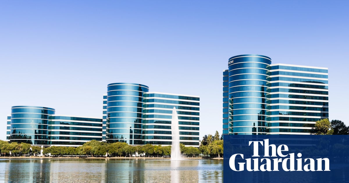
Oracle became an important tech player creating software for Fortune 500 firms around the world, but more recently found strength in cloud computing, having become the fastest-growing competitor to Amazon, Microsoft and Google. The surge in AI has also been a boon to the company, which has entered lucrative deals with the likes of OpenAI, the maker of ChatGPT.
However, there are also growing concerns about how reliant companies are becoming on each other’s financing within the AI ecosystem. Oracle said overnight that its measure of revenue from customer contracts rose by 440% over the past year, but analysts were wary when it emerged that the contracts were driven by new commitments from Meta and Amazon.
“Although these are two solid customers, it will not placate fears that big tech’s AI investments are becoming circular, which leaves it vulnerable to a loss of investor confidence,” Kathleen Brooks, a research director at XTB, said.
“Overall, strong contract growth was not enough to placate fears about AI and the huge amount of [capital expenditure] spending required by companies to build AI infrastructure.”
Other AI and tech-related stocks also slid in after-hours trading after the Oracle results. Nvidia’s share price fell by 1.3%, while Google owner Alphabet fell by 0.3%. In Japan, AI investor SoftBank’s shares fell by 7.7% on Thursday.