- Yen bid, dollar steadies as investors look for safety from global selloff Reuters
- Dollar hits fresh 9-1/2-month high vs yen Business Recorder
- Yen on defensive, dollar firms as traders dial back Fed rate cut bets Dunya News
- Japanese Yen struggles near multi-month low against firmer USD FXStreet
- The USDJPY is extending its gains-Analysis-18-11-2025 Economies.com
Category: 3. Business
-
Yen bid, dollar steadies as investors look for safety from global selloff – Reuters
-

Workplace Safety in Australia: A Strategic Guide for Regulated Sector Employers : Clyde & Co
Australia’s work health and safety (WHS) laws impose strict duties on employers, officers, and workers to ensure the health and safety of everyone in the workplace. Understanding these duties and applying them through robust governance frameworks is essential – particularly in high-risk environments such as aviation, shipping, mining, and construction, or in highly regulated industries such as banking, insurance, and superannuation.
Regulatory Framework
Australia operates under a harmonised WHS regime in most jurisdictions, based on the Model Work Health and Safety Act and Regulations. However, key differences exist in Victoria and Western Australia, which retain separate legislative schemes.
Primary federal legislation (harmonised jurisdictions):
- Work Health and Safety Act 2011 (Cth) — applies to Commonwealth agencies and certain national workplaces (including Comcare-regulated entities).
- Equivalent state/territory WHS Acts — largely aligned with the model WHS laws in NSW, QLD, SA, TAS, ACT, and NT.
Non-harmonised jurisdictions:
- Occupational Health and Safety Act 2004 (Vic)
- Work Health and Safety Act 2020 (WA) — partially harmonised but with notable variations.
Duties and Responsibilities
For Employers
A person conducting a business or undertaking (PCBU) must:
- Ensure, so far as reasonably practicable, the health and safety of workers and others affected by work.
- Implement safe systems of work, provide training, maintain safe plant and structures, and control workplace risks.
For Officers (Directors, Executives)
- Exercise due diligence to ensure the PCBU complies with WHS obligations, including:
- Staying informed about WHS matters
- Ensuring appropriate resources and processes for risk management
- Verifying compliance
- For officers and boards operating in banking, insurance, and superannuation industries, additional obligations may arise under the Australian Prudential Regulatory Authority (APRA) Prudential Standards: CPS 220 (Risk Management) and CPS 510 (Governance).
For Workers
- Take reasonable care for their own safety
- Follow lawful and reasonable instructions
- Use personal protective equipment (PPE) and report hazards
State and Federal Legislative Comparison Table
Jurisdiction Primary Legislation Notable Differences
Commonwealth (Comcare) WHS Act 2011 (Cth) Applies to Commonwealth agencies & self-insured licensees; strong focus on officer due diligence
NSW, QLD, SA, TAS, ACT, NT WHS Acts based on Model WHS Law Mostly uniform; high penalties for Category 1 offences
VIC OHS Act 2004 (Vic) No “PCBU” concept; duties owed by “employers” and “self-employed”; industrial manslaughter provisions apply
WA WHS Act 2020 (WA) Retains unique elements for mining and petroleum; transitional arrangements still in effect
Recommendations for Avoiding WHS Breaches
- Integrate WHS into Board Governance — align WHS KPIs with prudential and insurance risk metrics.
- Regular Safety Audits — tailored to industry-specific hazards (e.g., confined spaces, aviation maintenance, port operations).
- Contractual Risk Allocation — ensure WHS obligations are clearly allocated and monitored in contracts.
- Training and Competency Verification — especially for high-risk tasks and remote operations.
- Crisis and Incident Response Planning — cross-functional plans covering WHS, regulatory notifications, and insurance claims.
Final word
WHS compliance is not just about avoiding prosecution — it is about safeguarding your workforce, your reputation, and your licence to operate. Embedding safety into your governance, culture, and operational processes will deliver long-term resilience and protect against regulatory and commercial risk.
Clyde & Co’s Corporate Advisory team is working with global clients to align contracts, policies, and operational practices with Australia’s new Right to Disconnect laws. We support legal compliance, deliver global communications training, and help manage cross-border cultural expectations in a post-reform environment.
For further advice, contact the team at Clyde & Co.
Continue Reading
-
Australian wage growth steadies in Q3 driven by public sector gains – Reuters
- Australian wage growth steadies in Q3 driven by public sector gains Reuters
- Live: Wages growth data quashes rate cut hopes Australian Broadcasting Corporation
- Australian Dollar holds losses following Q3 Wage Price Index data FXStreet
- Wages And Salaries Reach Seasonal Peak Of $108.8 Billion: Australia Mirage News
- Wages growth running out of steam after inflation spike The Armidale Express
Continue Reading
-

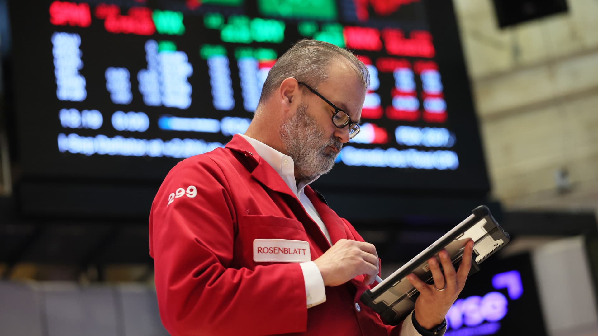
The flow of money in AI appears one-way at this point
The Anthropic website on Friday, Aug. 22, 2025.
Gabby Jones | Bloomberg | Getty Images
Money keeps flowing into artificial intelligence companies but out of AI stocks.
In what looks like — once again — a scenario of the left hand scratching the right, Microsoft and Nvidia will be investing a combined $15 billion into Anthropic, while the OpenAI competitor has committed to buying compute power from its two newest stakeholders. At this point, it seems as if a big proportion of AI news can be summarized as: "Company X invests in Company Y, and Company Y will buy things from Company X."
Okay, that's unfair. There are a lot of developments in the AI world that are not about investments but, well, development. Google unveiled the third version of Gemini, its AI model, which Demis Hassabis, CEO of Google's AI unit DeepMind, said "will be "trading cliché and flattery for genuine insight." (But I still want an AI chatbot to compliment me on my curiosity when I ask how to cut a pear, so I'm not sure if that's a pro for me.)
Investors, however, still appear skeptical about AI. Major names such as Nvidia, Amazon and Microsoft tumbled Tuesday stateside, giving the S&P 500 its fourth straight session in the red — the longest decline since August.
And if Nvidia — "the top company within the top industry within the top sector," as CFRA's chief investment strategist Sam Stovall puts it — fails to satisfy investors' expectations when it reports earnings Wednesday, we might be seeing the S&P 500's slide extend.
What you need to know today
The S&P 500 falls for a fourth consecutive day. Other major indexes also moved lower Tuesday stateside, while bitcoin prices dropped below $90,000 before recovering. Europe's regional Stoxx 600 sank 1.72% and touched its lowest level in a month.
Anthropic signs deal with Microsoft and Nvidia. Microsoft announced Tuesday it will invest up to $5 billion in the startup, while Nvidia will put in up to $10 billion. That puts Anthropic's valuation around $350 billion, according to a source.
Google announces its latest AI model Gemini 3. Alphabet CEO Sundar Pichai said Tuesday it will require "less prompting" for desired answers. The update comes eight months after Google introduced Gemini 2.5, and will be rolled out in the coming weeks.
U.S. Senators urge investigation into Trump-linked crypto firm. World Liberty Finance, heavily owned and run by the Trump family, sold tokens to a North Korean hacking organization, an Iranian crypto exchange and others, according to a corporate watchdog.
[PRO] Potentially resilient stocks amid AI slump. There are some global stocks and non-equity assets that could weather the turbulence in U.S. tech names happening recently, strategists told CNBC.
And finally...
Oleksii Liskonih | Istock | Getty Images
Diplomatic spat between Tokyo and Beijing threatens Japan's already fragile economy
Miffed over Japanese Prime Minister Sanae Takaichi's comments related to Taiwan, China on Friday advised its citizens against travelling to the country. Japanese tourism-exposed stocks fell in the aftermath of that warning, while experts caution the impact could be more severe over a longer duration.
Takahide Kiuchi, executive economist at Nomura Research Institute, said tensions between the two Asian powers could result in a 1.79 trillion yen drop in Japan's GDP over the course of one year — a 0.29% decline in the country's GDP.
— Lim Hui Jie
Continue Reading
-

Why it’s not too late to buy Alphabet’s stock, according to the newest Google bull
By Emily Bary
Alphabet’s stock has already doubled off its 2025 lows, but an analyst is still upbeat about Google’s resilience in search and its growing presence in semiconductors
Alphabet’s cloud business can dramatically outgrow Amazon’s through 2027, according to a Loop Capital analyst.
The hottest “Magnificent Seven” stock of the year could keep climbing, according to an analyst who just turned bullish on Alphabet shares.
Rob Sanderson of Loop Capital said Alphabet (GOOG) (GOOGL) has efficiently climbed the “wall of worry,” putting to bed fears that dogged the stock earlier this year. Sanderson is admittedly late to the game, with Alphabet’s stock roughly doubling off its 2025 lows and leading the Magnificent Seven group of megacap tech names with its 51% year-to-date gain, but he sees plenty of catalysts that could drive further price appreciation.
For one, the Google search business looks “as healthy as ever” – despite worries earlier this year about the threat posed by OpenAI’s ChatGPT.
Don’t miss: These 13 tech stocks have grown profits rapidly – and they’re still on sale
“Continued strength is disproving concerns (or at least postponing concerns) that AI chatbots are encroaching on Google as a primary starting point for the information seeking journey of users,” Sanderson wrote. “Strength is impressive considering Amazon pulled out of search ads in July and outsized growth in insurance as a category was expected to normalize” against tougher comparisons.
And Alphabet certainly isn’t sitting still when it comes to its own artificial-intelligence efforts, including with its Gemini large language model. “Through the year, Gemini has closed or overtaken leading modelbenchmarks in key areas and the company is now flexing its distribution muscle,” Sanderson noted.
Now excitement is building for Gemini 3.0, the next iteration of the model, which launched Tuesday.
See more: Google’s Gemini 3 is finally here. Can it power Alphabet’s stock even higher?
That said, there are risks to Alphabet’s moves in AI. Even its CEO sees some “irrationality” in the current AI mania. “I think no company is going to be immune, including us,” he told BBC when asked how Google would fare if the AI bubble burst.
So far, AI has been a boon to Alphabet’s cloud business, which is another element of the Google story that excites Sanderson. He thinks that unit alone is worth more than $1 trillion to Google. The business “has been on a big upswing,” with its backlog more than doubling over a five-quarter period, he noted.
Google Cloud should be able to “meaningfully outgrow” Amazon’s (AMZN) AWS cloud business through 2027, according to Sanderson, though he acknowledged that Alphabet’s performance is coming off a revenue base less than half the size of its major rival’s.
He is also upbeat about Alphabet’s custom chips known as tensor processing units, which he sees “becoming a big deal” for the company and within the industry, while helping Google stand out to cloud customers.
“Impressive price-performance benchmarks are making the Google TPU a differentiated driver for the cloud business as the company makes this internal advantage on compute economics available to third-party customers,” Sanderson wrote.
He moved to a buy rating from a prior hold stance on Alphabet’s stock. Sanderson’s $320 target price is 12% above current levels.
Read: One of Warren Buffett’s last moves as Berkshire CEO was to buy this ‘Magnificent Seven’ tech stock
-Emily Bary
This content was created by MarketWatch, which is operated by Dow Jones & Co. MarketWatch is published independently from Dow Jones Newswires and The Wall Street Journal.
(END) Dow Jones Newswires
11-18-25 1947ET
Copyright (c) 2025 Dow Jones & Company, Inc.
Continue Reading
-

Gold Steadies as Investors Weigh Stock Jitters, Rate-Cut Outlook
Gold steadied as investors weighed a decline in global equities, unease over lofty tech valuations and fading expectations of an interest-rate cut in the US.
Bullion was trading around $4,070 an ounce, having ended the previous session up 0.6%. A high-stakes earnings report from Nvidia Corp. due Wednesday will test investors’ nerves over stocks linked to artificial-intelligence developments. While gold often performs well when investors seek refuge from market turmoil, it can also suffer in the short term as traders are forced to unwind leveraged positions.
Continue Reading
-
Complex and Interconnected Risk: The BCI Horizon Scan 2025
The Business Continuity Institute (BCI) in association with Conducttr, has published its Horizon Scan report 2025, the eagerly anticipated annual study analyzing the top risks and threats that organizations have faced over the past 12 months, and those that are expected to sit at the top of the agenda over the coming years.
Organizations are facing risks and disruptions that are more complex and interconnected than ever before, spanning digital, environmental, operational, and human dimensions, often occurring simultaneously and compounding one another.
Along with cybersecurity threats, technological failures, climate-related events, and supply chain vulnerabilities these all have the potential to disrupt operations, affect service delivery, and erode stakeholder confidence.
The report examines these trends, combining findings from both quantitative survey data and qualitative interviews with practitioners on the front lines of business continuity and resilience. Their insights highlight that efforts towards improved response measures and better compliance should be considerate of the health and safety of the workforce.
Safety and IT
In the past 12 months safety incidents and IT-related disruptions rank very highly, a trend that has continued for the past five years. Safety incidents come top, closely followed by cyberattacks, fraud/attempted fraud, security incidents, and IT and telecom outages. Interestingly, extreme weather is the single largest cause of disruption over the past 12 months for the first time since 2017.
Looking at the next 12 months, the top five concerns also have a distinct technology flavour. In order, they are: cyberattacks, extreme weather events, IT and telecom outages, data breaches, and third-party failure / critical infrastructure failure.
This mix of disruptions have a broad impact, affecting operational performance, staff morale and customer experience. Consequences affect both internal and external stakeholders, with growing concerns over staff mental health.
The near future outlook
The future threat landscape is a complex combination of digital challenges, climate risk and geopolitical uncertainty. In many cases, the adoption of digital solutions represents both an opportunity and a potential risk for organizations. The top concerns for organizations over the next five to 10 years are cyber security: 63.6%, climate risk: 40.7%, the role of AI: 30.5%, geopolitical changes: 28.8%, and supply chain issues: 26.3%.
Trend analysis
Trend analysis remains a key tool for understanding emerging threats and guiding resilience planning. Just under half of organizations conduct trend analyses by a central corporate function (47.9%), with 23,5% having a decentralized approach across departments. A fifth (21.1%) don’t conduct these at all.
To conduct trend analysis, organizations rely on a combination of internal assessments and external insights to inform their risk landscape understanding. Interpersonal interaction remains a valuable option to practitioners. Internal risk and threat assessments came in at 87.2%, with external reports and industry insight (such as this report) a close second (75.2%). In the interpersonal interaction bucket come participation in industry events and conferences at (55.6%) and collaboration with peers at 52.1%.
The report also covers some of the legislation approaches – the relationship of BCM programmes to ISO 22301, the benefits of ISO 22301 Certification, and takes a look at investment levels in business continuity & resilience programmes.
Belen Santa-Olalla, Chief Creative Officer at Conductrr, commented, ‘We are proud to support the BCI Horizon Scan Report 2025. Its findings echo what we see every crisis: resilience depends on people, not paperwork. When pressure rises, it is human clarity, confidence, and connection that carry organizations through. That is why we focus on helping teams rehearse real situations in safe, authentic and realistic ways. This report is a reminder that preparation only becomes powerful when people are given space to practise, learn, and grow together.’
Download the report
More on
Continue Reading
-

UK pay settlements rise to highest in 2025, Brightmine says
LONDON, Nov 19 (Reuters) – Median pay settlements granted by British employers in the three months to the end of October rose to their highest so far this year at 3.3%, up from 3% in the three months to September.
Brightmine said the move reflected higher public sector pay deals which took effect in August and September.
Sign up here.
“Early indications suggest that 2026 pay awards are likely to remain steady – and potentially edge lower – as cost pressures continue to weigh on employers,” Brightmine manager Sheila Attwood said.
- Median public sector pay award 3.8% versus 3% in private sector
- 53% of pay awards were below 2024 levels, while 13% were above
- Survey based on 24 pay awards covering over 460,000 employees which took effect between August 1 and October 31
- 44% of employers said pay awards fell short of employees’ expectations
- The Bank of England is closely monitoring wage growth for signs of inflation pressure in the economy
Reporting by David Milliken; editing by Suban Abdulla
Our Standards: The Thomson Reuters Trust Principles.
Continue Reading
-
The request could not be satisfied
ERROR: The request could not be satisfied
The request could not be satisfied.
Request blocked.
We can’t connect to the server for this app or website at this time. There might be too much traffic or a configuration error. Try again later, or contact the app or website owner.
If you provide content to customers through CloudFront, you can find steps to troubleshoot and help prevent this error by reviewing the CloudFront documentation.
Generated by cloudfront (CloudFront)
Request ID: uBHgqv_idXmM99VUHVLiibLxWv1kFvtY7nd32GDNA_2F38bcZUQaKQ==
Continue Reading