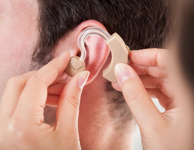
Category: 8. Health

  • Understanding how mRNA stability influences immune diseases

    Understanding how mRNA stability influences immune diseases

    A pizza shop with 30 delivery people ought to be able to deliver a lot of pizzas – if their cars don’t break down on the way. Likewise, genes that produce a lot of messenger RNA (mRNA) molecules can build a lot of proteins – if these molecules don’t fall apart before the job gets done.

    Inside almost every human cell is DNA, a comprehensive instruction manual for building and maintaining the body. Genes in that manual contain the instructions for making proteins. But those instructions must travel from the cell’s nucleus, where the DNA lives, to the outer region of the cell – the cytoplasm – where proteins are actually made.

    That’s where mRNA comes in. Like a messenger, it copies the instructions from the DNA in the nucleus and carries them out to the protein-making machinery. More mRNA typically means more protein – unless the mRNA is unstable and breaks down too quickly. 

    “Every mRNA has to die in the end,” says Xinshu Xiao, a professor of integrative biology and physiology at UCLA and senior author of a new paper published in Nature Genetics. “It’s produced, it does its job, and then it’s destroyed. But most research has focused on how mRNA is made. Much less attention has been paid towards how fast it’s degraded – and that’s just as important.”

    Both the production and stability of mRNA can be affected by mutations in the DNA, which are commonly referred to as genetic variants. These variants can affect how much protein a cell makes, and in turn, influence a person’s risk of disease. But figuring out whether a variant affects how much mRNA is made – or how long it survives – has been a major challenge.

    Led by UCLA doctoral student Elaine Huang, Xiao’s team developed a computational tool called RNAtracker, which is freely available. The software allows researchers to pinpoint whether a gene is being regulated through changes in mRNA production or in mRNA stability. In pizza terms: Is the problem that not enough pizzas are being made or that the delivery cars are breaking down? RNAtracker helps scientists trace the breakdown.

    The researchers applied RNAtracker to a publicly available dataset of 16 human cell lines, in which newly made mRNAs had been chemically labeled and tracked over time. This allowed them to identify genes whose stability varies due to specific mutations. Many of these genes were involved in immune system function – especially the innate immune system, the body’s first line of defense against infections.

    The team also found that several of the genetic variants linked to unstable mRNA had already been associated with autoimmune diseases in large-scale genetic studies.

    One insight from this project is that some disease-associated variants may be acting through effects on mRNA stability.”


    Xinshu Xiao, professor of integrative biology and physiology, UCLA 

    Using additional modeling, the researchers linked expression levels of these stability-regulated genes to diseases including allergic rhinitis, lupus, diabetes mellitus and multiple sclerosis. The findings suggest that mRNA stability – long overlooked – may be a key mechanism behind many immune-related diseases.

    “Basic research like ours shifts the paradigm of what people focus on,” said Huang. “For drug developers or researchers working on treatments, you can’t target what you don’t know is important. We are trying to bring attention to genetic variants that affect mRNA stability, which hasn’t gotten the spotlight it deserves.”

    The research, which received funding from the National Institutes of Health, used publicly available data generated by ENCODE, an NIH-supported consortium.

    “The NIH plays a critical role by supporting large-scale efforts like ENCODE,” said Xiao. “They make it possible for researchers around the world to access massive datasets and make discoveries like ours.”

    Additional authors include: Ting Fu, Ling Zhang, Guan’ao Yan, Ryo Yamamoto, Sari Terrazas, Thuy Linh Nguyen, Carlos Gonzalez-Figueroa, Armen Khanbabaei, Jae Hoon Bahn, Rajagopal Varada, Kofi Amoah, Jonatan Hervoso, Michelle Paulsen, Brian Magnuson, Mats Ljungman and Jingyi Jessica Li.

    Source:

    University of California – Los Angeles

    Journal reference:

    Huang, E., et al. (2025) Genetic variants affecting RNA stability influence complex traits and disease risk. Nature Genetics. doi.org/10.1038/s41588-025-02326-8

    Continue Reading

  • Just 1 dose of LSD could relieve anxiety for months, trial finds

    Just 1 dose of LSD could relieve anxiety for months, trial finds

    A single dose of the psychedelic drug LSD may ease generalized anxiety disorder for months, a clinical trial has found.

    The trial results, published Thursday (Sept. 4) in JAMA, include data from 194 people with moderate to severe anxiety across the U.S. The study compared these participants’ responses to different doses of LSD against a placebo treatment. It found that the drug alleviated symptoms in many patients for at least three months after just one exposure.

    Continue Reading

  • How mechanical changes in lung tissue trigger fibrosis

    How mechanical changes in lung tissue trigger fibrosis

    Fibrosis of the lungs is often a silent disease until it’s too late. By the time patients are diagnosed, the scarring of their lung tissue is already advanced, and current treatments offer little more than a slowing of the inevitable. But what if we could understand the very first steps of this disease before irreversible damage sets in?

    That’s the question Claudia Loebel, Reliance Industries Term Assistant Professor in Bioengineering, and Donia Ahmed, a doctoral student in Loebel’s lab, set out to answer. Their Nature Materials paper, a collaborative study spanning the University of Pennsylvania, the University of Michigan and Drexel University, explores how subtle changes in the mechanical environment of lung tissue might set off the chain reaction that leads to fibrosis.

    A fresh approach to an intractable disease

    Lung fibrosis is notoriously difficult to diagnose and treat. 

    Once it’s diagnosed, patients only have two FDA-approved drugs, and both just slow down the disease. They don’t stop it or reverse it. What’s worse is that we often don’t know what caused it in the first place, so we also don’t have a clear idea of how to prevent it.”


    Claudia Loebel, Reliance Industries Term Assistant Professor in Bioengineering

    Much of the research to date has focused on the later stages of the disease, when tissue has already stiffened and scarred. Loebel and Ahmed decided to flip the script, examining instead what happens right at the onset. Specifically, they looked at how tissue stiffness alone might influence cell behavior in the lungs, offering a new window into fibrosis as it unfolds.

    Lighting up the problem

    Using a technique called photochemical cross-linking, the researchers exposed lung tissue to blue light, which triggered the extracellular matrix – the fibrous scaffolding surrounding cells – to stiffen. Unlike traditional UV light, blue light is gentler on living cells, making it ideal for studying live tissue.

    With these flashes of blue light, the team was able to localize the stiffening of tissue in both healthy mouse and human lung tissue.

    “Think of the extracellular matrix like loose hair in a ponytail,” says Ahmed. “With light-triggered cross-linking, we braid it, stiffening the tissue just enough to mimic the kind of micro-injuries that might trigger fibrosis.”

    What makes this approach unique is that the team didn’t use engineered gels or decellularized tissue. Instead, they worked with intact, living tissue samples. That preservation of natural cellular and matrix interactions makes their technique a powerful tool for understanding real-time responses to mechanical changes in the lung.

    Cells that sense and get stuck

    As the tissue stiffened under the light, Ahmed observed that cells began to stretch out, changing shape. And, it wasn’t just cosmetic. This physical stretching was a sign that the cells were transitioning into a different cell type. 

    But then they stalled.

    “These cells were caught in a sort of identity crisis,” says Ahmed. “They were stuck between types, unable to perform either role well. And that’s a problem.”

    These “transitional” cells have been spotted before in fibrotic tissue samples, both in mice and humans. What hasn’t been understood, until now, is how they get there.

    Loebel and Ahmed’s model suggests that changes in tissue stiffness alone can prompt cells to begin transitioning, and when they get stuck, they contribute to the very stiffness that triggered them – setting up a potential feedback loop that accelerates disease.

    Imagine a child’s play tunnel: when it’s soft and flexible, it’s easy to crawl through. But once it becomes rigid, movement and communication amongst individuals become difficult. Similarly, in a stiffened extracellular matrix, cells can get trapped, lose their function and change shape. Worse, they may attract other “bad” cells that thrive in rigid environments, compounding the damage.

    A mechanical problem with biological consequences

    While the biology of fibrosis has long been studied, this project reframes the disease as a problem of mechanics. 

    “I love thinking about this from a mechanical engineering perspective,” says Ahmed. “It’s not just about chemical signals. The physical environment matters deeply.”

    To measure just how stiff the tissue had become, the team used a nanoindenter, an engineering tool typically reserved for testing materials like plastics or metals. They applied it to biological tissue, providing precise data about how stiffness changes in real time.

    “We are uniquely positioned to tackle this problem due to our expertise in both engineering and biology,” says Matthew Lee Tan, co-first author and former postdoctoral fellow at the University of Michigan. “This lets us identify opportunities to apply engineering tools to study disease and uncover new biological insights.”

    This interdisciplinary approach, combining tools from engineering, insights from biology and models built on real human tissue, reflects the collaborative spirit of Penn Engineering’s scientific ecosystem.

    Where to from here?

    The team’s leading hypothesis is that these early-responding cells, once stuck in a transitional state, lay the groundwork for fibrosis to progress. They not only lose their original function but actively stiffen the tissue around them, making the environment more attractive to fibrosis-promoting cells.

    And while this study focused on epithelial cells – those at the interface between lung tissue and air – the researchers plan to expand their work to other key players in fibrosis: macrophages, fibroblasts and neutrophils.

    “This is just the first step,” says Loebel. “Now that we’ve built this tool, we can use it to look at cell-specific contributions to fibrosis, not just in the lungs, but potentially in other organs like the liver or skin, where fibrosis also causes major health problems.”

    A blueprint for future therapies

    Ultimately, the hope is that by understanding how stiffness affects cells in the earliest phases of fibrosis, scientists and doctors can better predict who’s at risk and when to intervene.

    “We’re not trying to recreate fibrosis in the lab,” Loebel says. “We’re identifying its starting point. If we can understand the first responders, we can work toward treatments that prevent the entire cascade from happening.”

    Source:

    University of Pennsylvania School of Engineering and Applied Science

    Journal reference:

    Ahmed, D.W., et al. (2025) Local photocrosslinking of native tissue matrix regulates lung epithelial cell mechanosensing and function. Nature Materials. doi.org/10.1038/s41563-025-02329-0

    Continue Reading

  • ‘Three-tailed’ lipid helps cells survive during heart attack, stroke

    ‘Three-tailed’ lipid helps cells survive during heart attack, stroke

    When starved of oxygen during a heart attack or stroke, cells unleash a flurry of emergency measures to protect themselves and the body. For decades, scientists have observed that the body’s production of a “three-tailed” fat molecule consistently surges during this trauma but have puzzled over why. Now, Cornell researchers have uncovered its surprising role in cellular survival: protecting against damage when oxygen runs out.

    The research shows that the fat molecule, N-acylphosphatidylethanolamine (NAPE), helps cells survive ischemia – oxygen loss from reduced blood flow – by driving lactic acid out of cells. This toxic byproduct builds up during emergency metabolism, and NAPE’s surge appears to be part of the body’s protective response. Though still in an early stage, the findings suggest that boosting or mimicking NAPE could one day help limit tissue damage in heart attack and stroke.

    The study, published Sept. 5 in the Journal of the American Chemical Society, was led by graduate student Din-Chi Chiu and Jeremy Baskin, associate professor and Nancy and Peter Meinig Family Investigator in the Life Sciences in the Department of Chemistry and Chemical Biology in the College of Arts and Sciences, and the Weill Institute for Cell and Molecular Biology.

    “During heart attack or stroke, when there is an interruption in blood flow, the cells in the affected tissue, whether it is the heart or the brain, have to scramble to be able to continue to produce energy to survive,” Baskin said.

    Under normal conditions, cells largely produce energy by a longer and much higher yielding process involving mitochondria.

    “However, when energy needs are imminent and oxygen is limited, such as when blood flow is restricted, a metabolic switch occurs to favor glycolysis, which is a quick and dirty way of generating energy,” he said. “But to keep glycolysis going unabated, lactate, or lactic acid, is built up, and because it can be toxic at high levels, cells need to export it to prevent it from building up inside cells to undesirable levels.”

    Because NAPE repels water and is short-lived in cells, studying it directly has been nearly impossible. The research team overcame this by designing and synthesizing a chemical “look-alike” probe that tagged NAPE’s protein partners under UV light, revealing its interactions.

    The researchers observed NAPE latching onto proteins that regulate lactate transport. In particular, it bound to two cell-surface proteins, CD147 and CD44, which control transport proteins that act like gates controlling how lactic acid moves in and out of cells. The team’s experiments showed that when NAPE levels rise, lactate transport ramps up, and blocking those transporters erased the effect.

    “The work reframes NAPE as a signaling molecule,” Baskin said. “Our finding that NAPE can stimulate lactate export supports a model in which the role of NAPE in pathological events such as heart attack or stroke is part of a protective response.”

    For now, the team is exploring whether different versions of NAPE, with different tail compositions, might fine-tune lactate regulation in unique ways. They are also interested in whether NAPE plays roles in other tissues beyond the heart and brain.

    “Future studies in heart and brain tissue will test this hypothesis more directly,” Baskin said. “If confirmed, the work could support the creation of therapies that boost NAPE levels as a way to limit tissue damage in cardiovascular emergencies.”

    Other contributors to the research were graduate student Yuan-Ting Cho, a member of Baskin’s lab at the Weill Institute, and Hening Lin, professor of medicine and chemistry at the University of Chicago.

    This research was supported by the National Institutes of Health and by the Howard Hughes Medical Institute.

    Stephen D’Angelo is the communications manager for biological systems at Cornell Research and Innovation.

    Continue Reading

  • COVID vaccine myth-busting can work, study suggests

    COVID vaccine myth-busting can work, study suggests

    A COVID-19 misinformation-busting messaging intervention that presented a myth followed by a fact among people who had completed the initial vaccine series strengthened their resolve to follow up with a booster dose, suggests a University of Pennsylvania study published last week in Vaccine.

    For the randomized controlled experiment, the researchers randomly assigned 892 racially diverse US adults with vaccine safety concerns to receive no message (control arm) or one of three message types: (1) a myth followed by fact, (2) a fact followed by a myth and the fact again, (3) or a fact only. The team then surveyed participants about their intent to receive the initial COVID-19 vaccine series or a booster in the next 3 and 12 months.

    Concerns about turning people off to vaccination

    Participants’ ages ranged from 18 to 79 years (average, 36 years), about half were men, a fifth were Hispanic, 40% were non-White, a quarter were Republicans, and a third were Democrats.

    The myths were: “The COVID-19 vaccine causes infertility,” “It’s safer to get COVID-19 than to get the COVID-19 vaccine,” and “The effectiveness and safety of the COVID-19 vaccine cannot be trusted.”

    “Attempts to correct misinformation often use one of three common message structures,” the researchers wrote. “The effectiveness of these message structures is unclear, and concerns have been raised that some can ‘backfire’ by weakening vaccination intentions.”

    For example, some experts believe that the traditional myth-fact approach used by many public health organizations throughout the COVID-19 pandemic may inadvertently reinforce the myth by repeating it. 

    “People may have difficulty recalling whether the information was categorized as the myth or fact because the context is forgotten,” the study authors wrote. “In addition, people show an increased liking for stimuli when they are exposed to them more often.”

    No impact on booster intent in unvaccinated 

    Of 531 participants who had received zero or one COVID-19 vaccine doses, 65% said they had no intention of receiving a dose in the next 12 months, 28% reported weak intentions, and 7% reported strong intentions. Of 361 recipients of two or more doses, 16% said they had no intention of getting a booster within the first 12 months it was available to them, 37% reported weak intentions, and 48% indicated strong intentions.

    Exposure to the myth-followed-by-fact message was associated with stronger booster intentions among participants who had received an initial COVID-19 vaccine dose than those among controls. Of those who hadn’t received the initial vaccine series, the strength of intent to begin vaccination was the same across all study groups. 

    Receiving one COVID-19 vaccine dose (compared with zero), higher financial stress, and a Democratic political affiliation were significantly related to stronger intentions to receive the vaccine in the next 12 months. 

    Among participants who had completed the initial two-shot series, the intention to receive a COVID-19 booster in the first 12 months it was available to them was stronger among those who viewed the myth followed by the fact than in those who received no message. The fact-myth-fact and fact-only messages didn’t significantly strengthen or weaken booster intentions. 

    Probably not reinforcing the myth

    “In this study, only one of the three debunking message structures was effective, but none had counterproductive consequences, despite fears that they ‘backfire,’” the researchers wrote. “Therefore, the results did not suggest the messages repeating the myth are reinforcing the myth.” 

    Continue Reading

  • Children with sickle cell disease face low dental care utilization despite elevated risks

    Children with sickle cell disease face low dental care utilization despite elevated risks

    Children with sickle cell disease face low dental care utilization despite elevated risks | Image Credit: © Wat.Pt – stock.adobe.com.

    Children with sickle cell disease (SCD) are more likely to experience dental complications, yet fewer than half of those enrolled in Michigan Medicaid received dental care in 2022, according to a new study published in JAMA Network Open.1,2

    The study, led by researchers at Michigan Medicine and the RAND Corporation, examined Medicaid dental claims for 1,096 children with SCD and compared them with claims from more than 1.18 million Medicaid-enrolled children in Michigan. Despite increased risk for oral health problems, children with SCD had dental care utilization rates comparable to the general Medicaid pediatric population.

    “Sickle cell disease is known to increase the risk of dental complications in children, which underscores the importance of preventive dental care for this population,” said senior author Sarah Reeves, PhD, MPH, an associate professor of pediatrics and epidemiology at the University of Michigan Medical School and the Susan B. Meister Child Health Evaluation and Research (CHEAR) Center.

    Study design and findings

    Using validated case definitions from the Michigan Sickle Cell Data Collection program, investigators analyzed dental claims for preventive services, treatment, and overall utilization. The analysis followed STROBE reporting guidelines and used Centers for Medicare & Medicaid Services data for comparison.

    Results showed that 42% of children with SCD received any dental services in 2022, compared with 44% of the general Medicaid population. Preventive dental care was received by 38% of children with SCD compared with 40% of peers without SCD. Treatment services were accessed by 15% of children with SCD versus 18% of other Medicaid-enrolled children.

    When analyzed by age group, some disparities emerged. For example, children aged 6 to 14 years with SCD were significantly less likely to receive any dental services (50% vs 54%) and preventive dental services (46% vs 51%) compared with their peers. In contrast, children aged 1 to 5 years with SCD had slightly higher utilization rates, although these differences were not statistically significant.

    Clinical significance

    The authors emphasized the importance of preventive dental care for children with SCD due to the disease’s reciprocal relationship with oral health. Dental infections can trigger or worsen SCD complications by causing inflammation and red blood cell sickling.

    “Oral infections are especially dangerous for people with sickle cell disease because they can trigger or worsen symptoms and serious complications,” Reeves said. “Preventative dental care helps reduce the risk of pain crises and hospitalizations.”

    Despite these risks, access barriers persist. Medicaid covers pediatric dental services, and guidelines recommend annual visits for children. However, both children with and without SCD in the study showed low utilization rates.

    Barriers and recommendations

    The study noted several barriers limiting dental care access for children with SCD, including limited participation of dentists in Medicaid programs and a lack of clear clinical guidance for treating these patients.

    “Our findings show that we need to take steps to make sure kids with this condition get the dental care they need—by helping dentists feel more prepared to treat them and making sure doctors know how important dental health is for these children,” Reeves said.

    Targeted interventions may include improved training for dental providers, stronger coordination between medical and dental professionals, and expanded research on structural barriers.

    “Given increased risk of oral health problems among patients with sickle cell disease we need focused efforts to expand access to dental services for them,” Reeves said. “There are many reasons why dental care rates are low in this group. Future research should look at what those barriers are and how we can overcome them to improve care for this vulnerable population.”

    The study highlights the need for improved strategies to ensure preventive dental care reaches children with SCD, underscoring its role in preventing serious health complications.

    References

    1. Michigan Medicine – University of Michigan. Children with sickle cell disease face higher risk of dental issues, yet many don’t receive needed care. Eurekalert. September 3, 2025. Accessed September 5, 2025. https://www.eurekalert.org/news-releases/1096502
    2. Kranz A, Peng HK, King AA, Clark SJ, Plegue MA, Reeves SL. Sickle Cell Disease and Dental Care Access Among Medicaid-Enrolled Youths. JAMA Network Open. 2025;8(9):e2529849-e2529849. doi:https://doi.org/10.1001/jamanetworkopen.2025.29849

    Continue Reading

  • 15 dead after new Ebola outbreak in the Democratic Republic of Congo – The Washington Post

    1. 15 dead after new Ebola outbreak in the Democratic Republic of Congo  The Washington Post
    2. Democratic Republic of the Congo declares Ebola virus disease outbreak in Kasai Province  WHO | Regional Office for Africa
    3. Caring for Patients in High-Consequence Infectious Disease Hot Zones  Contagion Live
    4. IOM Sends Team to Aid DRC Ebola Response  Mirage News
    5. Ebola Outbreak in Congo Tests Global Response After US Aid Cuts  Bloomberg.com

    Continue Reading

  • Learn to Use Remote Sensing for Climate-Sensitive Infectious Diseases

    Learn to Use Remote Sensing for Climate-Sensitive Infectious Diseases

    Many infectious diseases are sensitive to certain climates and thrive in others. Consider malaria, which is rife in warm, humid, wet climates, and practically nonexistent in areas with cold temperatures or high altitudes. Given the fact that many of these climate variables can be observed from space, this allows users to predict where certain disease outbreaks may occur.

    On October 7 and 9, 2025, NASA’s Applied Remote Sensing Training Program (ARSET) is offering a two-part, live, online training on the use of NASA satellite data for tracking climate-sensitive vector-borne disease outbreaks. Those who attend will learn about general approaches when applying satellite remote sensing data to studying or forecasting climate-sensitive infectious diseases. These will be illustrated with a case study example showing how remote sensing has been used to forecast malaria outbreaks. 

    This training will also present some common, freely available NASA remote sensing datasets used in these applications, as well as where and how to access them, and how to decide which datasets are fit for this purpose.

    This training is open to the public and is recommended for biostatisticians, medical students, vector ecologists, biologists studying disease vector organisms, public health officials, and non-governmental organizations (NGOs) tasked with monitoring and preparing for infectious disease outbreaks.

    Continue Reading

  • Health news round-up: cardiovascular research special – The Pharmaceutical Journal

    1. Health news round-up: cardiovascular research special  The Pharmaceutical Journal
    2. Drugs, diet and AI: the ‘gamechanger’ new findings on tackling heart conditions  The Guardian
    3. ESC Congress 2025 Recap: 8 Trials to Know  HCPLive
    4. ESC Congress broke new record with over 33,000 attendees  Cardiovascular Business
    5. The biggest stories out of ESC 2025  MassDevice

    Continue Reading

  • This Habit May Raise the Risk of Breaking a Bone

    This Habit May Raise the Risk of Breaking a Bone

    • Skipping breakfast raised fracture risk by 18% in nearly one million adults.
    • Eating dinner within two hours of bed was tied to an 8% higher fracture risk.
    • Simple habits—like a morning meal and earlier dinners—may help protect bones.

    Your breakfast routine—or lack of one—may have lasting consequences for your bones.

    A massive new Japanese study found that skipping breakfast and eating late dinners were each linked to a higher risk of osteoporotic fractures. The findings add to growing evidence from the field of chrononutrition—which explores how the timing of meals interacts with the body’s internal clock—suggesting that when you eat may matter for long-term health, not just what you eat. For example, researchers have linked eating earlier in the day to better cholesterol levels, lower insulin resistance and less body fat, all of which support the idea of eating in line with your natural circadian rhythm.

    That potential connection matters because fragile bones are already a widespread problem. Roughly 13% of U.S. adults age 50 and older have osteoporosis, a disease that weakens bones and makes them more likely to break. Another 43% in the same age group have low bone mass, often called osteopenia. Taken together, more than half of older Americans are living with reduced bone strength and a higher risk of fractures.

    Lifestyle habits like exercise, alcohol use and smoking are well known to influence fracture risk. What hasn’t been studied much is whether the timing of meals makes a difference. This study is one of the first to look at how skipping breakfast or eating late dinners might influence fracture risk. The results were published in the Journal of the Endocrine Society.

    How Was the Study Conducted?

    Researchers analyzed health records from nearly 1 million Japanese adults who took part in routine checkups. The study followed people age 20 and older, linking their lifestyle questionnaires with medical records from a large national claims database. On average, participants were tracked for about 2.6 years.

    They focused on two self-reported eating habits: skipping breakfast and having a late dinner. The team tracked 4 types of osteoporotic fractures—hip, forearm, spine and upper arm—and compared people who reported these behaviors and who did not.

    What Did the Study Find?

    During the study, researchers logged just over 28,000 major fractures. People who skipped breakfast more than three times a week were about 18% more likely to break a bone than those who ate it regularly. Eating dinner within two hours of bedtime more than three times a week was linked to an 8% higher fracture risk.

    The study also reinforced what health professionals already know—women, people with lower body weight and older adults are more prone to fractures. It also showed that meal timing habits often traveled with other choices like smoking, drinking, getting less exercise and sleeping less. Put together, the results suggest that lifestyle plays a big role in bone health—and that something as simple as when you eat may add to the risk.

    How Does This Apply to Real Life?

    Meal timing alone won’t make or break your bones, but it may be worth paying attention to. Starting the day with breakfast gives you a steady supply of nutrients your bones can use. Even simple options like yogurt with fruit, eggs on whole-grain toast or a smoothie made with milk and leafy greens provide protein and calcium without much effort.

    Dinner habits matter too. Leaving two to three hours between your last meal and bedtime gives your body time to digest and aligns eating with your natural rhythms. Pairing that with regular weight-bearing exercise—like light strength training—and getting enough sleep helps reinforce bone strength over time.

    And if you’re concerned about protecting your bone health, try to incorporate nutrients like calcium, vitamin D, protein and healthy fats into your diet. (Canned salmon can be a great source of all four.) 

    Our Expert Take

    This recent study in the Journal of the Endocrine Society doesn’t prove that skipping breakfast or eating late dinners directly cause fractures, but it highlights how everyday routines may add up in ways we’re only starting to understand. The foundation for bone health is still the same—nutrient-rich foods, physical activity, quality sleep and limited smoking and alcohol—but meal timing may be another piece of the prevention puzzle.

    Continue Reading