ROFLUMILAST for psoriasis improved Global Assessment scores and was well tolerated versus placebo by Week 8 in a systematic review.
Why Roflumilast for Psoriasis Was Studied
Psoriasis is a chronic inflammatory skin disease that can…

ROFLUMILAST for psoriasis improved Global Assessment scores and was well tolerated versus placebo by Week 8 in a systematic review.
Psoriasis is a chronic inflammatory skin disease that can…

Microplastics are everywhere, from the highest mountains to the deepest seas and even in rainwater, as recently detected in Jakarta, Indonesia. Now, for the first time, scientists have confirmed the presence of…
Allard, M. W. et al. Genomics of food-borne pathogens for microbial food safety. Curr. Opin. Biotechnol. 49(2), 224–229. https://doi.org/10.1016/j.copbio.2017.11.002 (2018).
Deng, L. & Wang,…
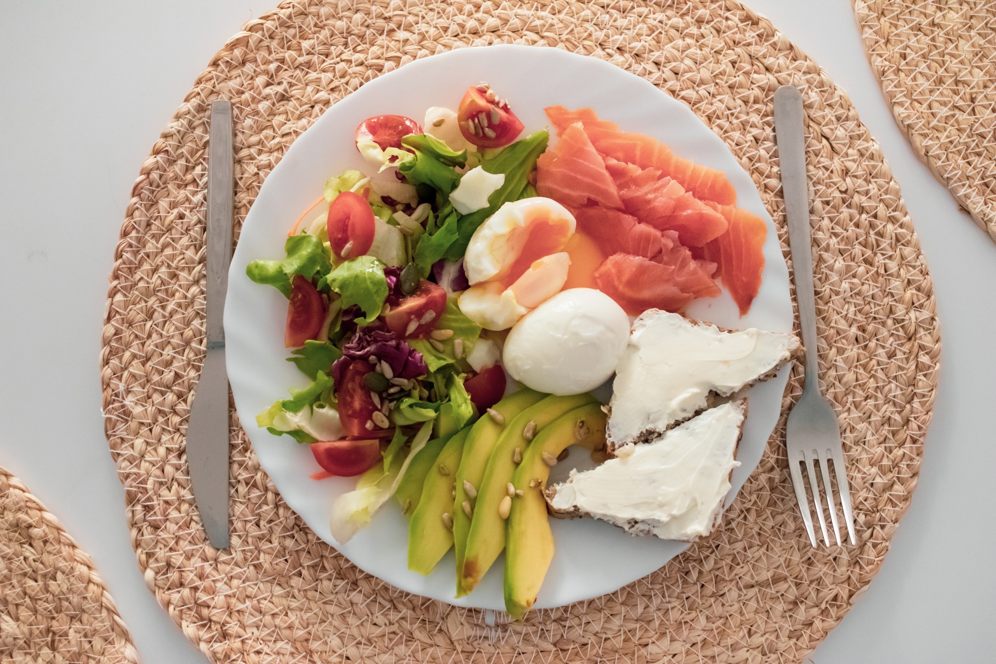
A controlled clinical trial shows that what you eat at breakfast, not just how much, may shape appetite, weight loss, and gut microbiota with implications for long term dietary strategies
Study: Big breakfast diet composition…

Large-scale U.S. population data suggest that red meat consumption patterns may be associated with diabetes prevalence, whereas alternative protein choices may confer metabolic advantages.
Study: Association Between Red Meat Intake…

Large international cohort data reveal that excess body fat may increase susceptibility to severe infections and substantially contribute to global infection mortality, underscoring prevention as a major public health priority.
Continue Reading

Researchers reveal why lung cancer in people who never smoked is increasing and explore how genetics, environmental exposures, and new screening strategies may help detect disease earlier and improve outcomes.
Opinion: Lung cancer…