The Cambodia Ministry of Health reports one case of human H5N1 avian influenza in a 30-year-old man from Meanrith village of Tuek Chhou district in Kampot province in southwest Cambodia.
The patient presented with symptoms of fever, cough and…

The Cambodia Ministry of Health reports one case of human H5N1 avian influenza in a 30-year-old man from Meanrith village of Tuek Chhou district in Kampot province in southwest Cambodia.
The patient presented with symptoms of fever, cough and…

When the CLEOPATRA trial results were first presented, they did more than show a statistical improvement. They reset expectations.
Before CLEOPATRA, HER2-positive metastatic breast cancer was considered aggressive but manageable….


NEOADJUVANT PEMBROLIZUMAB achieved a 71% pathological complete response rate in patients with resectable desmoplastic melanoma, meeting the prespecified primary endpoint in the phase 2 SWOG S1512 trial.

A TEAM of researchers has unveiled new insights into the functional and ecological diversity of the vaginal microbiome using a gene-based algorithm called VISTA (Vaginal Inference of Subspecies and Typing Algorithm). By linking metagenomic…
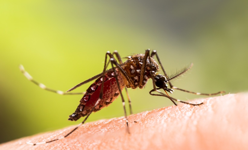
DENGUE infection raised two-year hospitalization risk and added measurable disability burden in Singapore’s linked national cohort.
A large retrospective cohort study used Singapore’s linked…
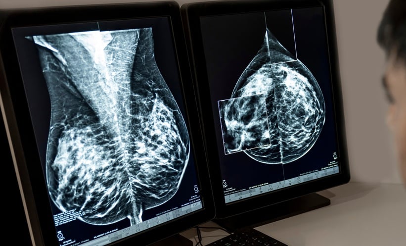
MAMMOGRAPHIC surveillance significantly lowers mortality among survivors of breast cancer and the use of adjunct imaging techniques may enhance early detection, a 2026 systematic review and meta-analysis has found.