Researchers have pinpointed a pressure-sensing switch on bone marrow stem cells that turns movement into new bone growth.
By targeting the switch, future medicines could strengthen fragile bones in people whose illness keeps them from moving much.
Researchers have pinpointed a pressure-sensing switch on bone marrow stem cells that turns movement into new bone growth.
By targeting the switch, future medicines could strengthen fragile bones in people whose illness keeps them from moving much.

How much do you know about living a long, healthy life? Most of us are aware of the basics… don’t smoke, don’t drink too much, eat plenty of fruit and veg (and avoid ultra-processed food), and get enough exercise. But researchers have just…
A woman has blamed Mounjaro for her ‘excruciating pain’ as she faces gallbladder removal surgery after using the weight loss drug.
In an article for The Telegraph, Catherine Hales from the UK opened up about how she had struggled with her weight…
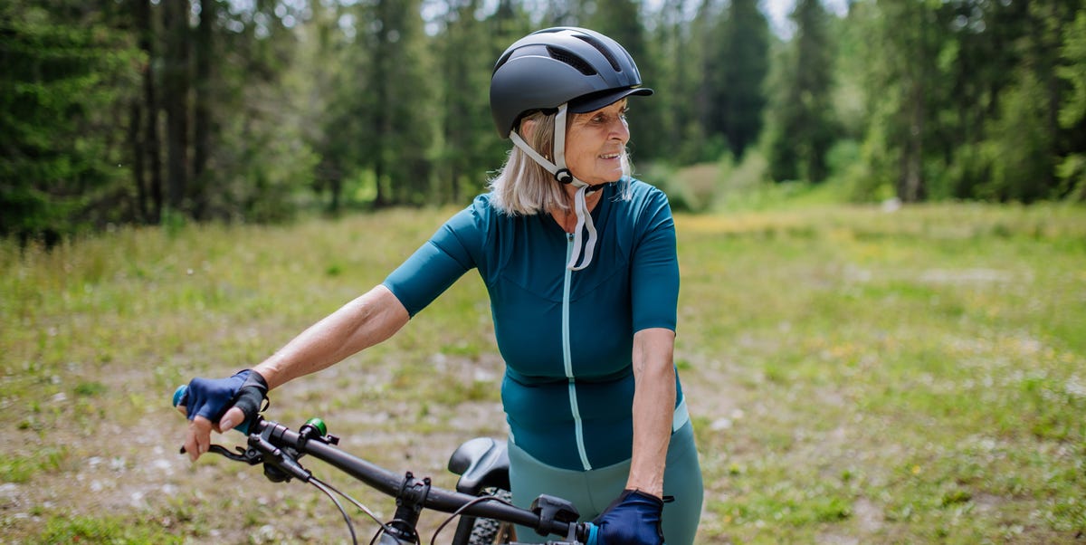
The benefits of strength training for women over 50 are vast; it is so important to maintain muscle mass with age. But strength training should be done alongside cardio for a well-rounded fitness routine. So, we asked fitness pros their top…

Lifestyle factors play a large role in living…

A research team led by scientists from several Japanese institutions has identified a compound called Mic-628 that directly influences the body’s internal timing system. The group included Emeritus Professor Tei H. (Kanazawa University),…
An expert has revealed the negative impacts not drinking enough water can have on your body, and it’ll make you want to ditch your morning coffee for a pint of water instead.
There’s a sweet spot of how much water you should be drinking a day,…