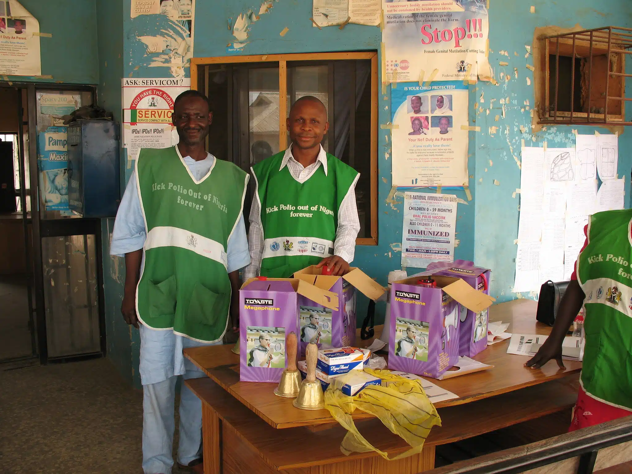
VANCOUVER, Canada — “We do this because we don’t want any child to go through what we did,” said Bello Dikko, the Chair of the Polio Survivors Association in Sokoto State. For him, the battle was personal. Locals like Dikko…
Category: 6. Health
-

The Most Comprehensive Genetic Risk Assessment, Combining Monogenic and Polygenic Insights Across Multiple Diseases
SAN FRANCISCO, Oct. 9, 2025 /PRNewswire/ — Allelica, the global leader in clinical polygenic risk score (PRS) testing, today announced the launch of AbsoluteDx, a comprehensive,…
Continue Reading
-

Somerset lung cancer self-referral trial extended
A scheme which allows patients to self-refer for a lung cancer X-ray is being extended after a successful trial.
Somerset NHS Foundation Trust said the service has been rolled out in the south of the county after being trialled in Bridgwater.
Continue Reading
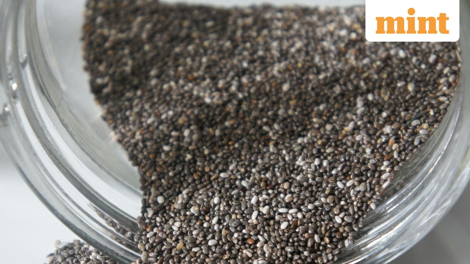
AIIMS-trained doctor explains what happens when you eat chia seeds for 14 days straight: ‘Gut health, slows digestion’
Chia seeds have become a staple in health-conscious diets worldwide, and now an AIIMS-trained gastroenterologist has explained why. In a recent video, Dr Saurabh Sethi, who has also trained at Harvard and Stanford Universities, broke down the…
Continue Reading
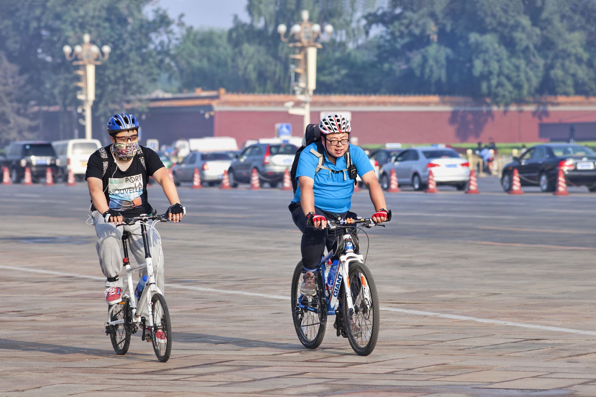
Air pollution tied to obesity and diabetes – AirQualityNews
Air pollution could make people more prone to obesity and diabetes, according to a new international study led by scientists at the University of Zurich (UZH) and Case Western Reserve University.
The…
Continue Reading
expert reaction to study on the health of the offspring of rats exposed to e-cigarette vapour
A study published in the Journal of Physiology looks at the health of the offspring of rats exposed to…
Continue Reading
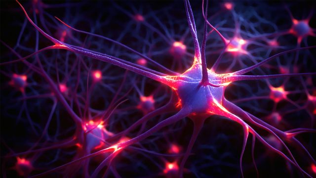
Seizures in MS Linked to Hippocampal Demyelination
A preclinical study by biomedical scientists at the University of California, Riverside, has shown why some people with multiple sclerosis, or MS, also suffer from seizures — a debilitating complication that can worsen cognitive outcomes…
Continue Reading