- National pharmacy stop-smoking service could support more than 200,000 quit attempts annually The Pharmaceutical Journal
- Fewer than half of councils fund pharmacy stop smoking services thepharmacist.co.uk
- Pharmacy-led smoking cessation service…
Category: 6. Health
-
National pharmacy stop-smoking service could support more than 200,000 quit attempts annually – The Pharmaceutical Journal
-

Veda Giri, MD, on strategies to advance prostate cancer genetic testing
In a recent interview with Urology Times®, Veda N. Giri, MD, outlined practical ways to improve prostate cancer genetic testing based on results from the PROGRESS Registry, which evaluated patient-reported outcomes from men regarding germline…
Continue Reading
-

Managing diabetes in primary care – key points for nurses
Continuing our series highlighting recent presentations from Nursing in Practice 365 events, Diabetes specialist nurse Anwen Jones provides an update on some key principles in management of diabetes in primary care
Currently an
Continue Reading
-

new discovery could reshape crop breeding and resilience
Published in PNAS, the study shows that plants use the hormone abscisic acid (ABA) as a fast‑track messaging system. When temperatures drop, maternal tissues increase ABA production and deliver it to the developing seed. This early hormonal…
Continue Reading
-

Expanded vaccine supply allows resumption of cholera prevention campaigns
Global cholera vaccine supply has now increased to a level sufficient to allow the resumption of life-saving preventive campaigns for the first time in over three years, Gavi, the Vaccine Alliance, UNICEF, and the World Health…
Continue Reading
-

Cold Spring Harbor Laboratory Researchers Find New Way to Slow Memory Loss in Alzheimer’s
COLD SPRING HARBOR, N.Y., Feb. 5, 2026 /PRNewswire/ — Alzheimer’s disease is often measured in statistics: millions affected worldwide, cases rising sharply, costs climbing into the trillions. For families, the disease is…
Continue Reading
-

Scientists Just Found a Way to Regrow Cartilage Without Stem Cells, And It Could Change Arthritis Treatment Forever
Scientists have identified a promising new method to regenerate cartilage by targeting a single enzyme linked to aging, a finding that could eventually change how osteoarthritis and joint damage are treated.
The study, published in…
Continue Reading
-
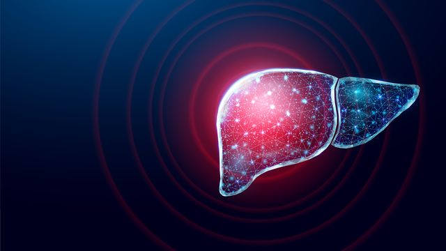
Liver Model Advances Drug Discovery for Fatty Liver Disease
More than 100 million people in the United States suffer from metabolic dysfunction-associated steatotic liver disease (MASLD), characterized by a buildup of fat in the liver. This condition can lead to the development of more severe liver…
Continue Reading
-

New AI tool predicts repeat heart attack risk for cancer patients
image: ©peterschreiber.media | iStock A new AI-powered tool can predict the risk of another heart attack in patients with cancer, helping doctors tailor care and improve outcomes, according to HDR UK and The Lancet
The…
Continue Reading