Scientists have discovered a hidden communication system inside bacteria that allows them to share genetic information and develop resistance to multiple antibiotics.

Biologists have identified a previously unknown mode of…
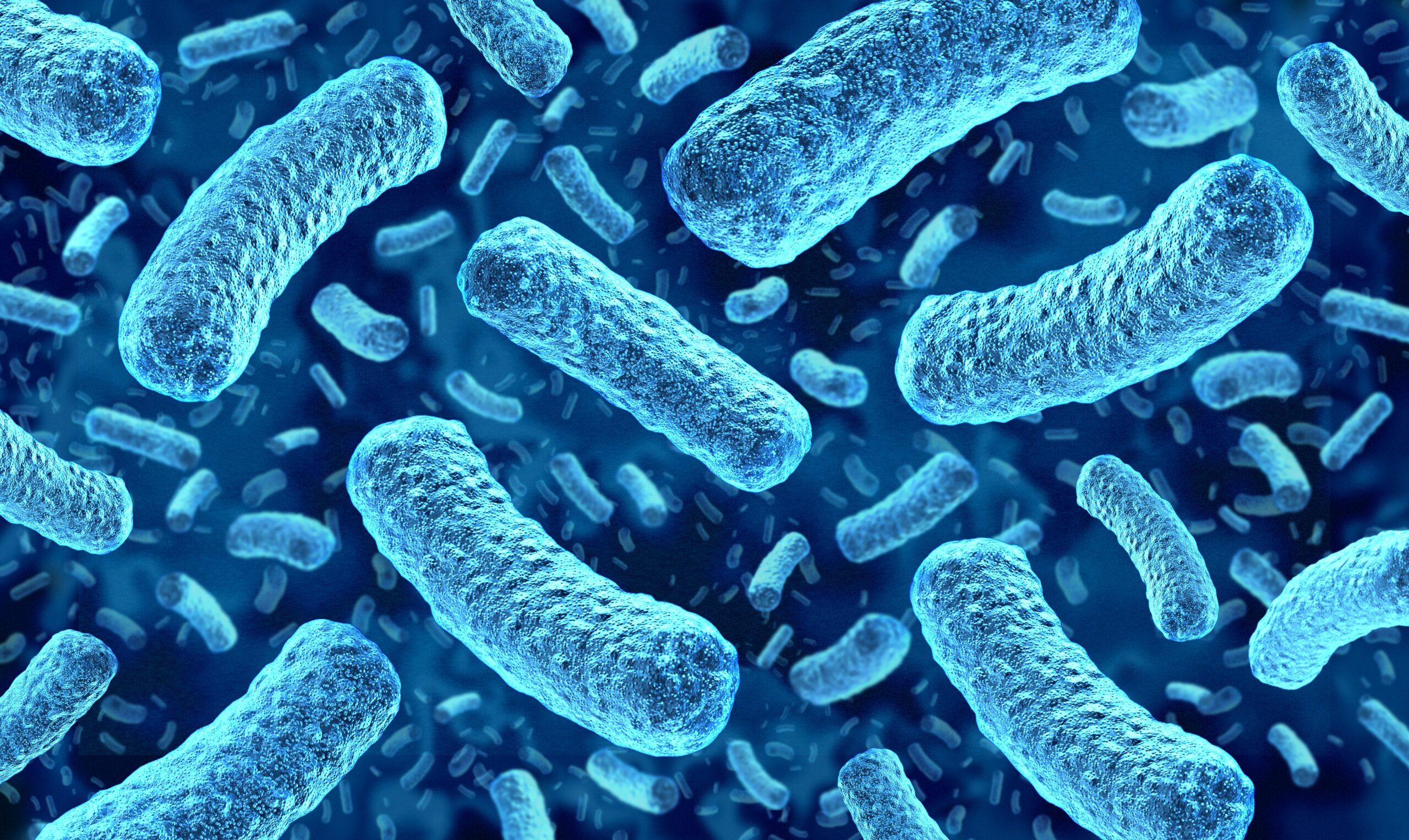
Scientists have discovered a hidden communication system inside bacteria that allows them to share genetic information and develop resistance to multiple antibiotics.
Biologists have identified a previously unknown mode of…

Emerging research in plant-based food and environmental health encourages people to consume less meat and fish. Researchers also highlight this diet’s benefits for lowering communicable disease risks.
Nutrition Insight examines findings…
Message from WHO Regional Director for Africa, Dr Mohamed Janabi
Cancer is no longer a silent crisis in Africa. It is a growing public health emergency that demands urgent, equitable and sustained action.
On World Cancer Day today, the World…
Lyon (France), Sao Paulo, (Brazil), February 3, 2026 – Valneva SE (Nasdaq: VALN; Euronext Paris: VLA), a specialty vaccine company and Instituto Butantan, one of the world’s largest biomedical research centers, today…

As we age, we begin to lose the connections that wire up our brains-and neuroscientists aren’t sure why.
It is increasingly clear, though, that the loss of synapses-the flexible and adaptive relay stations central to our brains’…
TOKYO, Feb. 2, 2026 /PRNewswire/ — The Global Health Innovative Technology (GHIT) Fund announced today a total investment of approximately JPY 1.39 billion (USD 8.8 million1) in…