Dementia is a progressive brain disorder caused due to physical damage to brain cells from diseases…
Category: 6. Health
-

Obesity’s Double Role in CKD as Comorbidity and Driver
As
obesity andchronic kidney disease (CKD) continue to rise as global clinical challenges, the intersection between excess weight and kidney function has become increasingly important to patient outcomes and disease…Continue Reading
-
What You Need To Know About Nipah Virus, Which Is Currently Surging In South Asia – Forbes
- What You Need To Know About Nipah Virus, Which Is Currently Surging In South Asia Forbes
- Why is India’s Nipah virus outbreak spooking the world? Al Jazeera
- Constant vigilance Dawn
- Nipah virus: Some Asia airports screen passengers after outbreak…
Continue Reading
-

Nipah virus outbreak in Malaysia traced from bats to pigs to humans
Summary
- According to international media reports, the virus was first identified in the Malaysian area of “Sungai Nipah” where workers associated with pig farms began showing cases of high fever and brain inflammation (encephalitis).
- Between…
Continue Reading
-
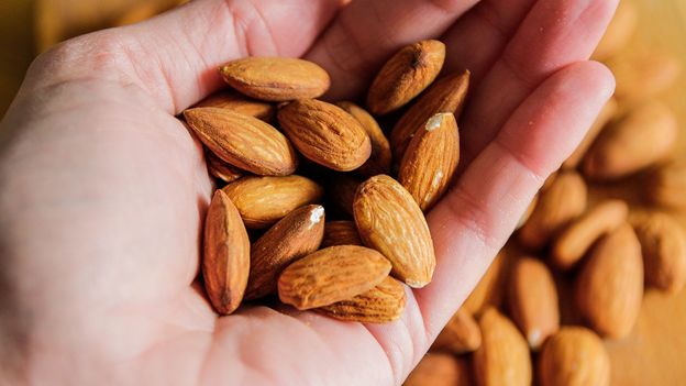
Seven foods you should be eating more of right now
More like this:
• The world’s most nutritious foods
• What should you eat at different life stages?
• The surprising foods that help you sleep better
Dandelion greens may not seem, at first glance, to be an obvious kitchen staple. But they are…
Continue Reading
-

Cambodia Heightens Medical Preparedness as Nipah Virus Raises Concerns in the Region
On Jan. 28, Minister of Health Chheang Ra led in Phnom Penh an inspection of the health screening and emergency response measures at Techo International Airport, accompanied by representatives from…
Continue Reading
-

New nasal spray vaccine efficiently prevents influenza
Researchers at WashU Medicine have developed a nasal vaccine against the highly pathogenic…
Continue Reading
-
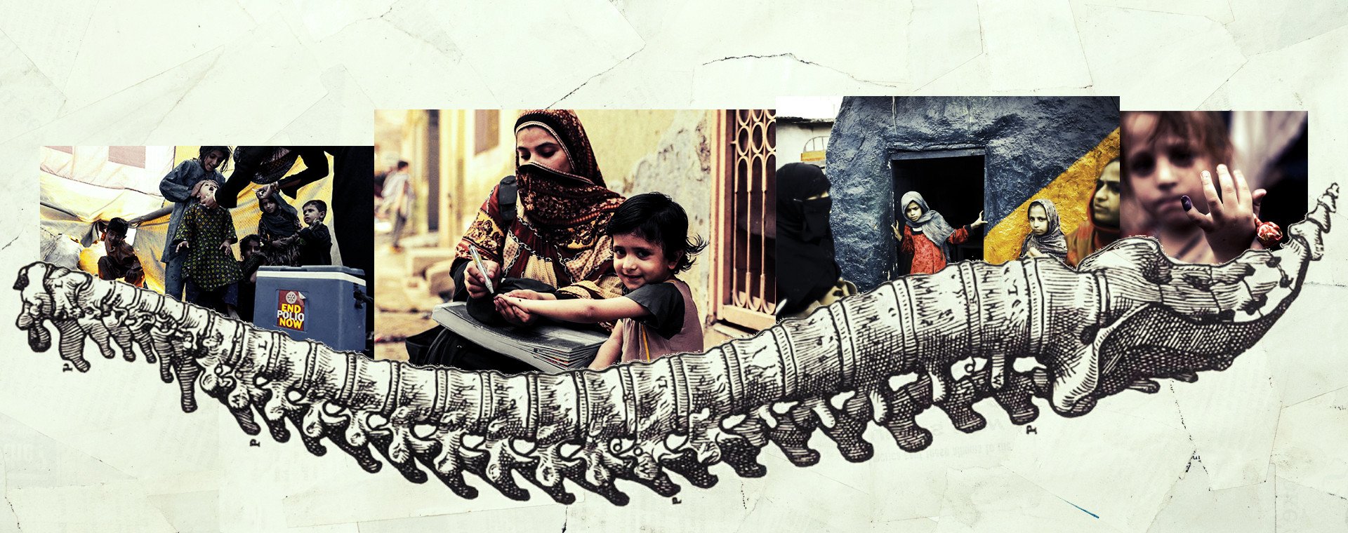
One KP father’s story challenges polio myths
PESHAWAR: Every evening, as the sun dips behind the snow-dusted Koh-e-Sufaid mountains, 45-year-old Sajjad Ahmad wheels his younger daughter to the edge of a dusty playground in village…
Continue Reading