- Pakistan is latest Asian country to step up checks for deadly Nipah virus Arab News
- Pakistan becomes latest Asian country to introduce checks for deadly Nipah virus Reuters
- Nipah virus: Some Asia airports screen passengers after outbreak in…
Category: 6. Health
-
Pakistan is latest Asian country to step up checks for deadly Nipah virus – Arab News
-
No immediate Nipah virus threat in Sri Lanka: health official-Xinhua
COLOMBO, Jan. 29 (Xinhua) — A senior Sri Lankan health official said Thursday that there is no immediate threat to the country from the Nipah virus, which has been reported in parts of neighboring India.
Deputy Health Minister Hansaka…
Continue Reading
-
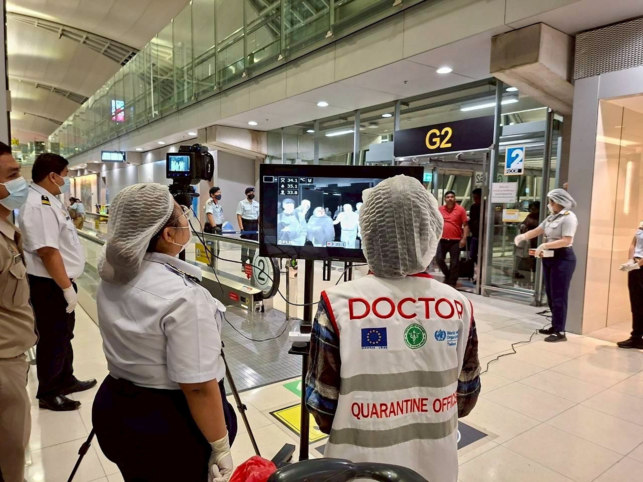
Pakistan becomes latest Asian country to introduce checks for deadly Nipah virus
LAHORE/HANOI, Jan 29 (Reuters) – Authorities in Pakistan have ordered enhanced screening of people entering the country for signs of infections of the deadly Nipah virus after India confirmed two cases, adding to the number of Asian countries…
Continue Reading
-
Page not found | Arab News PK
The requested page “/node/2630998/world” could not be found.
Continue Reading
-

Richer social connections linked to better brain health
A new study by an interdisciplinary team from McGill University and Université Laval sheds light on the relationship between social factors and cognitive health among aging adults.
While previous research had…
Continue Reading
-

Islam’s Teachings About Alcohol and Modern Science
© Shutterstock
Sinwan Basharat, Canada
It is not surprising that the new year coincides with the sharpest increase in sales of self-help books. People are eager to make improvements in life, and improving one’s health often tops the list.
The Pew…
Continue Reading
-
4 Parkinson’s disease symptoms that can show up decades before a diagnosis – The Washington Post
- 4 Parkinson’s disease symptoms that can show up decades before a diagnosis The Washington Post
- Many people think of a tremor as the quintessential warning sign of Parkinson’s disease. But other symptoms — many of them not involving changes…
Continue Reading
-
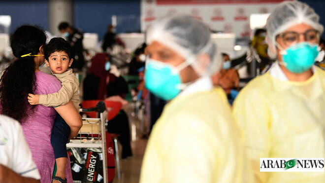
Pakistan orders screening of travelers at entry points amid Nipah virus threat
BARA, Pakistan: More than 70,000 people, mostly women and children, have fled a remote region in northwestern Pakistan bordering Afghanistan over uncertainty of a military operation against the Pakistani Taliban,…Continue Reading
-
Pakistan orders screening of travelers at entry points amid Nipah virus threat – Arab News
- Pakistan orders screening of travelers at entry points amid Nipah virus threat Arab News
- Nipah virus: Some Asia airports screen passengers after outbreak in India BBC
- Pakistan orders screening of travellers in view of Nipah virus threat Dawn
- What…
Continue Reading
-

Young Brits are no longer drinking – so what will a Saturday night look like for future generations? | Emma Brockes
It’s November 2024 and my puritanical American children are attending their first autumn fair at their new English primary school. There’s a laser show and hotdogs and a raffle. There’s also a bar for the parents, which makes my two pull up…
Continue Reading