Quantum Biopharma has completed dosing in two toxicology studies requested by the U.S. Food and Drug Administration (FDA) that aim to support the launch of clinical studies of Lucid-MS, an experimental treatment for multiple sclerosis (MS)…


Key takeaways:
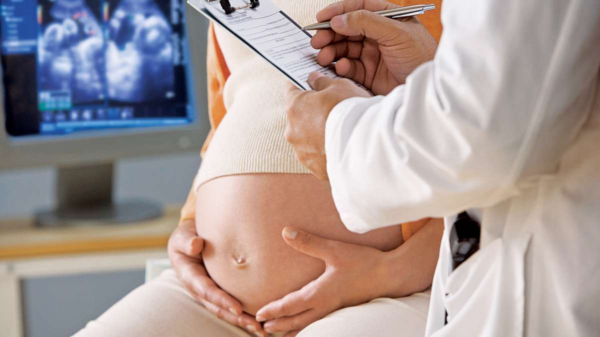
- COVID-19 vaccination during pregnancy was associated with significantly lower risks of hospitalization, ICU admission, and severe illness.
- Protective effects of vaccination were consistent across both the Delta and Omicron COVID-19…