
Category: 6. Health
-
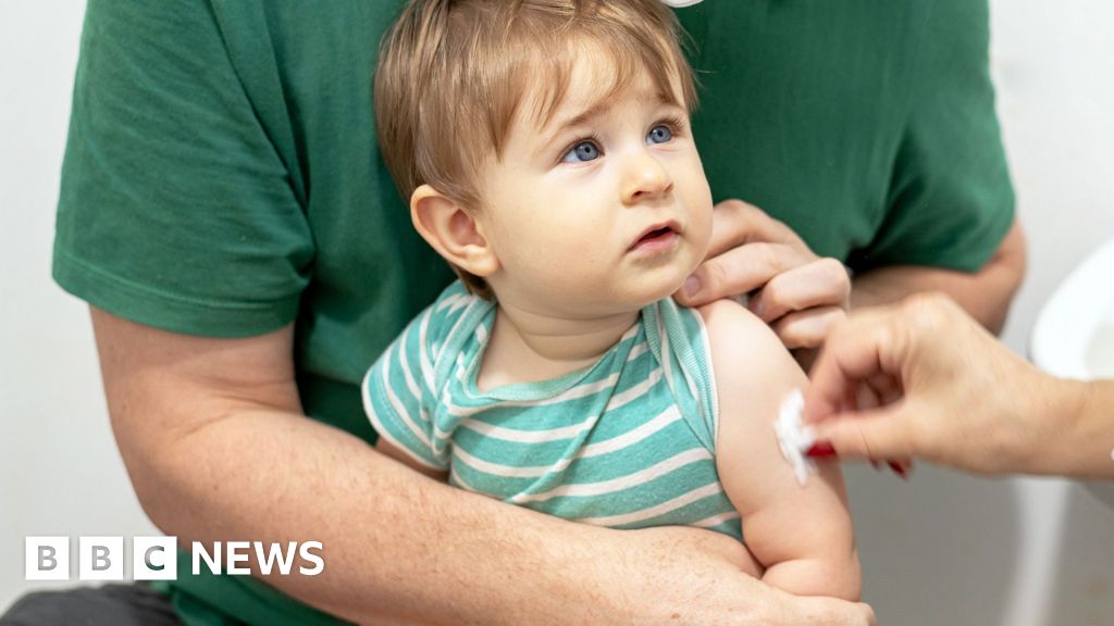
Chickenpox vaccines for children on NHS starts across UK
Philippa Roxby and Smitha MundasadHealth reporters
 Getty Images
All young children in the UK can now be offered protection against chickenpox for the first time on the NHS.
The vaccine will be combined with the existing MMR jab, given at 12 and 18…
Continue Reading
-
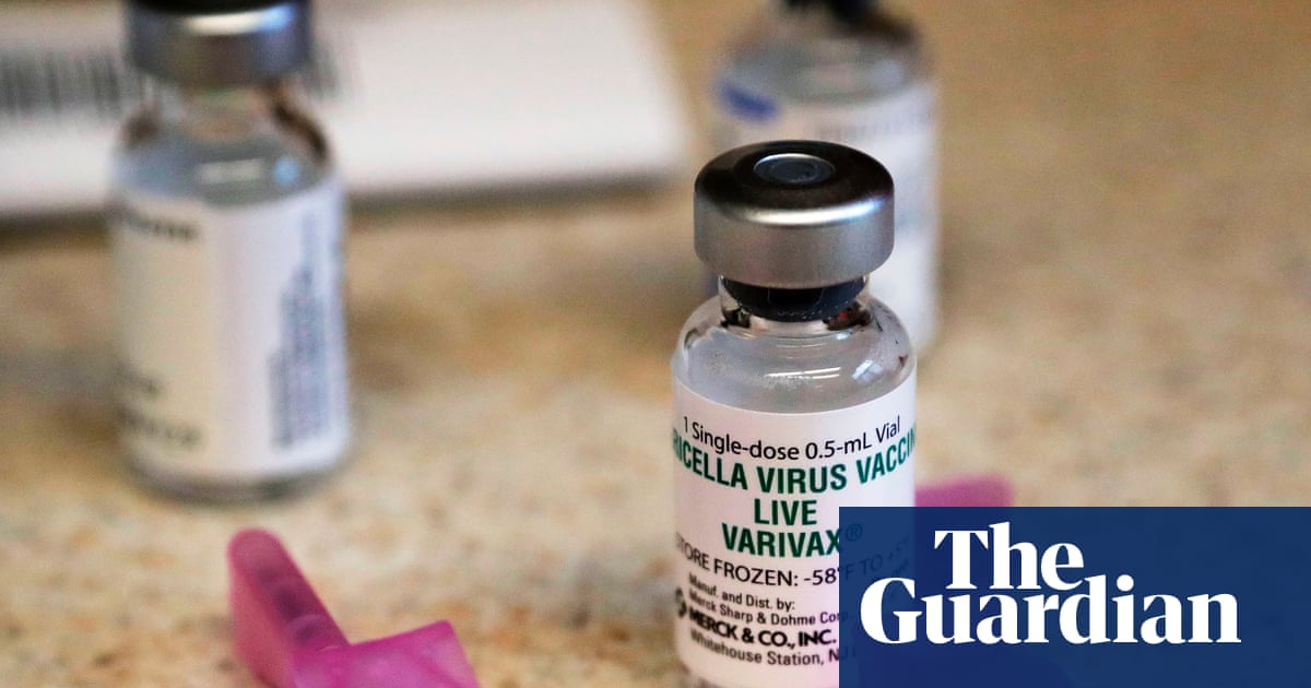
UK children to get chickenpox vaccine with measles, mumps and rubella jab | Vaccines and immunisation
Children in the UK are to be immunised against chickenpox at the same time as measles, mumps and rubella.
The NHS across the UK’s four home nations will administer a combined vaccine to young children to protect them against all four diseases…
Continue Reading
-

Depression at Midlife Can Raise Risk of Dementia Later; and More News
Long-Term Study Links Specific Midlife Symptoms of Depression to Later Dementia Risk
A 23-year study from researchers in the United Kingdom has found a connection between specific symptoms of depression in midlife and the…
Continue Reading
-
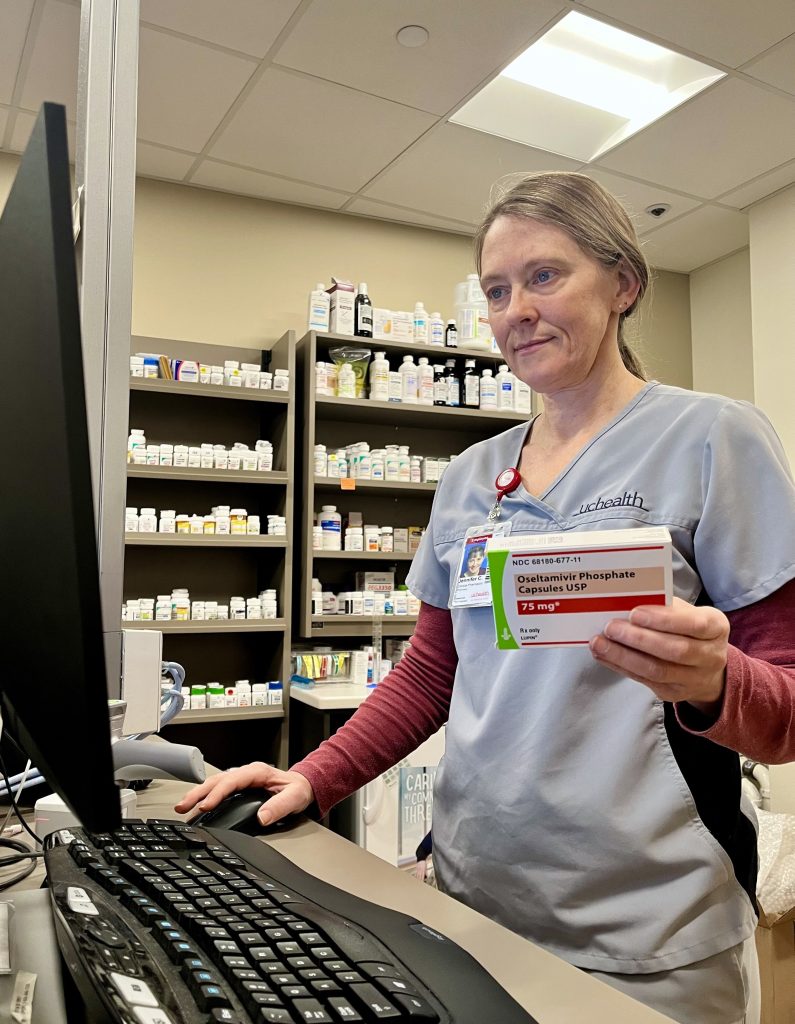
Influenza hitting Routt County earlier this season
Influenza is hitting Routt County earlier this year than last flu season, according to Routt County Public Health, and the number of positive flu cases is 71% higher now than the same time last year, according to UCHealth Yampa Valley Medical…
Continue Reading
-
Bringing closer and faster testing and treatment to First Nations communities
Date published:
Point of care testing takes place where the patient is seeking care. This could be in a clinic, a community health centre, or even in other…
Continue Reading
-

Maryland doctor predicts rough flu season will only get worse
Hospitals in the Greater Baltimore region have been dealing with the brunt of a tough flu season, especially with crowding in emergency rooms.
It follows a nationwide trend as flu numbers are up all across the country.
One of the leaders at…
Continue Reading
-

Carle Health adds visitor restrictions amid rise in respiratory illnesses – IPM Newsroom
- Carle Health adds visitor restrictions amid rise in respiratory illnesses IPM Newsroom
- As flu numbers increase, doctor reinforces healthy habits to stop the spread WAND-TV
- Hospitals across the region restrict visitors due to flu WHIO TV
- Temporary…
Continue Reading
-

At 51, I’m about to start work as a junior doctor. It’s been a wild ride to get here | Ben Collins
For the past four years I’ve been breaking every rule of appropriate, normal behaviour. I’ve poked, probed, inserted, stabbed and cut people in their most intimate body parts. I’ve examined dissected bodies and riffled through a bucket of…
Continue Reading
-

This tiny nerve may help keep the heart young
New research suggests that one of the most important keys to a healthier and more youthful heart may be the vagus nerve. A study coordinated by the Sant’Anna School of Advanced Studies in Pisa and published in Science Translational Medicine found…
Continue Reading