Category: 6. Health
-
A decades-old drug is helping people drink less alcohol—without giving it up completely
Kate Carbonari has been drinking since she was 15 years old. By her early 60s, she began to rethink her relationship with alcohol. “I’ve gone through a lot of different drinking cycles in my life, and I feel like now, finally, as a society,…
Continue Reading
-

Family support is key for people with mental illness but caregivers need help too : NPR
After caring for his brother, who has schizophrenia, for many years Mitul Desai started a company to support caregivers…
Continue Reading
-

Poor Sleep Quality Accelerates Brain Aging
While the link between poor sleep and dementia has long been known, it was unclear whether poor sleep habits could cause dementia or whether poor sleep was an early symptom of dementia. However, new research has revealed that sleep quality may…
Continue Reading
-

Duke Health to limit visitors due to rise in respiratory illness cases, joins other hospital systems :: WRAL.com
Duke Health will limit hospital visitors starting Jan. 6, 2026 out of concern due to the rise of respiratory illnesses.
The Duke University Health System stated in a press…
Continue Reading
-
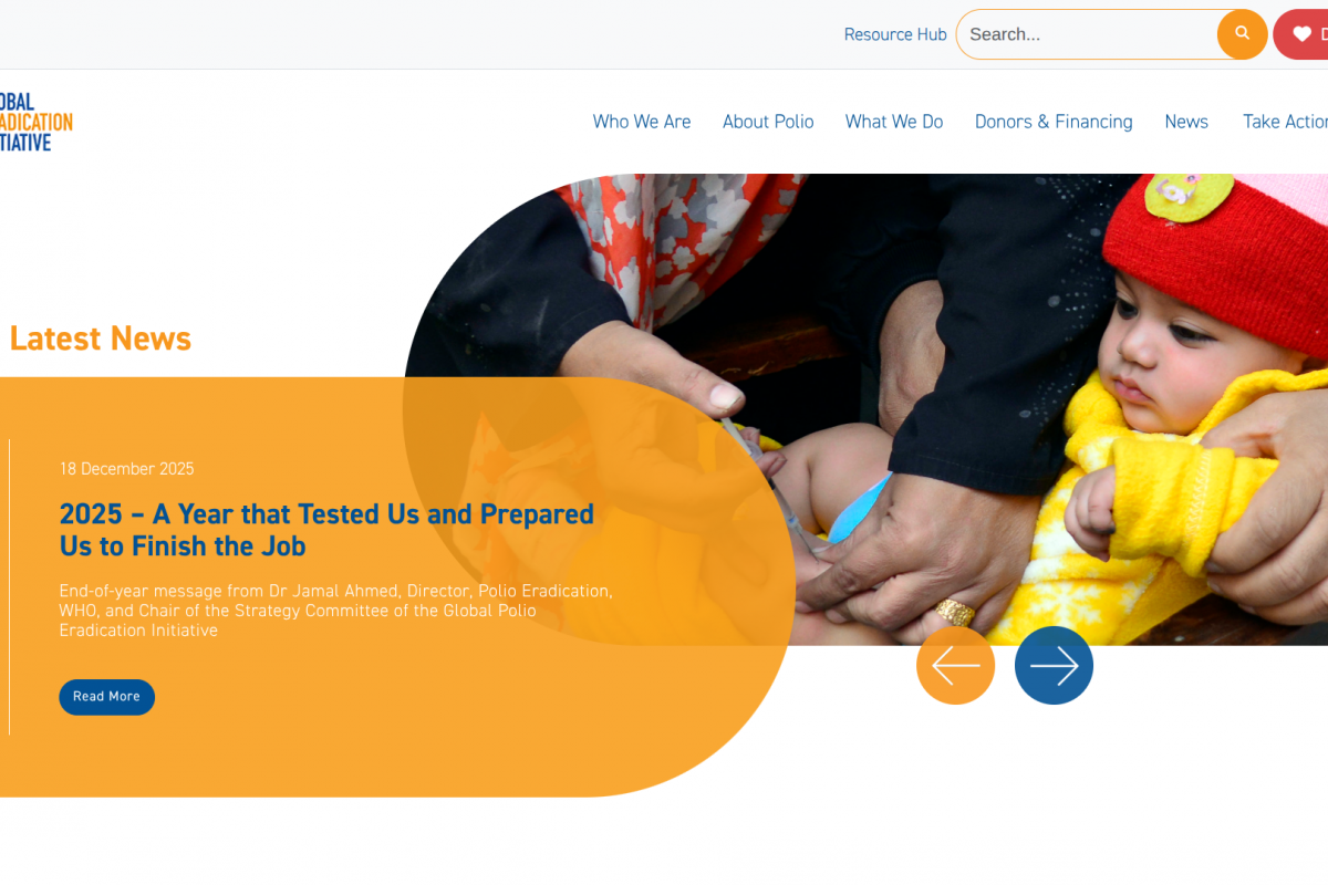
Two Billion nOPV2 Polio Vaccines Deployed — Vax-Before-Travel
(Vax-Before-Travel News)As 2025 draws to a close, I want to begin by first thanking the millions of health workers, vaccinators, surveillance officers, laboratory scientists, social mobilizers, and community volunteers who carried polio…
Continue Reading
-

New trial for Walsall heart failure patients
Heart failure patients are being sought by Walsall researchers to take part in a trial to test a new device aimed at preventing deterioration and helping them avoid hospital admission.
The ME-HF trial, working alongside Heartfelt…
Continue Reading
-
NeuroVoices: David A. Hafler, MD, FANA, on Understanding MS as an Autoimmune and Neurodegenerative Disease | NeurologyLive
Multiple sclerosis (MS) is a complex, genetically mediated autoimmune disease of the central nervous system in which anti–CD20–mediated B-cell depletion has demonstrated substantial efficacy, particularly in early disease. Although prior…
Continue Reading
-

The perfect evening routine: how to prepare for bed – from blue light to baths | Sleep
After a hard day at work, the last thing you want to do is fritter away your precious downtime slumped on the sofa in a dazed doomscroll. Yet, in the absence of a better plan, it happens with depressing ease. How we spend the hours between…
Continue Reading