Introduction
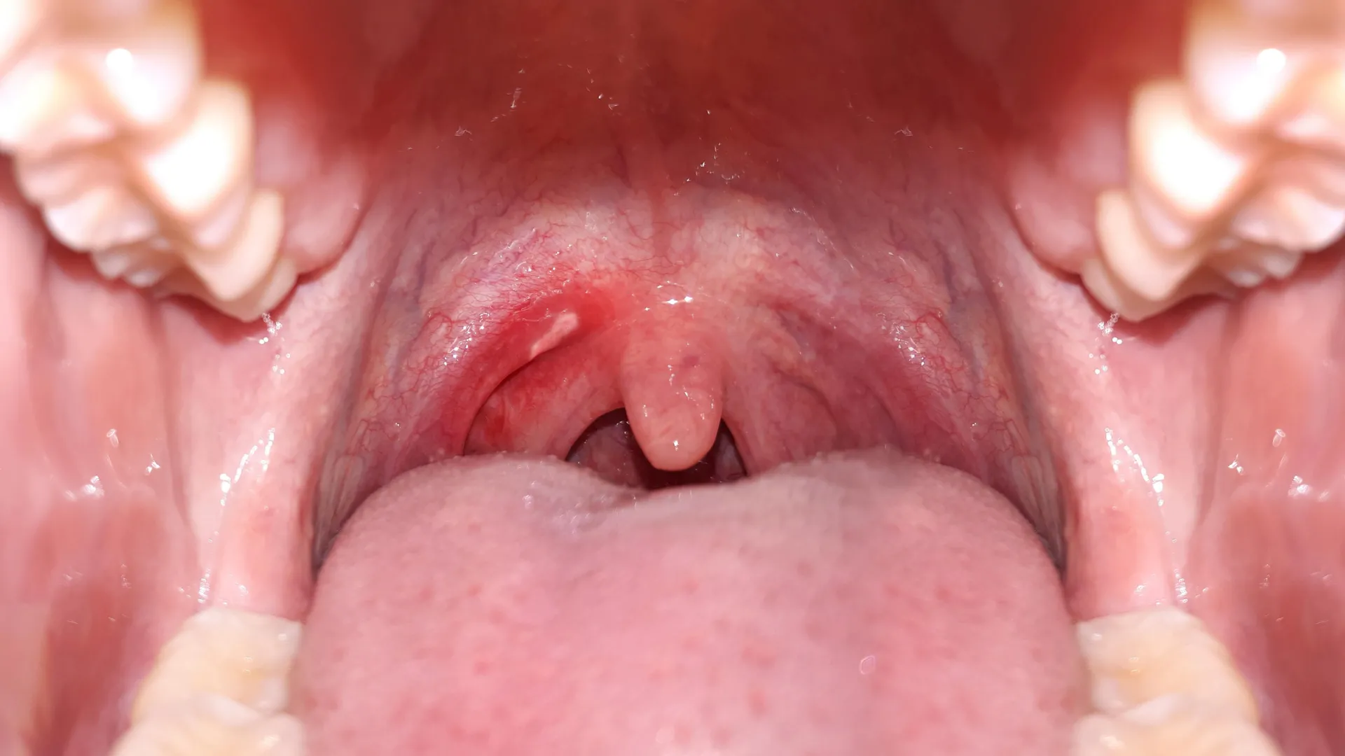
Ramsay Hunt Syndrome is a neurological condition caused by the reactivation of the varicella-zoster virus (VZV) in the facial nerve’s geniculate ganglion (cranial nerve VII).1 Vesicular eruptions in the mouth or ear, ear pain, and…
Ramsay Hunt Syndrome is a neurological condition caused by the reactivation of the varicella-zoster virus (VZV) in the facial nerve’s geniculate ganglion (cranial nerve VII).1 Vesicular eruptions in the mouth or ear, ear pain, and…
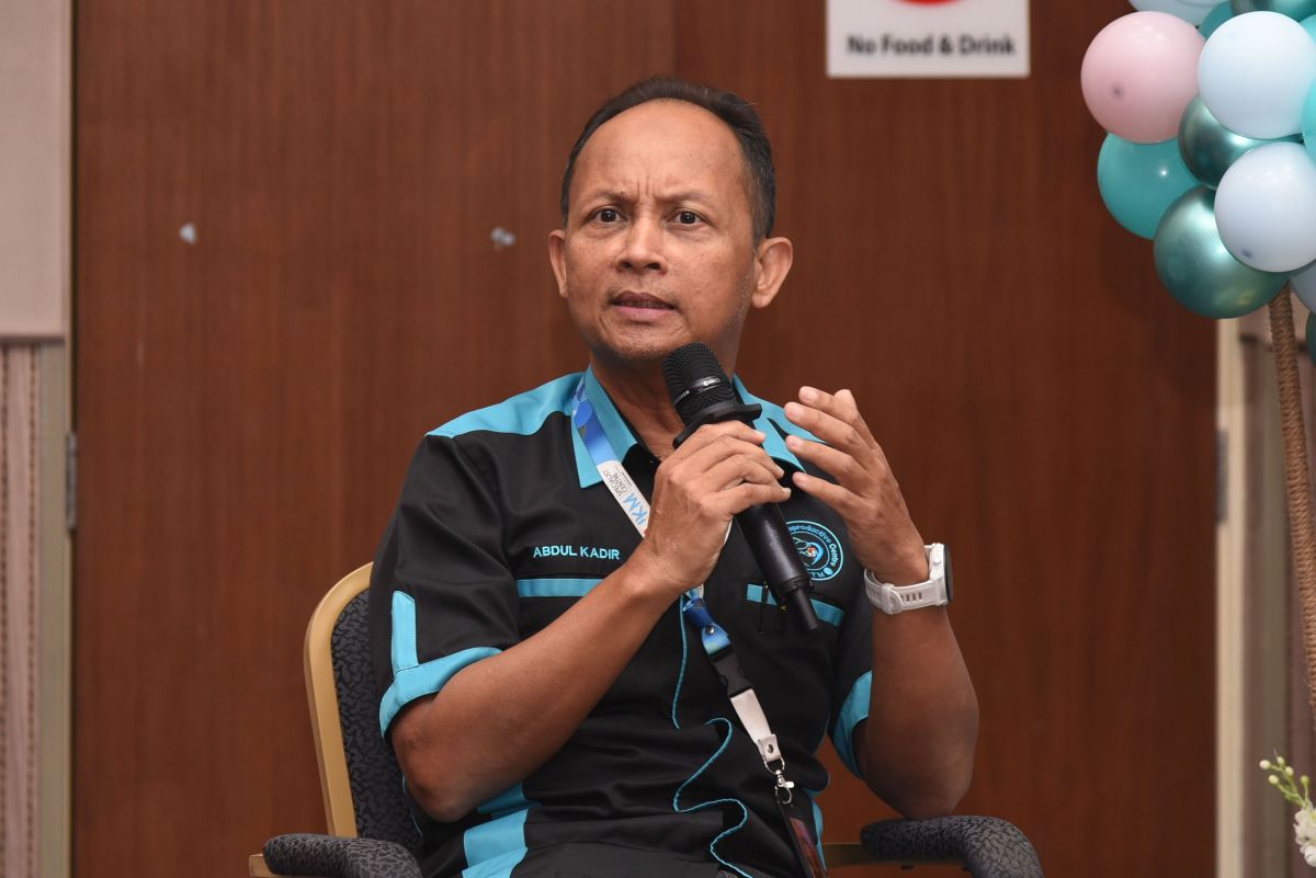
KUALA LUMPUR, Dec 31 — Male infertility is still treated as a taboo in many Malaysian households, but clinicians at Hospital Canselor Tuanku Muhriz (HCTM) UKM say more men have begun seeking assessment in…
A large comparative study published online in the open access journal BMJ Global Health has found that even low daily alcohol consumption is linked to a much higher risk of mouth cancer in India. Drinking just 9 g of alcohol per day, about the…

The fungal species Candida auris is spreading across the globe, and gaining in virulence, according to a new review by a Hackensack Meridian Center for Discovery and Innovation (CDI) scientist and colleagues.
But there are…

NYT The virus, sometimes called the stomach bug, is incredibly contagious. Here’s how to stay safe this season. Every year, there’s a surge in norovirus cases between November and April, and this holiday season is no…
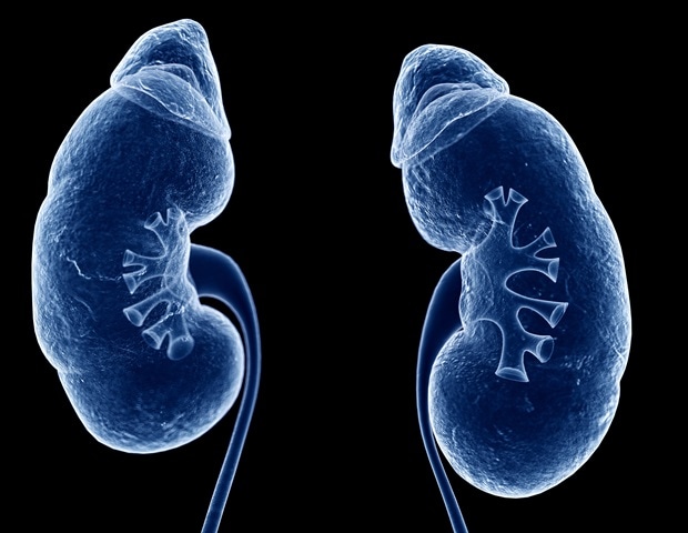
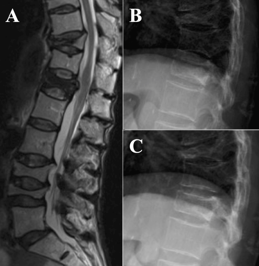
Diabetic kidney disease affects a growing proportion of people with diabetes and remains the leading cause of end-stage renal disease. Clinically, the condition is marked by persistent proteinuria and a gradual decline in kidney…

Appendix cancer is a rare type of cancer that begins when cells in the appendix grow out of control. The appendix is a small pouch attached to the first part of the large intestine. Because appendix…

Millions of people are turning to A&E departments in England for minor ailments including coughs, blocked noses and hiccups, according to data that health leaders say lays bare a failure to give patients prompt access to primary care.
Emergency…