A dual in vitro and in vivo study reveals promising effects of Ascophyllum nodosum and Fucus vesiculosus on oxidative stress modulation.
As livestock production systems evolve under the dual pressures of sustainability and performance,…
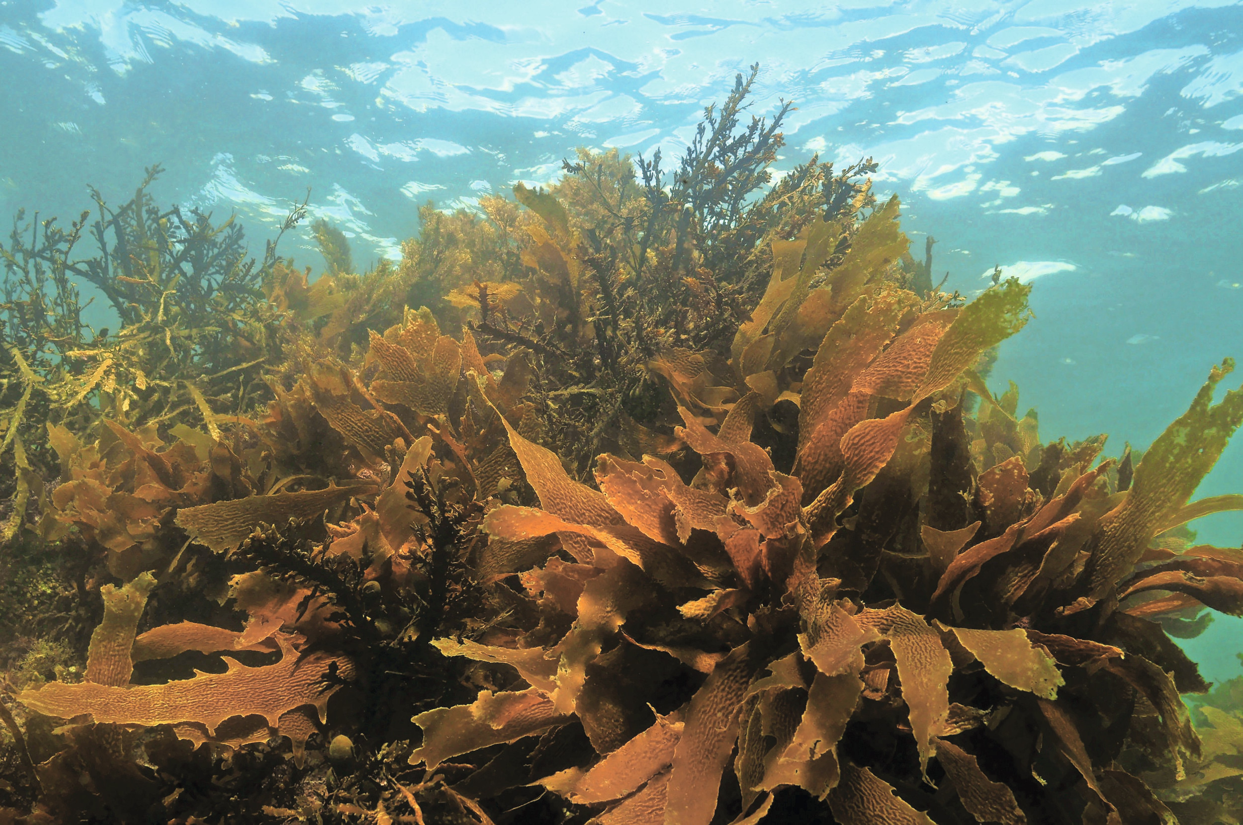
A dual in vitro and in vivo study reveals promising effects of Ascophyllum nodosum and Fucus vesiculosus on oxidative stress modulation.
As livestock production systems evolve under the dual pressures of sustainability and performance,…

OSTEOPATHIC manipulative treatment (OMT) may improve pulmonary function in asthma, though current evidence remains limited and methodologically inconsistent.
Asthma is a common obstructive airway disease…

Hong Kong women navigating menopause have often felt the need to “soldier on”, their hot flushes dismissed by others – and themselves – as stress, their mood swings blamed on “having teenagers” and their fatigue chalked up to ageing….

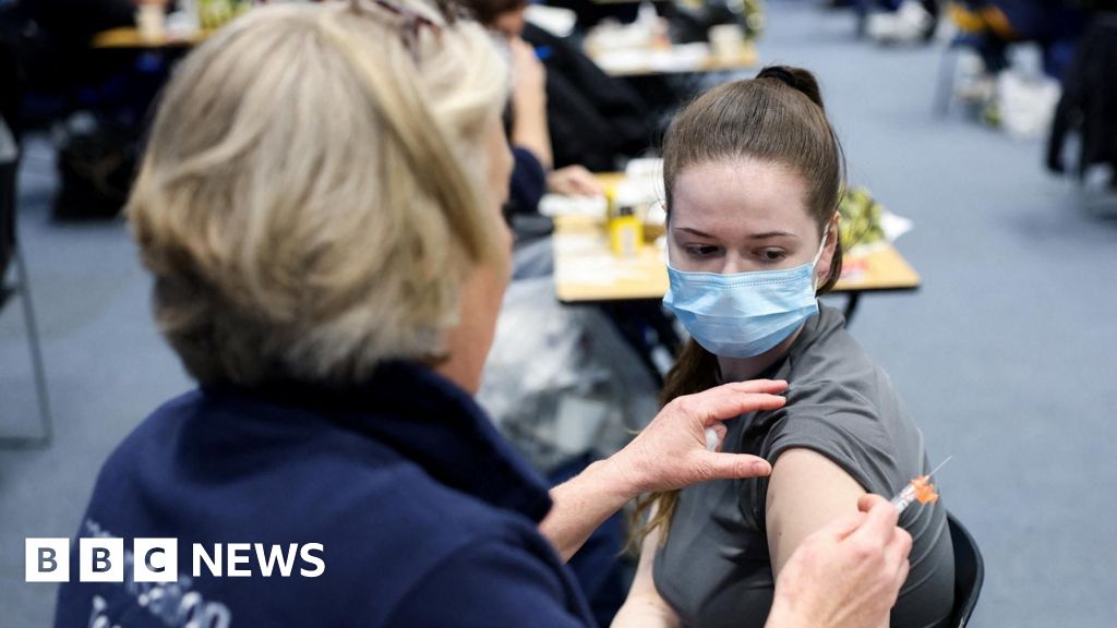
Tyra Skinner had already been violently sick three times when doctors at Kent’s William Harvey hospital realised something was badly wrong. The 20-year-old was rushed into critical care, racked with a pounding headache, a stiff neck and…

New research published in the Journal of the Endocrine Society suggests that exposure to per- and polyfluoroalkyl substances (PFAS) during early life could influence how children’s bones develop during adolescence.
PFAS are man-made chemicals…
We use the term ‘rabid’ as a byword for madness and extremely violent behaviour. The very word rabies – Latin for ‘rage’ – conjures horror images of wide-eyed insanity, victims foaming at the mouth and a prolonged excruciating death –…
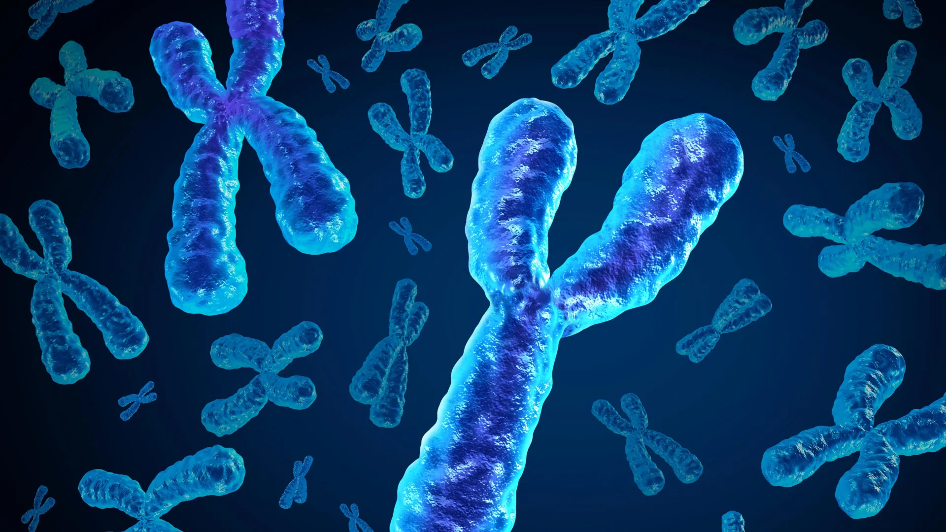
As men grow older, some of their cells gradually lose the Y chromosome. For a long time, scientists assumed this change would have little impact. The Y chromosome contains relatively few genes beyond those involved in male development, so its…
As men grow older, some of their cells gradually lose the Y chromosome. For a long time, scientists assumed this change would have little impact. The Y chromosome contains relatively few genes beyond those involved in male development, so its…