Newswise — AI model using deep transfer learning – the most advanced form of machine learning – predicted with 92% accuracy spoken language outcomes at one-to-three years after cochlear implants (implanted…
Category: 6. Health
-
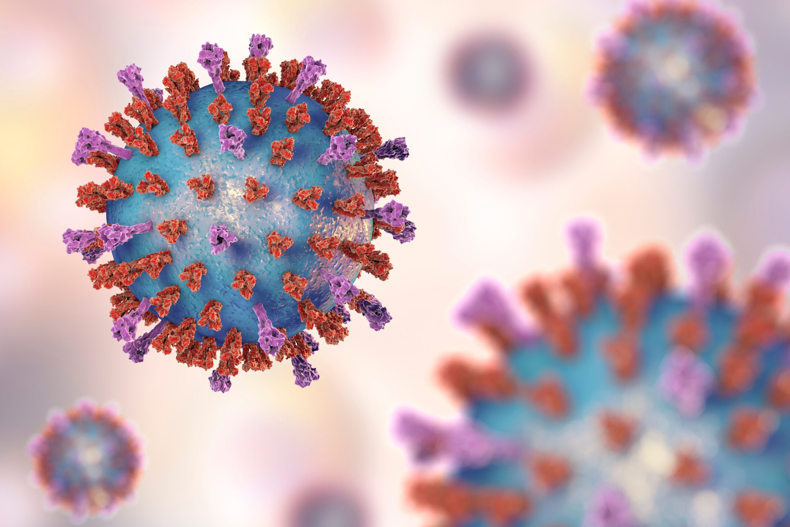
RSV Hospitalization Associated With Persistent Symptoms and Reduced Quality of Life in Adults
New study findings led by researchers from the University of Michigan School of Public Health found that respiratory syncytial virus (RSV) could have long-lasting effects among adults who have been hospitalized.1,2
The study authors, who…
Continue Reading
-

Mesenchymal stem cell therapy in osteoarthritis and rheumatoid arthrit
Introduction
Osteoarthritis (OA) and rheumatoid arthritis (RA), the most common types of arthritis, are chronic inflammatory, autoimmune, and age-related osteoarticular diseases. They are characterized by the degeneration of cartilage and the…
Continue Reading
-
Identification of risky prognostic factors for interventional treatmen
Introduction
Diabetes is a systemic metabolic disorder induced by defective insulin secretion or impaired biological function, and represents one of the most prevalent chronic diseases in China. According to estimates, the global prevalence of…
Continue Reading
-
Study reveals how melanoma “decoys” immune cells to escape attack-Xinhua
JERUSALEM, Dec. 29 (Xinhua) — Scientists have uncovered a stealth tactic used by melanoma, the most serious type of skin cancer, to disable the body’s immune defenses, according to a new international study published recently in the journal…
Continue Reading
-
Psychoeducational Needs of Sundanese Indonesian Family Caregivers Cari
Introduction
Schizophrenia is a chronic and severe mental disorder characterized by a constellation of symptoms, including positive symptoms (eg, hallucinations, delusions) and negative symptoms (eg, social withdrawal, affective flattening) that…
Continue Reading
-

Research Recap From the European College of Neuropsychopharmacology Congress
Phase 3 Data Support CT-155 as a Novel Digital Therapeutic for Negative Symptoms in Schizophrenia at ECNP
Data from the CONVOKE study showcased positive results with CT-155, a digital therapeutic for treatment of negative symptoms in…
Continue Reading
-

Top 5 Most-Read HIV Articles of 2025
Our top
HIV content focused primarily on domestic policies, as cuts to the President’s Emergency Plan for AIDS Relief (PEPFAR) had consequences in South Africa, and cuts to vaccine research for HIV came amid other challenges to treatment. The…Continue Reading