Joseph’s Coat, a plant long used in Brazilian folk medicine, reduced inflammation and joint damage in lab models of arthritis. (Getty Images)
A plant used in Brazilian medicine has scientific potential to help people with arthritis…
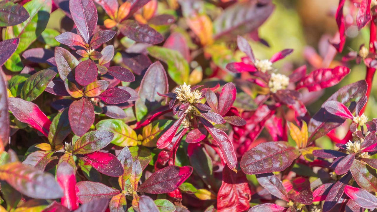
Joseph’s Coat, a plant long used in Brazilian folk medicine, reduced inflammation and joint damage in lab models of arthritis. (Getty Images)
A plant used in Brazilian medicine has scientific potential to help people with arthritis…