
Bear Grylls has once again claimed that meat-heavy diets are healthier and better for the planet than plant-based ones. However, a nutritionist has thoroughly debunked the comments and concluded that they are “not…
Category: 6. Health
-

AMPLITUDE Trial: Niraparib Plus Abiraterone in HRR-Mutated mCSPC
The AMPLITUDE trial represents a major step forward in the evolution of precision medicine for prostate cancer, specifically addressing whether PARP inhibition can improve outcomes when introduced early in the disease course….
Continue Reading
-

Australia issues advisory on counterfeit anti-rabies vaccine in India; pharma refutes charges
Australia advised its travellers who have been administered with Anti-Rabies vaccine – Abhayrab in India after November 1, 2023, to consider the vaccination invalid and initiate a new course of vaccination.
…Continue Reading
-

Is poor sleep responsible for your winter blues? Find out
Have you ever woken up feeling drowsy even though you’ve gotten enough hours of sleep?…
Continue Reading
-

Dads talk about ‘crippling’ condition
 How’s Dad?
Aled (right) says many men benefit from speaking to other dads going through the same experiences After suffering “stress and anxiety” while trying for a baby, Aled Edwards and his wife Sophie were delighted when they found out she was…
Continue Reading
-
Access Restricted
Access Restricted
Continue Reading
-
Prognostic relevance of geriatric nutritional risk index and the prognostic nutritional index in geriatric extensive stage small cell lung cancer
Cittolin-Santos, G. F. et al. The changing landscape of small cell lung cancer. Cancer 130 (14), 2453–2461. https://doi.org/10.1002/cncr.35281 (2024).
Felice, Y. et al. Enrollment of older…
Continue Reading
-

How eating takeout may heighten risk
Share on Pinterest Research suggests that higher takeout food consumption may increase a person’s risk of heart disease. Image credit: Luis Alvarez/Getty Images - There appears to be an association between diets high in takeout food and systemic…
Continue Reading
-

Symptom Experience and Management Needs of Breast Cancer Patients unde
Introduction
Breast cancer is the most common and fatal malignant tumor among women worldwide, surpassing lung cancer to become the top cancer globally.1,2 The treatment and care of breast cancer patients have brought serious economic and…
Continue Reading
-
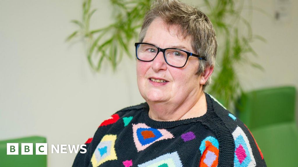
Cambridge breast cancer survivor’s plea after screening
 Cambridge University Hospitals
Shirley, 69, will undergo radiotherapy as part of her treatment A breast cancer patient who says a mammogram likely saved her life has urged people to attend routine screening appointments.
Shirley, 69, from Cambridge,…
Continue Reading